|
|
|
|
 Description
|
|
Compounds
|
||||||||||||||||||||
Chains, Units
Summary Information (see also Sequences/Alignments below) |
Ligands, Modified Residues, Ions (2, 6)| Asymmetric Unit (2, 6) Biological Unit 1 (2, 3) Biological Unit 2 (2, 3) |
Sites (6, 6)
Asymmetric Unit (6, 6)
|
SS Bonds (0, 0)| (no "SS Bond" information available for 256B) |
Cis Peptide Bonds (0, 0)| (no "Cis Peptide Bond" information available for 256B) |
SAPs(SNPs)/Variants (0, 0)| (no "SAP(SNP)/Variant" information available for 256B) |
PROSITE Motifs (0, 0)| (no "PROSITE Motif" information available for 256B) |
Exons (0, 0)| (no "Exon" information available for 256B) |
Sequences/Alignments
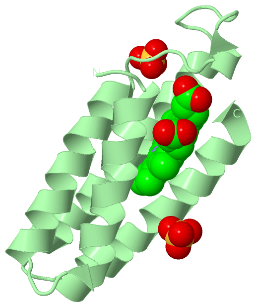
Asymmetric UnitChain A from PDB Type:PROTEIN Length:106 aligned with C562_ECOLX | P0ABE7 from UniProtKB/Swiss-Prot Length:128 Alignment length:106 32 42 52 62 72 82 92 102 112 122 C562_ECOLX 23 ADLEDNMETLNDNLKVIEKADNAAQVKDALTKMRAAALDAQKATPPKLEDKSPDSPEMKDFRHGFDILVGQIDDALKLANEGKVKEAQAAAEQLKTTRNAYHQKYR 128 SCOP domains d256ba_ A: Cytochrome b562 SCOP domains CATH domains 256bA00 A:1-106 [code=1.20.120.10, no name defined] CATH domains Pfam domains ---------------------------------------------------------------------------------------------------------- Pfam domains SAPs(SNPs) ---------------------------------------------------------------------------------------------------------- SAPs(SNPs) PROSITE ---------------------------------------------------------------------------------------------------------- PROSITE Transcript ---------------------------------------------------------------------------------------------------------- Transcript 256b A 1 ADLEDNMETLNDNLKVIEKADNAAQVKDALTKMRAAALDAQKATPPKLEDKSPDSPEMKDFRHGFDILVGQIDDALKLANEGKVKEAQAAAEQLKTTRNAYHQKYR 106 10 20 30 40 50 60 70 80 90 100 Chain B from PDB Type:PROTEIN Length:106 aligned with C562_ECOLX | P0ABE7 from UniProtKB/Swiss-Prot Length:128 Alignment length:106 32 42 52 62 72 82 92 102 112 122 C562_ECOLX 23 ADLEDNMETLNDNLKVIEKADNAAQVKDALTKMRAAALDAQKATPPKLEDKSPDSPEMKDFRHGFDILVGQIDDALKLANEGKVKEAQAAAEQLKTTRNAYHQKYR 128 SCOP domains d256bb_ B: Cytochrome b562 SCOP domains CATH domains 256bB00 B:1-106 [code=1.20.120.10, no name defined] CATH domains Pfam domains ---------------------------------------------------------------------------------------------------------- Pfam domains SAPs(SNPs) ---------------------------------------------------------------------------------------------------------- SAPs(SNPs) PROSITE ---------------------------------------------------------------------------------------------------------- PROSITE Transcript ---------------------------------------------------------------------------------------------------------- Transcript 256b B 1 ADLEDNMETLNDNLKVIEKADNAAQVKDALTKMRAAALDAQKATPPKLEDKSPDSPEMKDFRHGFDILVGQIDDALKLANEGKVKEAQAAAEQLKTTRNAYHQKYR 106 10 20 30 40 50 60 70 80 90 100
|
||||||||||||||||||||
SCOP Domains (1, 2)
Asymmetric Unit
 |
CATH Domains (1, 2)
Asymmetric Unit
|
Pfam Domains (0, 0)| (no "Pfam Domain" information available for 256B) |
Gene Ontology (7, 7)|
Asymmetric Unit(hide GO term definitions) Chain A,B (C562_ECOLX | P0ABE7)
|
||||||||||||||||||||||||||||||||||||||||||||||||||||||||||||
Interactive Views
|
|||||||||||||||||||||||||||||||||||||||||||||||||||||||||||||||||||||||||||||||||||||||||||||||||||||||||||||||||||||||||||||||||||||||||||||||||||||||||||||||||||||||||||||||||||||||
Still Images
|
||||||||||||||||
Databases
|
||||||||||||||||||||||||||||||||||||||||||||||||||||||||||||||||||||||||||||||||||||||||||||||||||||||||||||||||||||||||||||||||||||||||||||||||||||||||||||||||
Analysis Tools
|
|||||||||||||||||||||||||||||||||||||||||||||||||||||||||||||
Entries Sharing at Least One Protein Chain (UniProt ID)
Related Entries Specified in the PDB File
|
|