Show PDB file:   
         Plain Text   HTML   (compressed file size)
QuickSearch:   
by PDB,NDB,UniProt,PROSITE Code or Search Term(s)  
(-)Asym./Biol. Unit - manually
(-)Asym./Biol. Unit
(-)Asym./Biol. Unit - sites
collapse expand < >
Image Asym./Biol. Unit - manually
Asym./Biol. Unit - manually  (Jmol Viewer)
Image Asym./Biol. Unit
Asym./Biol. Unit  (Jmol Viewer)
Image Asym./Biol. Unit - sites
Asym./Biol. Unit - sites  (Jmol Viewer)

(-) Description

Title :  STRUCTURE OF PECTENOTOXIN-2 AND LATRUNCULIN B BOUND TO ACTIN
 
Authors :  J. S. Allingham, C. O. Miles, I. Rayment
Date :  22 May 07  (Deposition) - 17 Jul 07  (Release) - 24 Feb 09  (Revision)
Method :  X-RAY DIFFRACTION
Resolution :  1.45
Chains :  Asym./Biol. Unit :  A
Keywords :  Pectenotoxin, Latrunculin B, Actin, Anti-Tumor, Filament Capping, Natural Product, Cytotoxin, Macrolide, Structural Protein (Keyword Search: [Gene Ontology, PubMed, Web (Google))
 
Reference :  J. S. Allingham, C. O. Miles, I. Rayment
A Structural Basis For Regulation Of Actin Polymerization By Pectenotoxins.
J. Mol. Biol. V. 371 959 2007
PubMed-ID: 17599353  |  Reference-DOI: 10.1016/J.JMB.2007.05.056
(for further references see the PDB file header)

(-) Compounds

Molecule 1 - ACTIN
    ChainsA
    Organism CommonRABBIT
    Organism ScientificORYCTOLAGUS CUNICULUS
    Organism Taxid9986
    Other DetailsSOURCE OF ACTIN PROTEIN
    SynonymALPHA-ACTIN-1

 Structural Features

(-) Chains, Units

  1
Asymmetric/Biological Unit A

Summary Information (see also Sequences/Alignments below)
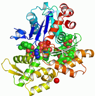
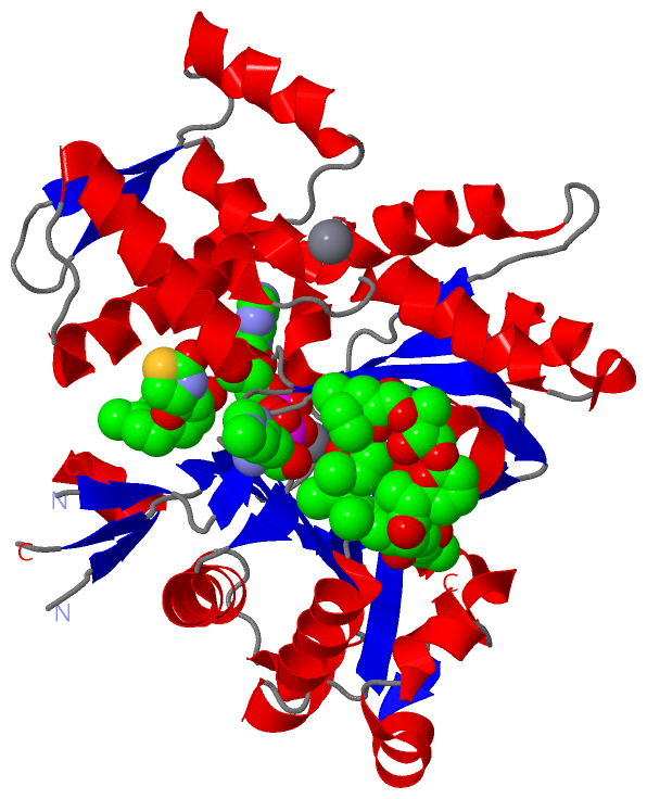
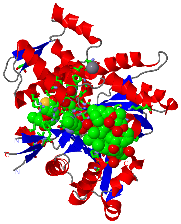
(-) Ligands, Modified Residues, Ions  (5, 6)

Asymmetric/Biological Unit (5, 6)
No.NameCountTypeFull Name
1ATP1Ligand/IonADENOSINE-5'-TRIPHOSPHATE
2CA2Ligand/IonCALCIUM ION
3HIC1Mod. Amino Acid4-METHYL-HISTIDINE
4LAB1Ligand/IonLATRUNCULIN B
5PXT1Ligand/IonPECTENOTOXIN-2

(-) Sites  (5, 5)

Asymmetric Unit (5, 5)
No.NameEvidenceResiduesDescription
1AC1SOFTWAREATP A:425 , HOH A:509 , HOH A:510 , HOH A:529 , HOH A:541BINDING SITE FOR RESIDUE CA A 400
2AC2SOFTWAREGLU A:241 , GLN A:263 , SER A:265 , HOH A:515 , HOH A:540 , HOH A:545 , HOH A:730BINDING SITE FOR RESIDUE CA A 501
3AC3SOFTWAREGLY A:13 , SER A:14 , GLY A:15 , LEU A:16 , LYS A:18 , GLY A:156 , ASP A:157 , GLY A:158 , VAL A:159 , GLY A:182 , ARG A:210 , LYS A:213 , GLU A:214 , GLY A:301 , GLY A:302 , THR A:303 , MET A:305 , TYR A:306 , LYS A:336 , CA A:400 , HOH A:506 , HOH A:510 , HOH A:529 , HOH A:548 , HOH A:568 , HOH A:597 , HOH A:616 , HOH A:729 , HOH A:754BINDING SITE FOR RESIDUE ATP A 425
4AC4SOFTWAREGLY A:15 , ILE A:34 , GLN A:59 , TYR A:69 , ASP A:157 , ARG A:183 , THR A:186 , ARG A:206 , GLU A:207 , ARG A:210 , HOH A:622BINDING SITE FOR RESIDUE LAB A 401
5AC5SOFTWAREASP A:56 , GLU A:57 , GLU A:107 , PRO A:109 , LEU A:110 , ASN A:111 , PRO A:112 , ARG A:116 , PRO A:172 , ILE A:175 , MET A:176 , ARG A:177 , LYS A:284 , HOH A:625 , HOH A:646 , HOH A:665 , HOH A:712 , HOH A:733 , HOH A:774 , HOH A:823BINDING SITE FOR RESIDUE PXT A 500

(-) SS Bonds  (0, 0)

(no "SS Bond" information available for 2Q0U)

(-) Cis Peptide Bonds  (0, 0)

(no "Cis Peptide Bond" information available for 2Q0U)

 Sequence-Structure Mapping

(-) SAPs(SNPs)/Variants  (0, 0)

(no "SAP(SNP)/Variant" information available for 2Q0U)

(-) PROSITE Motifs  (3, 3)

Asymmetric/Biological Unit (3, 3)
 PROSITEUniProtKBPDB
No.IDACDescriptionIDLocationCountLocation
1ACTINS_1PS00406 Actins signature 1.ACTS_RABIT55-65  1A:53-61
2ACTINS_ACT_LIKEPS01132 Actins and actin-related proteins signature.ACTS_RABIT106-118  1A:104-116
3ACTINS_2PS00432 Actins signature 2.ACTS_RABIT358-366  1A:356-364

(-) Exons   (0, 0)

(no "Exon" information available for 2Q0U)

(-) Sequences/Alignments

Asymmetric/Biological Unit
   Reformat: Number of residues per line =  ('0' or empty: single-line sequence representation)
  Number of residues per labelling interval =   
  UniProt sequence: complete  aligned part    
   Show mapping: SCOP domains CATH domains Pfam domains Secondary structure (by author)
SAPs(SNPs) PROSITE motifs Exons
(details for a mapped element are shown in a popup box when the mouse pointer rests over it)
Chain A from PDB  Type:PROTEIN  Length:352
 aligned with ACTS_RABIT | P68135 from UniProtKB/Swiss-Prot  Length:377

    Alignment length:368
                                    18        28        38        48        58        68        78        88        98       108       118       128       138       148       158       168       178       188       198       208       218       228       238       248       258       268       278       288       298       308       318       328       338       348       358       368        
           ACTS_RABIT     9 ALVCDNGSGLVKAGFAGDDAPRAVFPSIVGRPRHQGVMVGMGQKDSYVGDEAQSKRGILTLKYPIEHGIITNWDDMEKIWHHTFYNELRVAPEEHPTLLTEAPLNPKANREKMTQIMFETFNVPAMYVAIQAVLSLYASGRTTGIVLDSGDGVTHNVPIYEGYALPHAIMRLDLAGRDLTDYLMKILTERGYSFVTTAEREIVRDIKEKLCYVALDFENEMATAASSSSLEKSYELPDGQVITIGNERFRCPETLFQPSFIGMESAGIHETTYNSIMKCDIDIRKDLYANNVMSGGTTMYPGIADRMQKEITALAPSTMKIKIIAPPERKYSVWIGGSILASLSTFQQMWITKQEYDEAGPSIVHRKC 376
               SCOP domains d2q0ua1 A:7-146 Actin                                                                                                                       d2q0ua2 A:147-374 Actin                                                                                                                                                                                                              SCOP domains
               CATH domains 2q0uA01 A:7-137,A:339-374  [code            =3.30.420.4    0, no name defined]                                                     2q0uA02 A:138-179,A:272-338               2q0uA03 A:180-271 Actin; Chain A, domain 4                                                  2q0uA02 A:138-179,A:272-338  [code=3.30.420.40, no name defined]   2q0uA01 A:7-137,A:339-374            CATH domains
               Pfam domains Actin-2q0uA01 A:7-374                                                                                                                                                                                                                                                                                                                                                            Pfam domains
         Sec.struct. author .eeeee...eeeeee.......eeee..ee..------------..eehhhhhh.----.ee..ee..ee.hhhhhhhhhhhhhhh....hhhhh.eeeee....hhhhhhhhhhhhhhh....eeeeeehhhhhhhhh....eeeeee....eeeeeee..ee.hhhheee..hhhhhhhhhhhhhhhhh....hhhhhhhhhhhhhhhh....hhhhhhhhhhhh....eeee.....eeee.hhhhhhhhhhhhhhhhh....hhhhhhhhhhhh.hhhhhhhhhh.eeeehhhhh..hhhhhhhhhhhhhh.......ee......hhhhhhhhhhhhhhhhh...eehhhhhhhh.hhhhhhh Sec.struct. author
                 SAPs(SNPs) -------------------------------------------------------------------------------------------------------------------------------------------------------------------------------------------------------------------------------------------------------------------------------------------------------------------------------------------------------------------------------- SAPs(SNPs)
                    PROSITE ----------------------------------------------ACTINS_1   ----------------------------------------ACTINS_ACT_LI-----------------------------------------------------------------------------------------------------------------------------------------------------------------------------------------------------------------------------------------------ACTINS_2 ---------- PROSITE
                 Transcript -------------------------------------------------------------------------------------------------------------------------------------------------------------------------------------------------------------------------------------------------------------------------------------------------------------------------------------------------------------------------------- Transcript
                 2q0u A   7 ALVCDNGSGLVKAGFAGDDAPRAVFPSIVGRP------------DSYVGDEAQSK----TLKYPIEhGIITNWDDMEKIWHHTFYNELRVAPEEHPTLLTEAPLNPKANREKMTQIMFETFNVPAMYVAIQAVLSLYASGRTTGIVLDSGDGVTHNVPIYEGYALPHAIMRLDLAGRDLTDYLMKILTERGYSFVTTAEREIVRDIKEKLCYVALDFENEMATAASSSSLEKSYELPDGQVITIGNERFRCPETLFQPSFIGMESAGIHETTYNSIMKCDIDIRKDLYANNVMSGGTTMYPGIADRMQKEITALAPSTMKIKIIAPPERKYSVWIGGSILASLSTFQQMWITKQEYDEAGPSIVHRKC 374
                                    16        26        36 |       -    |   56    |   66      | 76        86        96       106       116       126       136       146       156       166       176       186       196       206       216       226       236       246       256       266       276       286       296       306       316       326       336       346       356       366        
                                                          38           51        61   66     73-HIC                                                                                                                                                                                                                                                                                                         

   Legend:   → Mismatch (orange background)
  - → Gap (green background, '-', border residues have a numbering label)
    → Modified Residue (blue background, lower-case, 'x' indicates undefined single-letter code, labelled with number + name)
  x → Chemical Group (purple background, 'x', labelled with number + name, e.g. ACE or NH2)
  extra numbering lines below/above indicate numbering irregularities and modified residue names etc., number ends below/above '|'

 Classification and Annotation

(-) SCOP Domains  (1, 2)

Asymmetric/Biological Unit

(-) CATH Domains  (2, 3)

Asymmetric/Biological Unit
(-)
Class: Alpha Beta (26913)

(-) Pfam Domains  (1, 1)

Asymmetric/Biological Unit

(-) Gene Ontology  (17, 17)

Asymmetric/Biological Unit(hide GO term definitions)
Chain A   (ACTS_RABIT | P68135)
molecular function
    GO:0005524    ATP binding    Interacting selectively and non-covalently with ATP, adenosine 5'-triphosphate, a universally important coenzyme and enzyme regulator.
    GO:0048306    calcium-dependent protein binding    Interacting selectively and non-covalently with any protein or protein complex (a complex of two or more proteins that may include other nonprotein molecules), in the presence of calcium.
    GO:0042802    identical protein binding    Interacting selectively and non-covalently with an identical protein or proteins.
    GO:0000166    nucleotide binding    Interacting selectively and non-covalently with a nucleotide, any compound consisting of a nucleoside that is esterified with (ortho)phosphate or an oligophosphate at any hydroxyl group on the ribose or deoxyribose.
    GO:0005515    protein binding    Interacting selectively and non-covalently with any protein or protein complex (a complex of two or more proteins that may include other nonprotein molecules).
biological process
    GO:0090131    mesenchyme migration    The process in which the population of cells that make up a mesenchyme undergo directed movement.
    GO:0010628    positive regulation of gene expression    Any process that increases the frequency, rate or extent of gene expression. Gene expression is the process in which a gene's coding sequence is converted into a mature gene product or products (proteins or RNA). This includes the production of an RNA transcript as well as any processing to produce a mature RNA product or an mRNA or circRNA (for protein-coding genes) and the translation of that mRNA or circRNA into protein. Protein maturation is included when required to form an active form of a product from an inactive precursor form.
    GO:0048741    skeletal muscle fiber development    The process whose specific outcome is the progression of the skeletal muscle fiber over time, from its formation to the mature structure. Muscle fibers are formed by the maturation of myotubes. They can be classed as slow, intermediate/fast or fast.
    GO:0030240    skeletal muscle thin filament assembly    The aggregation, arrangement and bonding together of proteins to form the actin-based thin filaments of myofibrils in skeletal muscle.
cellular component
    GO:0005884    actin filament    A filamentous structure formed of a two-stranded helical polymer of the protein actin and associated proteins. Actin filaments are a major component of the contractile apparatus of skeletal muscle and the microfilaments of the cytoskeleton of eukaryotic cells. The filaments, comprising polymerized globular actin molecules, appear as flexible structures with a diameter of 5-9 nm. They are organized into a variety of linear bundles, two-dimensional networks, and three dimensional gels. In the cytoskeleton they are most highly concentrated in the cortex of the cell just beneath the plasma membrane.
    GO:0044297    cell body    The portion of a cell bearing surface projections such as axons, dendrites, cilia, or flagella that includes the nucleus, but excludes all cell projections.
    GO:0005737    cytoplasm    All of the contents of a cell excluding the plasma membrane and nucleus, but including other subcellular structures.
    GO:0005856    cytoskeleton    Any of the various filamentous elements that form the internal framework of cells, and typically remain after treatment of the cells with mild detergent to remove membrane constituents and soluble components of the cytoplasm. The term embraces intermediate filaments, microfilaments, microtubules, the microtrabecular lattice, and other structures characterized by a polymeric filamentous nature and long-range order within the cell. The various elements of the cytoskeleton not only serve in the maintenance of cellular shape but also have roles in other cellular functions, including cellular movement, cell division, endocytosis, and movement of organelles.
    GO:0030175    filopodium    Thin, stiff, actin-based protrusion extended by the leading edge of a motile cell such as a crawling fibroblast or amoeba, or an axonal or dendritic growth cone, or a dendritic shaft.
    GO:0030027    lamellipodium    A thin sheetlike process extended by the leading edge of a migrating cell or extending cell process; contains a dense meshwork of actin filaments.
    GO:0001725    stress fiber    A contractile actin filament bundle that consists of short actin filaments with alternating polarity, cross-linked by alpha-actinin and possibly other actin bundling proteins, and with myosin present in a periodic distribution along the fiber.
    GO:0005865    striated muscle thin filament    Filaments formed of actin and associated proteins; attached to Z discs at either end of sarcomeres in myofibrils.

 Visualization

(-) Interactive Views

Asymmetric/Biological Unit
  Complete Structure
    Jena3D(integrated viewing of ligand, site, SAP, PROSITE, SCOP information)
    WebMol | AstexViewer[tm]@PDBe
(Java Applets, require no local installation except for Java; loading may be slow)
    STRAP
(Java WebStart application, automatic local installation, requires Java; full application with system access!)
    RasMol
(require local installation)
    Molscript (VRML)
(requires installation of a VRML viewer; select preferred view via VRML and generate a mono or stereo PDF format file)
 
  Ligands, Modified Residues, Ions
    ATP  [ RasMol | Jena3D ]  +environment [ RasMol | Jena3D ]
    CA  [ RasMol | Jena3D ]  +environment [ RasMol | Jena3D ]
    HIC  [ RasMol | Jena3D ]  +environment [ RasMol | Jena3D ]
    LAB  [ RasMol | Jena3D ]  +environment [ RasMol | Jena3D ]
    PXT  [ RasMol | Jena3D ]  +environment [ RasMol | Jena3D ]
 
  Sites
    AC1  [ RasMol ]  +environment [ RasMol ]
    AC2  [ RasMol ]  +environment [ RasMol ]
    AC3  [ RasMol ]  +environment [ RasMol ]
    AC4  [ RasMol ]  +environment [ RasMol ]
    AC5  [ RasMol ]  +environment [ RasMol ]
 
  Cis Peptide Bonds
(no "Cis Peptide Bonds" information available for 2q0u)
 

(-) Still Images

Jmol
  protein: cartoon or spacefill or dots and stick; nucleic acid: cartoon and stick; ligands: spacefill; active site: stick
Molscript
  protein, nucleic acid: cartoon; ligands: spacefill; active site: ball and stick

 Databases and Analysis Tools

(-) Databases

Access by PDB/NDB ID
  2q0u
    Family and Domain InformationProDom | SYSTERS
    General Structural InformationGlycoscienceDB | MMDB | NDB | OCA | PDB | PDBe | PDBj | PDBsum | PDBWiki | PQS | PROTEOPEDIA
    Orientation in MembranesOPM
    Protein SurfaceSURFACE
    Secondary StructureDSSP (structure derived) | HSSP (homology derived)
    Structural GenomicsGeneCensus
    Structural NeighboursCE | VAST
    Structure ClassificationCATH | Dali | SCOP
    Validation and Original DataBMRB Data View | BMRB Restraints Grid | EDS | PROCHECK | RECOORD | WHAT_CHECK
 
Access by UniProt ID/Accession number
  ACTS_RABIT | P68135
    Comparative Protein Structure ModelsModBase
    Genomic InformationEnsembl
    Protein-protein InteractionDIP
    Sequence, Family and Domain InformationInterPro | Pfam | SMART | UniProtKB/SwissProt
 
Access by Enzyme Classificator   (EC Number)
  (no 'Enzyme Classificator' available)
    General Enzyme InformationBRENDA | EC-PDB | Enzyme | IntEnz
    PathwayKEGG | MetaCyc
 
Access by Disease Identifier   (MIM ID)
  (no 'MIM ID' available)
    Disease InformationOMIM
 
Access by GenAge ID
  (no 'GenAge ID' available)
    Age Related InformationGenAge

(-) Analysis Tools

Access by PDB/NDB ID
    Domain InformationXDom
    Interatomic Contacts of Structural UnitsCSU
    Ligand-protein ContactsLPC
    Protein CavitiescastP
    Sequence and Secondary StructurePDBCartoon
    Structure AlignmentSTRAP(Java WebStart application, automatic local installation, requires Java; full application with system access!)
    Structure and Sequence BrowserSTING
 
Access by UniProt ID/Accession number
  ACTS_RABIT | P68135
    Protein Disorder PredictionDisEMBL | FoldIndex | GLOBPLOT (for more information see DisProt)

 Related Entries

(-) Entries Sharing at Least One Protein Chain (UniProt ID)

UniProtKB/Swiss-Prot
        ACTS_RABIT | P681351alm 1atn 1eqy 1esv 1h1v 1ijj 1j6z 1kxp 1lcu 1lot 1m8q 1ma9 1mvw 1nwk 1o18 1o19 1o1a 1o1b 1o1c 1o1d 1o1e 1o1f 1o1g 1p8z 1qz5 1qz6 1rdw 1rfq 1rgi 1s22 1sqk 1t44 1uy5 1wua 1y64 1yxq 2a3z 2a40 2a41 2a42 2a5x 2asm 2aso 2asp 2d1k 2ff3 2ff6 2fxu 2gwj 2gwk 2hmp 2pav 2pbd 2q0r 2q1n 2q31 2q36 2q97 2v51 2v52 2vcp 2vyp 2w49 2w4u 2y83 2yje 2yjf 2zwh 3b5u 3buz 3cjb 3cjc 3daw 3ffk 3g37 3hbt 3j4k 3j8a 3j8i 3j8j 3j8k 3jbi 3jbj 3jbk 3m1f 3m3n 3m6g 3mfp 3mn5 3sjh 3tpq 3tu5 3u8x 3u9z 3ue5 4a7f 4a7h 4a7l 4a7n 4b1v 4b1w 4b1x 4b1y 4b1z 4eah 4gy2 4h03 4h0t 4h0v 4h0x 4h0y 4k41 4k42 4k43 4pkg 4pkh 4pki 4pl8 4v0u 4wyb 4z94 5h53 5jlf 5kg8

(-) Related Entries Specified in the PDB File

1esv 1ijj 1qz5 1yxq 2asm 2fxu