Show PDB file:   
         Plain Text   HTML   (compressed file size)
QuickSearch:   
by PDB,NDB,UniProt,PROSITE Code or Search Term(s)  
(-)Asymmetric Unit
(-)Biological Unit 1
collapse expand < >
Image Asymmetric Unit
Asymmetric Unit  (Jmol Viewer)
Image Biological Unit 1
Biological Unit 1  (Jmol Viewer)

(-) Description

Title :  CYCLIC DI-GMP SENSING VIA THE INNATE IMMUNE SIGNALING PROTEIN STING
 
Authors :  V. Kabaleeswaran, H. Wu
Date :  18 May 12  (Deposition) - 25 Jul 12  (Release) - 01 Jan 14  (Revision)
Method :  X-RAY DIFFRACTION
Resolution :  2.75
Chains :  Asym. Unit :  A
Biol. Unit 1:  A  (2x)
Keywords :  Sting, Eris, Mita, Stimulator Of Interferon Genes Protein, Innate Immunity 5Helix And 5 Beta Strand, Protein Binding (Keyword Search: [Gene Ontology, PubMed, Web (Google))
 
Reference :  Q. Yin, Y. Tian, V. Kabaleeswaran, X. Jiang, D. Tu, M. J. Eck, Z. J. Chen, H. Wu
Cyclic Di-Gmp Sensing Via The Innate Immune Signaling Protein Sting.
Mol. Cell V. 46 735 2012
PubMed-ID: 22705373  |  Reference-DOI: 10.1016/J.MOLCEL.2012.05.029

(-) Compounds

Molecule 1 - TRANSMEMBRANE PROTEIN 173
    ChainsA
    EngineeredYES
    Expression SystemESCHERICHIA COLI
    Expression System StrainBL21(DE3)
    Expression System Taxid469008
    FragmentC-TERMINAL DOMAIN (UNP RESIDUES 139-379)
    GeneTMEM173, ERIS, MITA, STING
    Organism CommonHUMAN
    Organism ScientificHOMO SAPIENS
    Organism Taxid9606
    SynonymENDOPLASMIC RETICULUM INTERFERON STIMULATOR, ERIS, MEDIATOR OF IRF3 ACTIVATION, HMITA, STIMULATOR OF INTERFERON GENES PROTEIN, HSTING

 Structural Features

(-) Chains, Units

  1
Asymmetric Unit A
Biological Unit 1 (2x)A

Summary Information (see also Sequences/Alignments below)

(-) Ligands, Modified Residues, Ions  (0, 0)

(no "Ligand,Modified Residues,Ions" information available for 4F9E)

(-) Sites  (0, 0)

(no "Site" information available for 4F9E)

(-) SS Bonds  (0, 0)

(no "SS Bond" information available for 4F9E)

(-) Cis Peptide Bonds  (0, 0)

(no "Cis Peptide Bond" information available for 4F9E)

 Sequence-Structure Mapping

(-) SAPs(SNPs)/Variants  (0, 0)

(no "SAP(SNP)/Variant" information available for 4F9E)

(-) PROSITE Motifs  (0, 0)

(no "PROSITE Motif" information available for 4F9E)

(-) Exons   (0, 0)

(no "Exon" information available for 4F9E)

(-) Sequences/Alignments

Asymmetric Unit
   Reformat: Number of residues per line =  ('0' or empty: single-line sequence representation)
  Number of residues per labelling interval =   
  UniProt sequence: complete  aligned part    
   Show mapping: SCOP domains CATH domains Pfam domains Secondary structure (by author)
SAPs(SNPs) PROSITE motifs Exons
(details for a mapped element are shown in a popup box when the mouse pointer rests over it)
Chain A from PDB  Type:PROTEIN  Length:173
                                                                                                                                                                                                             
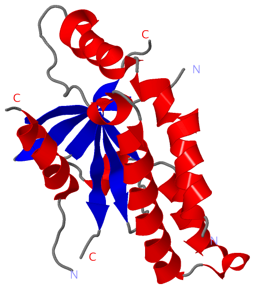
               SCOP domains d4f9ea_ A: Tyrosinase cofactor MelC1                                                                                                                                          SCOP domains
               CATH domains ----------------------------------------------------------------------------------------------------------------------------------------------------------------------------- CATH domains
               Pfam domains ----------------------------------------------------------------------------------------------------------------------------------------------------------------------------- Pfam domains
         Sec.struct. author .hhhhhhhhhhhhhhhhhh.hhhhhhhhhhhhh...........eeeeee.......hhhhh...eeeeee........eeeeeee..eeeeeeeeee..hhhhhhhhhhhhhhh.hhhhhhhhhhhhhhhhhhhhhh..hhhhheeeeee.......hhhhhhhhhhhhh.. Sec.struct. author
                 SAPs(SNPs) ----------------------------------------------------------------------------------------------------------------------------------------------------------------------------- SAPs(SNPs)
                    PROSITE ----------------------------------------------------------------------------------------------------------------------------------------------------------------------------- PROSITE
                 Transcript ----------------------------------------------------------------------------------------------------------------------------------------------------------------------------- Transcript
                 4f9e A 154 NVAHGLAWSYYIGYLRLILPELQARIRTYNQHYNNLLRGAVSQRLYILLPLDCGVPDNLSMADPNIRFLDKLPQQVYSNSIYELLENGQRAGTCVLEYATPLQTLFAMSQYSQAGFSREDRLEQAKLFCRTLEDILADAPESQNNCRLIAYQEPDSSFSLSQEVLRHLRQEEK 338
                                   163       173       183       193       203       213       223    || 243       253       263       273       283       293       303       313   ||  325       335   
                                                                                                    228|                                                                           317|                  
                                                                                                     239                                                                            320                  

   Legend:   → Mismatch (orange background)
  - → Gap (green background, '-', border residues have a numbering label)
    → Modified Residue (blue background, lower-case, 'x' indicates undefined single-letter code, labelled with number + name)
  x → Chemical Group (purple background, 'x', labelled with number + name, e.g. ACE or NH2)
  extra numbering lines below/above indicate numbering irregularities and modified residue names etc., number ends below/above '|'

 Classification and Annotation

(-) SCOP Domains  (1, 1)

Asymmetric Unit

(-) CATH Domains  (0, 0)

(no "CATH Domain" information available for 4F9E)

(-) Pfam Domains  (0, 0)

(no "Pfam Domain" information available for 4F9E)

(-) Gene Ontology  (36, 36)

Asymmetric Unit(hide GO term definitions)

 Visualization

(-) Interactive Views

Asymmetric Unit
  Complete Structure
    Jena3D(integrated viewing of ligand, site, SAP, PROSITE, SCOP information)
    WebMol | AstexViewer[tm]@PDBe
(Java Applets, require no local installation except for Java; loading may be slow)
    STRAP
(Java WebStart application, automatic local installation, requires Java; full application with system access!)
    RasMol
(require local installation)
    Molscript (VRML)
(requires installation of a VRML viewer; select preferred view via VRML and generate a mono or stereo PDF format file)
 
  Ligands, Modified Residues, Ions
(no "Ligands, Modified Residues, Ions" information available for 4f9e)
 
  Sites
(no "Sites" information available for 4f9e)
 
  Cis Peptide Bonds
(no "Cis Peptide Bonds" information available for 4f9e)
 
Biological Unit
  Complete Structure
    Biological Unit 1  [ Jena3D ]

(-) Still Images

Jmol
  protein: cartoon or spacefill or dots and stick; nucleic acid: cartoon and stick; ligands: spacefill; active site: stick
Molscript
  protein, nucleic acid: cartoon; ligands: spacefill; active site: ball and stick

 Databases and Analysis Tools

(-) Databases

Access by PDB/NDB ID
  4f9e
    Family and Domain InformationProDom | SYSTERS
    General Structural InformationGlycoscienceDB | MMDB | NDB | OCA | PDB | PDBe | PDBj | PDBsum | PDBWiki | PQS | PROTEOPEDIA
    Orientation in MembranesOPM
    Protein SurfaceSURFACE
    Secondary StructureDSSP (structure derived) | HSSP (homology derived)
    Structural GenomicsGeneCensus
    Structural NeighboursCE | VAST
    Structure ClassificationCATH | Dali | SCOP
    Validation and Original DataBMRB Data View | BMRB Restraints Grid | EDS | PROCHECK | RECOORD | WHAT_CHECK
 
Access by UniProt ID/Accession number
  STING_HUMAN | Q86WV6
    Comparative Protein Structure ModelsModBase
    Genomic InformationEnsembl
    Protein-protein InteractionDIP
    Sequence, Family and Domain InformationInterPro | Pfam | SMART | UniProtKB/SwissProt
 
Access by Enzyme Classificator   (EC Number)
  (no 'Enzyme Classificator' available)
    General Enzyme InformationBRENDA | EC-PDB | Enzyme | IntEnz
    PathwayKEGG | MetaCyc
 
Access by Disease Identifier   (MIM ID)
  (no 'MIM ID' available)
    Disease InformationOMIM
 
Access by GenAge ID
  (no 'GenAge ID' available)
    Age Related InformationGenAge

(-) Analysis Tools

Access by PDB/NDB ID
    Domain InformationXDom
    Interatomic Contacts of Structural UnitsCSU
    Ligand-protein ContactsLPC
    Protein CavitiescastP
    Sequence and Secondary StructurePDBCartoon
    Structure AlignmentSTRAP(Java WebStart application, automatic local installation, requires Java; full application with system access!)
    Structure and Sequence BrowserSTING
 
Access by UniProt ID/Accession number
  STING_HUMAN | Q86WV6
    Protein Disorder PredictionDisEMBL | FoldIndex | GLOBPLOT (for more information see DisProt)

 Related Entries

(-) Entries Sharing at Least One Protein Chain (UniProt ID)

UniProtKB/Swiss-Prot
        STING_HUMAN | Q86WV64ef4 4ef5 4emt 4emu 4f5d 4f5e 4f5w 4f5y 4f9g 4ksy 4loh 4loi 4qxo 4qxp 4qxq 4qxr 5bqx 5jej

(-) Related Entries Specified in the PDB File

4f9g