Show PDB file:   
         Plain Text   HTML   (compressed file size)
QuickSearch:   
by PDB,NDB,UniProt,PROSITE Code or Search Term(s)  
(-)Asymmetric Unit
(-)Asym. Unit - sites
(-)Biological Unit 1
(-)Biol. Unit 1 - sites
(-)Biological Unit 2
collapse expand < >
Image Asymmetric Unit
Asymmetric Unit  (Jmol Viewer)
Image Asym. Unit - sites
Asym. Unit - sites  (Jmol Viewer)
Image Biological Unit 1
Biological Unit 1  (Jmol Viewer)
Image Biol. Unit 1 - sites
Biol. Unit 1 - sites  (Jmol Viewer)
Image Biological Unit 2
Biological Unit 2  (Jmol Viewer)

(-) Description

Title :  CRYSTAL STRUCTURE OF HUMAN STING BOUND TO C-DI-GMP
 
Authors :  P. Li
Date :  12 Apr 12  (Deposition) - 13 Jun 12  (Release) - 25 Jul 12  (Revision)
Method :  X-RAY DIFFRACTION
Resolution :  1.50
Chains :  Asym. Unit :  A,B
Biol. Unit 1:  A,B  (1x)
Biol. Unit 2:  A,B  (2x)
Keywords :  Alpha/Beta Fold, Innate Immune Sensor, C-Di-Gmp, C-Di-Amp, Membrane Protein (Keyword Search: [Gene Ontology, PubMed, Web (Google))
 
Reference :  C. Shu, G. Yi, T. Watts, C. C. Kao, P. Li
Structure Of Sting Bound To Cyclic Di-Gmp Reveals The Mechanism Of Cyclic Dinucleotide Recognition By The Immune System.
Nat. Struct. Mol. Biol. V. 19 722 2012
PubMed-ID: 22728658  |  Reference-DOI: 10.1038/NSMB.2331

(-) Compounds

Molecule 1 - TRANSMEMBRANE PROTEIN 173
    ChainsA, B
    EngineeredYES
    Expression SystemESCHERICHIA COLI
    Expression System Taxid562
    FragmentUNP RESIDUES 155 - 341
    GeneTMEM173, ERIS, MITA, STING
    Organism CommonHUMAN
    Organism ScientificHOMO SAPIENS
    Organism Taxid9606
    SynonymENDOPLASMIC RETICULUM INTERFERON STIMULATOR, ERIS, MEDIATOR OF IRF3 ACTIVATION, HMITA, STIMULATOR OF INTERFERON GENES PROTEIN, HSTING

 Structural Features

(-) Chains, Units

  12
Asymmetric Unit AB
Biological Unit 1 (1x)AB
Biological Unit 2 (2x)AB

Summary Information (see also Sequences/Alignments below)

(-) Ligands, Modified Residues, Ions  (3, 6)

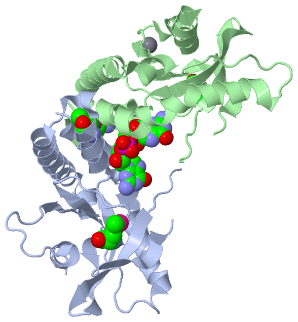
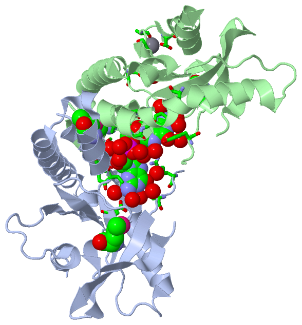
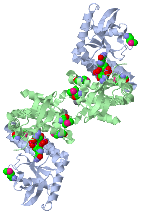
Asymmetric Unit (3, 6)
No.NameCountTypeFull Name
1C2E1Ligand/Ion9,9'-[(2R,3R,3AS,5S,7AR,9R,10R,10AS,12S,14AR)-3,5,10,12-TETRAHYDROXY-5,12-DIOXIDOOCTAHYDRO-2H,7H-DIFURO[3,2-D:3',2'-J][1,3,7,9,2,8]TETRAOXADIPHOSPHACYCLODODECINE-2,9-DIYL]BIS(2-AMINO-1,9-DIHYDRO-6H-PURIN-6-ONE)
2CA1Ligand/IonCALCIUM ION
3MSE4Mod. Amino AcidSELENOMETHIONINE
Biological Unit 1 (2, 5)
No.NameCountTypeFull Name
1C2E1Ligand/Ion9,9'-[(2R,3R,3AS,5S,7AR,9R,10R,10AS,12S,14AR)-3,5,10,12-TETRAHYDROXY-5,12-DIOXIDOOCTAHYDRO-2H,7H-DIFURO[3,2-D:3',2'-J][1,3,7,9,2,8]TETRAOXADIPHOSPHACYCLODODECINE-2,9-DIYL]BIS(2-AMINO-1,9-DIHYDRO-6H-PURIN-6-ONE)
2CA-1Ligand/IonCALCIUM ION
3MSE4Mod. Amino AcidSELENOMETHIONINE
Biological Unit 2 (2, 10)
No.NameCountTypeFull Name
1C2E2Ligand/Ion9,9'-[(2R,3R,3AS,5S,7AR,9R,10R,10AS,12S,14AR)-3,5,10,12-TETRAHYDROXY-5,12-DIOXIDOOCTAHYDRO-2H,7H-DIFURO[3,2-D:3',2'-J][1,3,7,9,2,8]TETRAOXADIPHOSPHACYCLODODECINE-2,9-DIYL]BIS(2-AMINO-1,9-DIHYDRO-6H-PURIN-6-ONE)
2CA-1Ligand/IonCALCIUM ION
3MSE8Mod. Amino AcidSELENOMETHIONINE

(-) Sites  (2, 2)

Asymmetric Unit (2, 2)
No.NameEvidenceResiduesDescription
1AC1SOFTWARESER A:162 , TYR A:163 , GLY A:166 , TYR A:167 , ARG A:238 , TYR A:240 , GLU A:260 , THR A:263 , PRO A:264 , HOH A:502 , HOH A:503 , HOH A:507 , HOH A:512 , HOH A:531 , HOH A:535 , HOH A:536 , HOH A:538 , HOH A:540 , HOH A:555 , HOH A:558 , HOH A:565 , HOH A:572 , HOH A:574 , HOH A:597 , HOH A:600 , SER B:162 , GLY B:166 , TYR B:167 , TYR B:240 , GLU B:260 , THR B:263 , PRO B:264 , HOH B:503 , HOH B:531 , HOH B:547BINDING SITE FOR RESIDUE C2E A 401
2AC2SOFTWAREASP B:205 , GLU B:316 , ALA B:318 , ASP B:320 , HOH B:502 , HOH B:558BINDING SITE FOR RESIDUE CA B 401

(-) SS Bonds  (0, 0)

(no "SS Bond" information available for 4EMT)

(-) Cis Peptide Bonds  (0, 0)

(no "Cis Peptide Bond" information available for 4EMT)

 Sequence-Structure Mapping

(-) SAPs(SNPs)/Variants  (0, 0)

(no "SAP(SNP)/Variant" information available for 4EMT)

(-) PROSITE Motifs  (0, 0)

(no "PROSITE Motif" information available for 4EMT)

(-) Exons   (0, 0)

(no "Exon" information available for 4EMT)

(-) Sequences/Alignments

Asymmetric Unit
   Reformat: Number of residues per line =  ('0' or empty: single-line sequence representation)
  Number of residues per labelling interval =   
  UniProt sequence: complete  aligned part    
   Show mapping: SCOP domains CATH domains Pfam domains Secondary structure (by author)
SAPs(SNPs) PROSITE motifs Exons
(details for a mapped element are shown in a popup box when the mouse pointer rests over it)
Chain A from PDB  Type:PROTEIN  Length:175
                                                                                                                                                                                                               
               SCOP domains d4emta_ A: Tyrosinase cofactor MelC1                                                                                                                                            SCOP domains
               CATH domains ------------------------------------------------------------------------------------------------------------------------------------------------------------------------------- CATH domains
               Pfam domains ------------------------------------------------------------------------------------------------------------------------------------------------------------------------------- Pfam domains
         Sec.struct. author .hhhhhhhhhhhhhhhhhhhhhhhhhhhhhhhh...........eeeeee.......hhhhhh..eeeeeee.......eeeeeeee..eeeeeeeeee.hhhhhhhhhhhhhhhh.hhhhhhhhhhhhhhhhhhhhhhhhhhhh.eeeeee.........hhhhhhhhhhh... Sec.struct. author
                 SAPs(SNPs) ------------------------------------------------------------------------------------------------------------------------------------------------------------------------------- SAPs(SNPs)
                    PROSITE ------------------------------------------------------------------------------------------------------------------------------------------------------------------------------- PROSITE
                 Transcript ------------------------------------------------------------------------------------------------------------------------------------------------------------------------------- Transcript
                 4emt A 154 SVAHGLAWSYYIGYLRLILPELQARIRTYNQHYNNLLRGAVSQRLYILLPLDCGVPDNLSmADPNIRFLDKLPQDRVYSNSIYELLENGQRAGTCVLEYATPLQTLFAmSQYSQAGFSREDRLEQAKLFCRTLEDILADAPESQNNCRLIAYQEPADDSSFSLSQEVLRHLRQEE 337
                                   163       173       183       193       203       213|      223   ||  242       252       262       272       282       292       302       312       322       332     
                                                                                      214-MSE      227|                               271-MSE                                                              
                                                                                                    237                                                                                                    

Chain B from PDB  Type:PROTEIN  Length:175
                                                                                                                                                                                                               
               SCOP domains d4emtb_ B: Tyrosinase cofactor MelC1                                                                                                                                            SCOP domains
               CATH domains ------------------------------------------------------------------------------------------------------------------------------------------------------------------------------- CATH domains
               Pfam domains ------------------------------------------------------------------------------------------------------------------------------------------------------------------------------- Pfam domains
         Sec.struct. author .hhhhhhhhhhhhhhhhhhhhhhhhhhhhhhhh...........eeeeee.......hhhhhh..eeeeee.........eeeeeee..eeeeeeeeee.hhhhhhhhhhhhhhhh.hhhhhhhhhhhhhhhhhhhhhhhhhhhhheeeeee....hhhhhhhhhhhhhhhhhh. Sec.struct. author
                 SAPs(SNPs) ------------------------------------------------------------------------------------------------------------------------------------------------------------------------------- SAPs(SNPs)
                    PROSITE ------------------------------------------------------------------------------------------------------------------------------------------------------------------------------- PROSITE
                 Transcript ------------------------------------------------------------------------------------------------------------------------------------------------------------------------------- Transcript
                 4emt B 154 SVAHGLAWSYYIGYLRLILPELQARIRTYNQHYNNLLRGAVSQRLYILLPLDCGVPDNLSmADPNIRFLDKLPQDRVYSNSIYELLENGQRAGTCVLEYATPLQTLFAmSQYSQAGFSREDRLEQAKLFCRTLEDILADAPESQNNCRLIAYQEPADDSSFSLSQEVLRHLRQEE 337
                                   163       173       183       193       203       213|      223   ||  242       252       262       272       282       292       302       312       322       332     
                                                                                      214-MSE      227|                               271-MSE                                                              
                                                                                                    237                                                                                                    

   Legend:   → Mismatch (orange background)
  - → Gap (green background, '-', border residues have a numbering label)
    → Modified Residue (blue background, lower-case, 'x' indicates undefined single-letter code, labelled with number + name)
  x → Chemical Group (purple background, 'x', labelled with number + name, e.g. ACE or NH2)
  extra numbering lines below/above indicate numbering irregularities and modified residue names etc., number ends below/above '|'

 Classification and Annotation

(-) SCOP Domains  (1, 2)

Asymmetric Unit

(-) CATH Domains  (0, 0)

(no "CATH Domain" information available for 4EMT)

(-) Pfam Domains  (0, 0)

(no "Pfam Domain" information available for 4EMT)

(-) Gene Ontology  (36, 36)

Asymmetric Unit(hide GO term definitions)

 Visualization

(-) Interactive Views

Asymmetric Unit
  Complete Structure
    Jena3D(integrated viewing of ligand, site, SAP, PROSITE, SCOP information)
    WebMol | AstexViewer[tm]@PDBe
(Java Applets, require no local installation except for Java; loading may be slow)
    STRAP
(Java WebStart application, automatic local installation, requires Java; full application with system access!)
    RasMol
(require local installation)
    Molscript (VRML)
(requires installation of a VRML viewer; select preferred view via VRML and generate a mono or stereo PDF format file)
 
  Ligands, Modified Residues, Ions
    C2E  [ RasMol | Jena3D ]  +environment [ RasMol | Jena3D ]
    CA  [ RasMol | Jena3D ]  +environment [ RasMol | Jena3D ]
    MSE  [ RasMol | Jena3D ]  +environment [ RasMol | Jena3D ]
 
  Sites
    AC1  [ RasMol ]  +environment [ RasMol ]
    AC2  [ RasMol ]  +environment [ RasMol ]
 
  Cis Peptide Bonds
(no "Cis Peptide Bonds" information available for 4emt)
 
Biological Units
  Complete Structure
    Biological Unit 1  [ Jena3D ]
    Biological Unit 2  [ Jena3D ]

(-) Still Images

Jmol
  protein: cartoon or spacefill or dots and stick; nucleic acid: cartoon and stick; ligands: spacefill; active site: stick
Molscript
  protein, nucleic acid: cartoon; ligands: spacefill; active site: ball and stick

 Databases and Analysis Tools

(-) Databases

Access by PDB/NDB ID
  4emt
    Family and Domain InformationProDom | SYSTERS
    General Structural InformationGlycoscienceDB | MMDB | NDB | OCA | PDB | PDBe | PDBj | PDBsum | PDBWiki | PQS | PROTEOPEDIA
    Orientation in MembranesOPM
    Protein SurfaceSURFACE
    Secondary StructureDSSP (structure derived) | HSSP (homology derived)
    Structural GenomicsGeneCensus
    Structural NeighboursCE | VAST
    Structure ClassificationCATH | Dali | SCOP
    Validation and Original DataBMRB Data View | BMRB Restraints Grid | EDS | PROCHECK | RECOORD | WHAT_CHECK
 
Access by UniProt ID/Accession number
  STING_HUMAN | Q86WV6
    Comparative Protein Structure ModelsModBase
    Genomic InformationEnsembl
    Protein-protein InteractionDIP
    Sequence, Family and Domain InformationInterPro | Pfam | SMART | UniProtKB/SwissProt
 
Access by Enzyme Classificator   (EC Number)
  (no 'Enzyme Classificator' available)
    General Enzyme InformationBRENDA | EC-PDB | Enzyme | IntEnz
    PathwayKEGG | MetaCyc
 
Access by Disease Identifier   (MIM ID)
  (no 'MIM ID' available)
    Disease InformationOMIM
 
Access by GenAge ID
  (no 'GenAge ID' available)
    Age Related InformationGenAge

(-) Analysis Tools

Access by PDB/NDB ID
    Domain InformationXDom
    Interatomic Contacts of Structural UnitsCSU
    Ligand-protein ContactsLPC
    Protein CavitiescastP
    Sequence and Secondary StructurePDBCartoon
    Structure AlignmentSTRAP(Java WebStart application, automatic local installation, requires Java; full application with system access!)
    Structure and Sequence BrowserSTING
 
Access by UniProt ID/Accession number
  STING_HUMAN | Q86WV6
    Protein Disorder PredictionDisEMBL | FoldIndex | GLOBPLOT (for more information see DisProt)

 Related Entries

(-) Entries Sharing at Least One Protein Chain (UniProt ID)

UniProtKB/Swiss-Prot
        STING_HUMAN | Q86WV64ef4 4ef5 4emu 4f5d 4f5e 4f5w 4f5y 4f9e 4f9g 4ksy 4loh 4loi 4qxo 4qxp 4qxq 4qxr 5bqx 5jej

(-) Related Entries Specified in the PDB File

4emu