Show PDB file:   
         Plain Text   HTML   (compressed file size)
QuickSearch:   
by PDB,NDB,UniProt,PROSITE Code or Search Term(s)  
(-)Asym./Biol. Unit
(-)Asym./Biol. Unit - sites
collapse expand < >
Image Asym./Biol. Unit
Asym./Biol. Unit  (Jmol Viewer)
Image Asym./Biol. Unit - sites
Asym./Biol. Unit - sites  (Jmol Viewer)

(-) Description

Title :  CRYSTAL STRUCTURE OF CYANOVIRIN-N COMPLEXED TO OLIGOMANNOSE-9 (MAN-9)
 
Authors :  I. Botos, B. R. O'Keefe, S. R. Shenoy, P. H. Seeberger, M. R. Boyd, A. Wlod
Date :  03 Apr 09  (Deposition) - 14 Apr 09  (Release) - 13 Jul 11  (Revision)
Method :  X-RAY DIFFRACTION
Resolution :  2.50
Chains :  Asym./Biol. Unit :  A,B
Keywords :  Cyanovirin-N, Hiv-Inactivating, Domain-Swapping, Gp120, Man-9, Oligosaccharide, Antiviral Protein, Disulfide Bond, Protein Synthesis Inhibitor (Keyword Search: [Gene Ontology, PubMed, Web (Google))
 
Reference :  I. Botos, B. R. O'Keefe, S. R. Shenoy, L. K. Cartner, D. M. Ratner, P. H. Seeberger, M. R. Boyd, A. Wlodawer
Structures Of The Complexes Of A Potent Anti-Hiv Protein Cyanovirin-N And High Mannose Oligosaccharides
J. Biol. Chem. V. 277 34336 2002
PubMed-ID: 12110688  |  Reference-DOI: 10.1074/JBC.M205909200

(-) Compounds

Molecule 1 - CYANOVIRIN-N
    ChainsA, B
    EngineeredYES
    Expression SystemESCHERICHIA COLI
    Expression System Taxid562
    Organism ScientificNOSTOC ELLIPSOSPORUM
    Organism Taxid45916
    SynonymCV-N

 Structural Features

(-) Chains, Units

  12
Asymmetric/Biological Unit AB

Summary Information (see also Sequences/Alignments below)

(-) Ligands, Modified Residues, Ions  (5, 16)

Asymmetric/Biological Unit (5, 16)
No.NameCountTypeFull Name
1BMA2Ligand/IonBETA-D-MANNOSE
2MAN10Ligand/IonALPHA-D-MANNOSE
3MG1Ligand/IonMAGNESIUM ION
4NAG2Ligand/IonN-ACETYL-D-GLUCOSAMINE
5NHE1Ligand/Ion2-[N-CYCLOHEXYLAMINO]ETHANE SULFONIC ACID

(-) Sites  (16, 16)

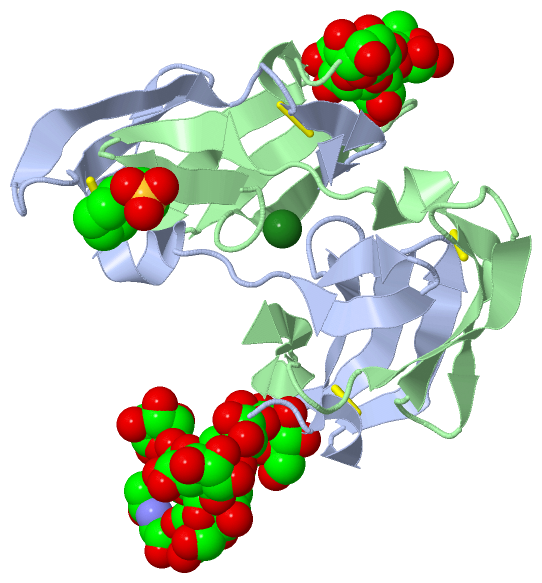
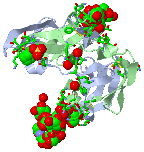
Asymmetric Unit (16, 16)
No.NameEvidenceResiduesDescription
01AC1SOFTWARENAG A:502 , GLU B:56 , CYS B:58BINDING SITE FOR RESIDUE NAG A 501
02AC2SOFTWARENAG A:501 , BMA A:503BINDING SITE FOR RESIDUE NAG A 502
03AC3SOFTWARENAG A:502 , MAN A:504 , MAN A:506 , MAN A:507BINDING SITE FOR RESIDUE BMA A 503
04AC4SOFTWAREGLU A:23 , THR A:25 , BMA A:503 , MAN A:505 , ASN B:93 , ASP B:95BINDING SITE FOR RESIDUE MAN A 504
05AC5SOFTWAREGLY A:2 , LYS A:3 , PHE A:4 , GLN A:6 , THR A:7 , MAN A:504 , MAN A:506 , ALA B:92 , ASN B:93BINDING SITE FOR RESIDUE MAN A 505
06AC6SOFTWARELEU A:1 , GLY A:2 , LYS A:3 , BMA A:503 , MAN A:505 , MAN A:507 , MAN A:510 , MAN A:511 , GLU B:101BINDING SITE FOR RESIDUE MAN A 506
07AC7SOFTWAREBMA A:503 , MAN A:506 , MAN A:508 , MAN A:510BINDING SITE FOR RESIDUE MAN A 507
08AC8SOFTWAREGLN A:14 , GLY A:15 , HOH A:117 , MAN A:507 , ASN B:60 , THR B:61BINDING SITE FOR RESIDUE MAN A 508
09AC9SOFTWAREMAN A:506 , MAN A:507 , MAN A:511BINDING SITE FOR RESIDUE MAN A 510
10BC1SOFTWARELEU A:1 , LYS A:3 , MAN A:506 , MAN A:510BINDING SITE FOR RESIDUE MAN A 511
11BC2SOFTWARELYS A:48 , TRP A:49 , ASN A:53 , LYS A:74 , THR A:75 , ARG A:76 , HOH A:116 , ASN B:42 , ASP B:44BINDING SITE FOR RESIDUE NHE A 301
12BC3SOFTWARESER A:38 , GLN A:50 , SER B:38 , GLN B:50 , HOH B:103 , HOH B:108BINDING SITE FOR RESIDUE MG A 102
13BC4SOFTWARETHR B:25 , MAN B:604BINDING SITE FOR RESIDUE BMA B 603
14BC5SOFTWAREASN A:93 , ASP A:95 , THR B:7 , GLU B:23 , ARG B:24 , THR B:25 , BMA B:603 , MAN B:605BINDING SITE FOR RESIDUE MAN B 604
15BC6SOFTWAREALA A:92 , ASN A:93 , GLY B:2 , LYS B:3 , PHE B:4 , GLN B:6 , THR B:7 , MAN B:604 , MAN B:606BINDING SITE FOR RESIDUE MAN B 605
16BC7SOFTWAREGLU A:101 , LEU B:1 , GLY B:2 , LYS B:3 , MAN B:605BINDING SITE FOR RESIDUE MAN B 606

(-) SS Bonds  (4, 4)

Asymmetric/Biological Unit
No.Residues
1A:8 -A:22
2A:58 -A:73
3B:8 -B:22
4B:58 -B:73

(-) Cis Peptide Bonds  (0, 0)

(no "Cis Peptide Bond" information available for 3GXZ)

 Sequence-Structure Mapping

(-) SAPs(SNPs)/Variants  (0, 0)

(no "SAP(SNP)/Variant" information available for 3GXZ)

(-) PROSITE Motifs  (0, 0)

(no "PROSITE Motif" information available for 3GXZ)

(-) Exons   (0, 0)

(no "Exon" information available for 3GXZ)

(-) Sequences/Alignments

Asymmetric/Biological Unit
   Reformat: Number of residues per line =  ('0' or empty: single-line sequence representation)
  Number of residues per labelling interval =   
  UniProt sequence: complete  aligned part    
   Show mapping: SCOP domains CATH domains Pfam domains Secondary structure (by author)
SAPs(SNPs) PROSITE motifs Exons
(details for a mapped element are shown in a popup box when the mouse pointer rests over it)
Chain A from PDB  Type:PROTEIN  Length:101
 aligned with CVN_NOSEL | P81180 from UniProtKB/Swiss-Prot  Length:101

    Alignment length:101
                                    10        20        30        40        50        60        70        80        90       100 
            CVN_NOSEL     1 LGKFSQTCYNSAIQGSVLTSTCERTNGGYNTSSIDLNSVIENVDGSLKWQPSNFIETCRNTQLAGSSELAAECKTRAQQFVSTKINLDDHIANIDGTLKYE 101
               SCOP domains d3gxza_ A: Cyanovirin-N                                                                               SCOP domains
               CATH domains 3gxzA00 A:1-101 HIV-inactivating Protein,Cyanovirin-n;                                                CATH domains
               Pfam domains ----------------------------------------------------------------------------------------------------- Pfam domains
         Sec.struct. author ..hhhh.eeeeeee..eeeeeee.....eeeeeee....eeee..eeee...hhhhheeeeeee...eeeeeee.....eeeeeee....eeee..eeee. Sec.struct. author
                 SAPs(SNPs) ----------------------------------------------------------------------------------------------------- SAPs(SNPs)
                    PROSITE ----------------------------------------------------------------------------------------------------- PROSITE
                 Transcript ----------------------------------------------------------------------------------------------------- Transcript
                 3gxz A   1 LGKFSQTCYNSAIQGSVLTSTCERTNGGYNTSSIDLNSVIENVDGSLKWQPSNFIETCRNTQLAGSSELAAECKTRAQQFVSTKINLDDHIANIDGTLKYE 101
                                    10        20        30        40        50        60        70        80        90       100 

Chain B from PDB  Type:PROTEIN  Length:101
 aligned with CVN_NOSEL | P81180 from UniProtKB/Swiss-Prot  Length:101

    Alignment length:101
                                    10        20        30        40        50        60        70        80        90       100 
            CVN_NOSEL     1 LGKFSQTCYNSAIQGSVLTSTCERTNGGYNTSSIDLNSVIENVDGSLKWQPSNFIETCRNTQLAGSSELAAECKTRAQQFVSTKINLDDHIANIDGTLKYE 101
               SCOP domains d3gxzb_ B: Cyanovirin-N                                                                               SCOP domains
               CATH domains 3gxzB00 B:1-101 HIV-inactivating Protein,Cyanovirin-n;                                                CATH domains
               Pfam domains ----------------------------------------------------------------------------------------------------- Pfam domains
         Sec.struct. author ..hhhh.eeeeeee..eeeeeee.....eeeeeee....eeee..eeee...hhhhheeeeeee...eeeeeee.....eeeeeee....eeee..eeee. Sec.struct. author
                 SAPs(SNPs) ----------------------------------------------------------------------------------------------------- SAPs(SNPs)
                    PROSITE ----------------------------------------------------------------------------------------------------- PROSITE
                 Transcript ----------------------------------------------------------------------------------------------------- Transcript
                 3gxz B   1 LGKFSQTCYNSAIQGSVLTSTCERTNGGYNTSSIDLNSVIENVDGSLKWQPSNFIETCRNTQLAGSSELAAECKTRAQQFVSTKINLDDHIANIDGTLKYE 101
                                    10        20        30        40        50        60        70        80        90       100 

   Legend:   → Mismatch (orange background)
  - → Gap (green background, '-', border residues have a numbering label)
    → Modified Residue (blue background, lower-case, 'x' indicates undefined single-letter code, labelled with number + name)
  x → Chemical Group (purple background, 'x', labelled with number + name, e.g. ACE or NH2)
  extra numbering lines below/above indicate numbering irregularities and modified residue names etc., number ends below/above '|'

 Classification and Annotation

(-) SCOP Domains  (1, 2)

Asymmetric/Biological Unit

(-) CATH Domains  (1, 2)

Asymmetric/Biological Unit
(-)
Class: Mainly Beta (13760)

(-) Pfam Domains  (0, 0)

(no "Pfam Domain" information available for 3GXZ)

(-) Gene Ontology  (2, 2)

Asymmetric/Biological Unit(hide GO term definitions)
Chain A,B   (CVN_NOSEL | P81180)
molecular function
    GO:0030246    carbohydrate binding    Interacting selectively and non-covalently with any carbohydrate, which includes monosaccharides, oligosaccharides and polysaccharides as well as substances derived from monosaccharides by reduction of the carbonyl group (alditols), by oxidation of one or more hydroxy groups to afford the corresponding aldehydes, ketones, or carboxylic acids, or by replacement of one or more hydroxy group(s) by a hydrogen atom. Cyclitols are generally not regarded as carbohydrates.
biological process
    GO:0050688    regulation of defense response to virus    Any process that modulates the frequency, rate or extent of the antiviral response of a cell or organism.

 Visualization

(-) Interactive Views

Asymmetric/Biological Unit
  Complete Structure
    Jena3D(integrated viewing of ligand, site, SAP, PROSITE, SCOP information)
    WebMol | AstexViewer[tm]@PDBe
(Java Applets, require no local installation except for Java; loading may be slow)
    STRAP
(Java WebStart application, automatic local installation, requires Java; full application with system access!)
    RasMol
(require local installation)
    Molscript (VRML)
(requires installation of a VRML viewer; select preferred view via VRML and generate a mono or stereo PDF format file)
 
  Ligands, Modified Residues, Ions
    BMA  [ RasMol | Jena3D ]  +environment [ RasMol | Jena3D ]
    MAN  [ RasMol | Jena3D ]  +environment [ RasMol | Jena3D ]
    MG  [ RasMol | Jena3D ]  +environment [ RasMol | Jena3D ]
    NAG  [ RasMol | Jena3D ]  +environment [ RasMol | Jena3D ]
    NHE  [ RasMol | Jena3D ]  +environment [ RasMol | Jena3D ]
 
  Sites
    AC1  [ RasMol ]  +environment [ RasMol ]
    AC2  [ RasMol ]  +environment [ RasMol ]
    AC3  [ RasMol ]  +environment [ RasMol ]
    AC4  [ RasMol ]  +environment [ RasMol ]
    AC5  [ RasMol ]  +environment [ RasMol ]
    AC6  [ RasMol ]  +environment [ RasMol ]
    AC7  [ RasMol ]  +environment [ RasMol ]
    AC8  [ RasMol ]  +environment [ RasMol ]
    AC9  [ RasMol ]  +environment [ RasMol ]
    BC1  [ RasMol ]  +environment [ RasMol ]
    BC2  [ RasMol ]  +environment [ RasMol ]
    BC3  [ RasMol ]  +environment [ RasMol ]
    BC4  [ RasMol ]  +environment [ RasMol ]
    BC5  [ RasMol ]  +environment [ RasMol ]
    BC6  [ RasMol ]  +environment [ RasMol ]
    BC7  [ RasMol ]  +environment [ RasMol ]
 
  Cis Peptide Bonds
(no "Cis Peptide Bonds" information available for 3gxz)
 

(-) Still Images

Jmol
  protein: cartoon or spacefill or dots and stick; nucleic acid: cartoon and stick; ligands: spacefill; active site: stick
Molscript
  protein, nucleic acid: cartoon; ligands: spacefill; active site: ball and stick

 Databases and Analysis Tools

(-) Databases

Access by PDB/NDB ID
  3gxz
    Family and Domain InformationProDom | SYSTERS
    General Structural InformationGlycoscienceDB | MMDB | NDB | OCA | PDB | PDBe | PDBj | PDBsum | PDBWiki | PQS | PROTEOPEDIA
    Orientation in MembranesOPM
    Protein SurfaceSURFACE
    Secondary StructureDSSP (structure derived) | HSSP (homology derived)
    Structural GenomicsGeneCensus
    Structural NeighboursCE | VAST
    Structure ClassificationCATH | Dali | SCOP
    Validation and Original DataBMRB Data View | BMRB Restraints Grid | EDS | PROCHECK | RECOORD | WHAT_CHECK
 
Access by UniProt ID/Accession number
  CVN_NOSEL | P81180
    Comparative Protein Structure ModelsModBase
    Genomic InformationEnsembl
    Protein-protein InteractionDIP
    Sequence, Family and Domain InformationInterPro | Pfam | SMART | UniProtKB/SwissProt
 
Access by Enzyme Classificator   (EC Number)
  (no 'Enzyme Classificator' available)
    General Enzyme InformationBRENDA | EC-PDB | Enzyme | IntEnz
    PathwayKEGG | MetaCyc
 
Access by Disease Identifier   (MIM ID)
  (no 'MIM ID' available)
    Disease InformationOMIM
 
Access by GenAge ID
  (no 'GenAge ID' available)
    Age Related InformationGenAge

(-) Analysis Tools

Access by PDB/NDB ID
    Domain InformationXDom
    Interatomic Contacts of Structural UnitsCSU
    Ligand-protein ContactsLPC
    Protein CavitiescastP
    Sequence and Secondary StructurePDBCartoon
    Structure AlignmentSTRAP(Java WebStart application, automatic local installation, requires Java; full application with system access!)
    Structure and Sequence BrowserSTING
 
Access by UniProt ID/Accession number
  CVN_NOSEL | P81180
    Protein Disorder PredictionDisEMBL | FoldIndex | GLOBPLOT (for more information see DisProt)

 Related Entries

(-) Entries Sharing at Least One Protein Chain (UniProt ID)

UniProtKB/Swiss-Prot
        CVN_NOSEL | P811801iiy 1j4v 1l5b 1l5e 1lom 1n02 2ezm 2ezn 2pys 2rdk 2rp3 2z21 3czz 3ezm 3gxy 3lhc 3s3y 3s3z 4j4c 4j4d 4j4e 4j4f 4j4g

(-) Related Entries Specified in the PDB File

1m5j THE SAME PROTEIN COMPLEXED TO SYNTHETIC HEXAMANNOSE
1m5m THE SAME PROTEIN COMPLEXED WITH MAN-9
3gxy