|
|
|
|
 Description
|
|
Compounds
|
||||||||||||||||||||||||||||||||||||||||||||||||||||||||||||||||||||||||||||||||||||||||||||||||||||||||||||||||||||||||||||||||||||||||||||||||||||
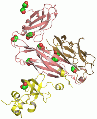
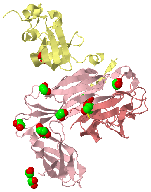
Chains, Units
Summary Information (see also Sequences/Alignments below) |
Ligands, Modified Residues, Ions (1, 12)
Asymmetric/Biological Unit (1, 12)
|
Sites (12, 12)
Asymmetric Unit (12, 12)
|
SS Bonds (2, 2)
Asymmetric/Biological Unit
|
||||||||||||
Cis Peptide Bonds (2, 2)
Asymmetric/Biological Unit
|
||||||||||||
SAPs(SNPs)/Variants (2, 2)
Asymmetric/Biological Unit (2, 2)
|
|||||||||||||||||||||||||||||||||||||||||||||||||||||||||||||||||||||||
PROSITE Motifs (1, 1)
Asymmetric/Biological Unit (1, 1)
|
||||||||||||||||||||||||
Exons (0, 0)| (no "Exon" information available for 1ZE3) |
Sequences/Alignments
Asymmetric/Biological UnitChain C from PDB Type:PROTEIN Length:205 aligned with FIMC_ECOLI | P31697 from UniProtKB/Swiss-Prot Length:241 Alignment length:205 46 56 66 76 86 96 106 116 126 136 146 156 166 176 186 196 206 216 226 236 FIMC_ECOLI 37 GVALGATRVIYPAGQKQEQLAVTNNDENSTYLIQSWVENADGVKDGRFIVTPPLFAMKGKKENTLRILDATNNQLPQDRESLFWMNVKAIPSMDKSKLTENTLQLAIISRIKLYYRPAKLALPPDQAAEKLRFRRSANSLTLINPTPYYLTVTELNAGTRVLENALVPPMGESTVKLPSDAGSNITYRTINDYGALTPKMTGVME 241 SCOP domains d1ze3c1 C:1-121 Periplasmic chaperone FimC d1ze3c2 C:122-205 FimC SCOP domains CATH domains -1ze3C01 C:2-118 PapD-like ---1ze3C02 C:122-205 [code=2.60.40.1070, no name defined] CATH domains Pfam domains ------------------------------------------------------------------------------------------------------------------------------------------------------------------------------------------------------------- Pfam domains SAPs(SNPs) -----------------V-----------------------------------------------------------------------------------------------------------------------------------------------------------A------------------------------- SAPs(SNPs) PROSITE --------------------------------------------------------------------------PILI_CHAPERONE ----------------------------------------------------------------------------------------------------------------- PROSITE Transcript ------------------------------------------------------------------------------------------------------------------------------------------------------------------------------------------------------------- Transcript 1ze3 C 1 GVALGATRVIYPAGQKQEQLAVTNNDENSTYLIQSWVENADGVKDGRFIVTPPLFAMKGKKENTLRILDATNNQLPQDRESLFWMNVKAIPSMDKSKLTENTLQLAIISRIKLYYRPAKLALPPDQAAEKLRFRRSANSLTLINPTPYYLTVTELNAGTRVLENALVPPMGESTVKLPSDAGSNITYRTINDYGALTPKMTGVME 205 10 20 30 40 50 60 70 80 90 100 110 120 130 140 150 160 170 180 190 200 Chain D from PDB Type:PROTEIN Length:116 aligned with FIMD_ECOLI | P30130 from UniProtKB/Swiss-Prot Length:878 Alignment length:125 55 65 75 85 95 105 115 125 135 145 155 165 FIMD_ECOLI 46 DLYFNPRFLADDPQAVADLSRFENGQELPPGTYRVDIYLNNGYMATRDVTFNTGDSEQGIVPCLTRAQLASMGLNTASVAGMNLLADDACVPLTTMVQDATAHLDVGQQRLNLTIPQAFMSNRAR 170 SCOP domains d1ze3d1 D :1-125 Outer membrane usher protein FimD SCOP domains CATH domains ----------------------------------------------------------------------------------------------------------------------------- CATH domains Pfam domains ----------------------------------------------------------------------------------------------------------------------------- Pfam domains SAPs(SNPs) ----------------------------------------------------------------------------------------------------------------------------- SAPs(SNPs) PROSITE ----------------------------------------------------------------------------------------------------------------------------- PROSITE Transcript ----------------------------------------------------------------------------------------------------------------------------- Transcript 1ze3 D 1 DLYFNPRFL---------LSRFENGQELPPGTYRVDIYLNNGYMATRDVTFNTGDSEQGIVPCLTRAQLASMGLNTASVAGMNLLADDACVPLTTMVQDATAHLDVGQQRLNLTIPQAFMSNRAR 125 |- 20 30 40 50 60 70 80 90 100 110 120 9 19 Chain H from PDB Type:PROTEIN Length:122 aligned with FIMH_ECOLI | P08191 from UniProtKB/Swiss-Prot Length:300 Alignment length:122 188 198 208 218 228 238 248 258 268 278 288 298 FIMH_ECOLI 179 TGGCDVSARDVTVTLPDYPGSVPIPLTVYCAKSQNLGYYLSGTTADAGNSIFTNTASFSPAQGVGVQLTRNGTIIPANNTVSLGAVGTSAVSLGLTANYARTGGQVTAGNVQSIIGVTFVYQ 300 SCOP domains d1ze3h_ H: Mannose-specific adhesin FimH SCOP domains CATH domains 1ze3H00 H:158-279 [code=2.60.40.1090, no name defined] CATH domains Pfam domains -------------------------------------------------------------------------------------------------------------------------- Pfam domains SAPs(SNPs) -------------------------------------------------------------------------------------------------------------------------- SAPs(SNPs) PROSITE -------------------------------------------------------------------------------------------------------------------------- PROSITE Transcript -------------------------------------------------------------------------------------------------------------------------- Transcript 1ze3 H 158 TGGCDVSARDVTVTLPDYPGSVPIPLTVYCAKSQNLGYYLSGTTADAGNSIFTNTASFSPAQGVGVQLTRNGTIIPANNTVSLGAVGTSAVSLGLTANYARTGGQVTAGNVQSIIGVTFVYQ 279 167 177 187 197 207 217 227 237 247 257 267 277
|
||||||||||||||||||||
SCOP Domains (4, 4)
Asymmetric/Biological Unit

|
CATH Domains (3, 3)
Asymmetric/Biological Unit
|
Pfam Domains (0, 0)| (no "Pfam Domain" information available for 1ZE3) |
Gene Ontology (18, 19)|
Asymmetric/Biological Unit(hide GO term definitions) Chain C (FIMC_ECOLI | P31697)
Chain D (FIMD_ECOLI | P30130)
Chain H (FIMH_ECOLI | P08191)
|
||||||||||||||||||||||||||||||||||||||||||||||||||||||||||||||||||||||||||||||||||||||||||||||||||||||||||||||||||||||||||||||||||||||||||||||||||||||||||||||||||||||||
Interactive Views
|
|||||||||||||||||||||||||||||||||||||||||||||||||||||||||||||||||||||||||||||||||||||||||||||||||||||||||||||||||||||||||||||||||||||||||||||||||||||||||||||||||||||||||||||||||||||||||||||||||||||||||||
Still Images
|
||||||||||||||||
Databases
|
||||||||||||||||||||||||||||||||||||||||||||||||||||||||||||||||||||||||||||||||||||||||||||||||||||||||||||||||||||||||||||||||||||||||||||||||||||||||||||||||||||||||||||||||||||||||||||||||||||||||||||||||||||
Analysis Tools
|
|||||||||||||||||||||||||||||||||||||||||||||||||||||||||||||||||||||||||||||||||||
Entries Sharing at Least One Protein Chain (UniProt ID)
Related Entries Specified in the PDB File
|
|