|
|
|
|
 Description
|
|
Compounds
|
||||||||||||||||||||||||||||||||||||||||||||||||||||
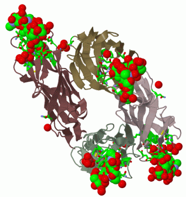
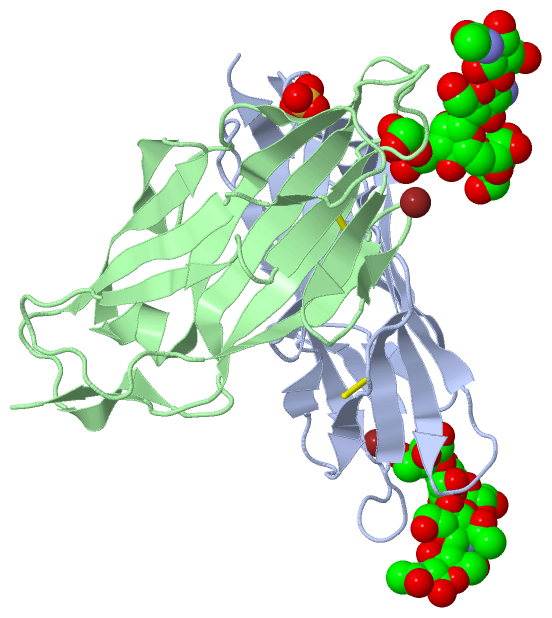
Chains, Units
Summary Information (see also Sequences/Alignments below) |
Ligands, Modified Residues, Ions (4, 13)| Asymmetric Unit (4, 13) Biological Unit 1 (3, 22) |
Sites (5, 5)
Asymmetric Unit (5, 5)
|
SS Bonds (2, 2)
Asymmetric Unit
|
||||||||||||
Cis Peptide Bonds (2, 2)
Asymmetric Unit
|
||||||||||||
SAPs(SNPs)/Variants (0, 0)| (no "SAP(SNP)/Variant" information available for 2VCO) |
PROSITE Motifs (0, 0)| (no "PROSITE Motif" information available for 2VCO) |
Exons (0, 0)| (no "Exon" information available for 2VCO) |
Sequences/Alignments
Asymmetric UnitChain A from PDB Type:PROTEIN Length:158 aligned with FIMH_ECOLI | P08191 from UniProtKB/Swiss-Prot Length:300 Alignment length:158 31 41 51 61 71 81 91 101 111 121 131 141 151 161 171 FIMH_ECOLI 22 FACKTANGTAIPIGGGSANVYVNLAPVVNVGQNLVVDLSTQIFCHNDYPETITDYVTLQRGSAYGGVLSNFSGTVKYSGSSYPFPTTSETPRVVYNSRTDKPWPVALYLTPVSSAGGVAIKAGSLIAVLILRQTNNYNSDDFQFVWNIYANNDVVVPT 179 SCOP domains d2vcoa_ A: Mannose-specific adhesin FimH SCOP domains CATH domains 2vcoA00 A:1-158 [code=2.60.40.1090, no name defined] CATH domains Pfam domains -------------------------------------------------------------------------------------------------------------------------------------------------------------- Pfam domains SAPs(SNPs) -------------------------------------------------------------------------------------------------------------------------------------------------------------- SAPs(SNPs) PROSITE -------------------------------------------------------------------------------------------------------------------------------------------------------------- PROSITE Transcript -------------------------------------------------------------------------------------------------------------------------------------------------------------- Transcript 2vco A 1 FACKTANGTAIPIGGGSANVYVNLAPVVNVGQNLVVDLSTQIFCHNDYPETITDYVTLQRGSAYGGVLSNFSGTVKYSGSSYPFPTTSETPRVVYNSRTDKPWPVALYLTPVSSAGGVAIKAGSLIAVLILRQTNNYNSDDFQFVWNIYANNDVVVPT 158 10 20 30 40 50 60 70 80 90 100 110 120 130 140 150 Chain B from PDB Type:PROTEIN Length:158 aligned with FIMH_ECOLI | P08191 from UniProtKB/Swiss-Prot Length:300 Alignment length:158 31 41 51 61 71 81 91 101 111 121 131 141 151 161 171 FIMH_ECOLI 22 FACKTANGTAIPIGGGSANVYVNLAPVVNVGQNLVVDLSTQIFCHNDYPETITDYVTLQRGSAYGGVLSNFSGTVKYSGSSYPFPTTSETPRVVYNSRTDKPWPVALYLTPVSSAGGVAIKAGSLIAVLILRQTNNYNSDDFQFVWNIYANNDVVVPT 179 SCOP domains d2vcob_ B: Mannose-specific adhesin FimH SCOP domains CATH domains 2vcoB00 B:1-158 [code=2.60.40.1090, no name defined] CATH domains Pfam domains (1) -------------------------------------------------------------------------------------------------------------------------------------------------Fimbrial-2vco Pfam domains (1) Pfam domains (2) -------------------------------------------------------------------------------------------------------------------------------------------------Fimbrial-2vco Pfam domains (2) Pfam domains (3) --FimH_man-bind-2vcoB03 B:3-147 ----------- Pfam domains (3) Pfam domains (4) --FimH_man-bind-2vcoB04 B:3-147 ----------- Pfam domains (4) SAPs(SNPs) -------------------------------------------------------------------------------------------------------------------------------------------------------------- SAPs(SNPs) PROSITE -------------------------------------------------------------------------------------------------------------------------------------------------------------- PROSITE Transcript -------------------------------------------------------------------------------------------------------------------------------------------------------------- Transcript 2vco B 1 FACKTANGTAIPIGGGSANVYVNLAPVVNVGQNLVVDLSTQIFCHNDYPETITDYVTLQRGSAYGGVLSNFSGTVKYSGSSYPFPTTSETPRVVYNSRTDKPWPVALYLTPVSSAGGVAIKAGSLIAVLILRQTNNYNSDDFQFVWNIYANNDVVVPT 158 10 20 30 40 50 60 70 80 90 100 110 120 130 140 150
|
||||||||||||||||||||
SCOP Domains (1, 2)
Asymmetric Unit
 |
CATH Domains (1, 2)
Asymmetric Unit
|
Pfam Domains (2, 4)
Asymmetric Unit
|
Gene Ontology (4, 4)|
Asymmetric Unit(hide GO term definitions) Chain A,B (FIMH_ECOLI | P08191)
|
||||||||||||||||||||||||||||||||||||||||||
Interactive Views
|
|||||||||||||||||||||||||||||||||||||||||||||||||||||||||||||||||||||||||||||||||||||||||||||||||||||||||||||||||||||||||||||||||||||||||||||||||||||||||||||||||||||||||||||||||||||||||||||||||
Still Images
|
||||||||||||||||
Databases
|
||||||||||||||||||||||||||||||||||||||||||||||||||||||||||||||||||||||||||||||||||||||||||||||||||||||||||||||||||||||||||||||||||||||||||||||||||||||||||||||||
Analysis Tools
|
|||||||||||||||||||||||||||||||||||||||||||||||||||||||||||||
Entries Sharing at Least One Protein Chain (UniProt ID)
Related Entries Specified in the PDB File
|
|