Show PDB file:   
         Plain Text   HTML   (compressed file size)
QuickSearch:   
by PDB,NDB,UniProt,PROSITE Code or Search Term(s)  
(-)Asymmetric Unit
(-)Biological Unit 1
(-)Biological Unit 2
collapse expand < >
Image Asymmetric Unit
Asymmetric Unit  (Jmol Viewer)
Image Biological Unit 1
Biological Unit 1  (Jmol Viewer)
Image Biological Unit 2
Biological Unit 2  (Jmol Viewer)

(-) Description

Title :  CRYSTAL STRUCTURE OF SH3:RGT COMPLEX
 
Authors :  R. Xiao, G. Meng
Date :  11 Nov 12  (Deposition) - 28 Nov 12  (Release) - 19 Mar 14  (Revision)
Method :  X-RAY DIFFRACTION
Resolution :  2.00
Chains :  Asym. Unit :  A,B,C
Biol. Unit 1:  A,C  (1x)
Biol. Unit 2:  B  (1x)
Keywords :  Integrin Beta-3, C-Src Kinase, Sh3 Domain, Rgt, Thrombosis, Cell Signaling, Blood Clotting, Signaling Protein (Keyword Search: [Gene Ontology, PubMed, Web (Google))
 
Reference :  R. Xiao, X. D. Xi, Z. Chen, S. J. Chen, G. Meng
Structural Framework Of C-Src Activation By Integrin Beta 3
Blood V. 121 700 2013
PubMed-ID: 23169783  |  Reference-DOI: 10.1182/BLOOD-2012-07-440644

(-) Compounds

Molecule 1 - PROTO-ONCOGENE TYROSINE-PROTEIN KINASE SRC
    ChainsA, B
    EC Number2.7.10.2
    EngineeredYES
    Expression SystemESCHERICHIA COLI
    Expression System PlasmidPET32A
    Expression System StrainBL21(DE3)
    Expression System Taxid562
    Expression System Vector TypePLASMID
    FragmentHUMAN C-SRC SH3 DOMAIN, UNP RESIDUES 87-144
    GeneSRC, SRC1
    Organism CommonHUMAN
    Organism ScientificHOMO SAPIENS
    Organism Taxid9606
    SynonymC-SRC, PROTO-ONCOGENE C-SRC, PP60C-SRC, P60-SRC
 
Molecule 2 - C-TERMINAL 3-MER PEPTIDE FROM INTEGRIN BETA-3
    ChainsC
    EngineeredYES
    Organism CommonHUMAN
    Organism ScientificHOMO SAPIENS
    Organism Taxid9606
    Other DetailsTHE RGT PEPTIDE WAS SYNTHESIZED IN THE SOLID PHASE
    SyntheticYES

 Structural Features

(-) Chains, Units

  123
Asymmetric Unit ABC
Biological Unit 1 (1x)A C
Biological Unit 2 (1x) B 

Summary Information (see also Sequences/Alignments below)

(-) Ligands, Modified Residues, Ions  (0, 0)

(no "Ligand,Modified Residues,Ions" information available for 4HXJ)

(-) Sites  (0, 0)

(no "Site" information available for 4HXJ)

(-) SS Bonds  (0, 0)

(no "SS Bond" information available for 4HXJ)

(-) Cis Peptide Bonds  (0, 0)

(no "Cis Peptide Bond" information available for 4HXJ)

 Sequence-Structure Mapping

(-) SAPs(SNPs)/Variants  (0, 0)

(no "SAP(SNP)/Variant" information available for 4HXJ)

(-) PROSITE Motifs  (0, 0)

(no "PROSITE Motif" information available for 4HXJ)

(-) Exons   (0, 0)

(no "Exon" information available for 4HXJ)

(-) Sequences/Alignments

Asymmetric Unit
   Reformat: Number of residues per line =  ('0' or empty: single-line sequence representation)
  Number of residues per labelling interval =   
  UniProt sequence: complete  aligned part    
   Show mapping: SCOP domains CATH domains Pfam domains Secondary structure (by author)
SAPs(SNPs) PROSITE motifs Exons
(details for a mapped element are shown in a popup box when the mouse pointer rests over it)
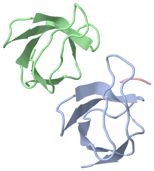
Chain A from PDB  Type:PROTEIN  Length:60
                                                                                            
               SCOP domains ------------------------------------------------------------ SCOP domains
               CATH domains ------------------------------------------------------------ CATH domains
               Pfam domains ------------------------------------------------------------ Pfam domains
         Sec.struct. author ...eeee..................eee........eeeeee.....eeeee...eee.. Sec.struct. author
                 SAPs(SNPs) ------------------------------------------------------------ SAPs(SNPs)
                    PROSITE ------------------------------------------------------------ PROSITE
                 Transcript ------------------------------------------------------------ Transcript
                 4hxj A  82 GSTTFVALYDYESRTETDLSFKKGERLQIVNNTEGDWWLAHSLSTGQTGYIPSNYVAPSD 141
                                    91       101       111       121       131       141

Chain B from PDB  Type:PROTEIN  Length:59
                                                                                           
               SCOP domains ----------------------------------------------------------- SCOP domains
               CATH domains ----------------------------------------------------------- CATH domains
               Pfam domains ----------------------------------------------------------- Pfam domains
         Sec.struct. author ...eee..................eeeeee.....eeeeee.....eeeee...eee.. Sec.struct. author
                 SAPs(SNPs) ----------------------------------------------------------- SAPs(SNPs)
                    PROSITE ----------------------------------------------------------- PROSITE
                 Transcript ----------------------------------------------------------- Transcript
                 4hxj B  83 STTFVALYDYESRTETDLSFKKGERLQIVNNTEGDWWLAHSLSTGQTGYIPSNYVAPSD 141
                                    92       102       112       122       132         

Chain C from PDB  Type:PROTEIN  Length:3
                                   
               SCOP domains --- SCOP domains
               CATH domains --- CATH domains
               Pfam domains --- Pfam domains
         Sec.struct. author ... Sec.struct. author
                 SAPs(SNPs) --- SAPs(SNPs)
                    PROSITE --- PROSITE
                 Transcript --- Transcript
                 4hxj C 760 RGT 762

   Legend:   → Mismatch (orange background)
  - → Gap (green background, '-', border residues have a numbering label)
    → Modified Residue (blue background, lower-case, 'x' indicates undefined single-letter code, labelled with number + name)
  x → Chemical Group (purple background, 'x', labelled with number + name, e.g. ACE or NH2)
  extra numbering lines below/above indicate numbering irregularities and modified residue names etc., number ends below/above '|'

 Classification and Annotation

(-) SCOP Domains  (0, 0)

(no "SCOP Domain" information available for 4HXJ)

(-) CATH Domains  (0, 0)

(no "CATH Domain" information available for 4HXJ)

(-) Pfam Domains  (0, 0)

(no "Pfam Domain" information available for 4HXJ)

(-) Gene Ontology  (219, 237)

Asymmetric Unit(hide GO term definitions)

 Visualization

(-) Interactive Views

Asymmetric Unit
  Complete Structure
    Jena3D(integrated viewing of ligand, site, SAP, PROSITE, SCOP information)
    WebMol | AstexViewer[tm]@PDBe
(Java Applets, require no local installation except for Java; loading may be slow)
    STRAP
(Java WebStart application, automatic local installation, requires Java; full application with system access!)
    RasMol
(require local installation)
    Molscript (VRML)
(requires installation of a VRML viewer; select preferred view via VRML and generate a mono or stereo PDF format file)
 
  Ligands, Modified Residues, Ions
(no "Ligands, Modified Residues, Ions" information available for 4hxj)
 
  Sites
(no "Sites" information available for 4hxj)
 
  Cis Peptide Bonds
(no "Cis Peptide Bonds" information available for 4hxj)
 
Biological Units
  Complete Structure
    Biological Unit 1  [ Jena3D ]
    Biological Unit 2  [ Jena3D ]

(-) Still Images

Jmol
  protein: cartoon or spacefill or dots and stick; nucleic acid: cartoon and stick; ligands: spacefill; active site: stick
Molscript
  protein, nucleic acid: cartoon; ligands: spacefill; active site: ball and stick

 Databases and Analysis Tools

(-) Databases

Access by PDB/NDB ID
  4hxj
    Family and Domain InformationProDom | SYSTERS
    General Structural InformationGlycoscienceDB | MMDB | NDB | OCA | PDB | PDBe | PDBj | PDBsum | PDBWiki | PQS | PROTEOPEDIA
    Orientation in MembranesOPM
    Protein SurfaceSURFACE
    Secondary StructureDSSP (structure derived) | HSSP (homology derived)
    Structural GenomicsGeneCensus
    Structural NeighboursCE | VAST
    Structure ClassificationCATH | Dali | SCOP
    Validation and Original DataBMRB Data View | BMRB Restraints Grid | EDS | PROCHECK | RECOORD | WHAT_CHECK
 
Access by UniProt ID/Accession number
  ITB3_HUMAN | P05106
    Comparative Protein Structure ModelsModBase
    Genomic InformationEnsembl
    Protein-protein InteractionDIP
    Sequence, Family and Domain InformationInterPro | Pfam | SMART | UniProtKB/SwissProt
  SRC_HUMAN | P12931
    Comparative Protein Structure ModelsModBase
    Genomic InformationEnsembl
    Protein-protein InteractionDIP
    Sequence, Family and Domain InformationInterPro | Pfam | SMART | UniProtKB/SwissProt
 
Access by Enzyme Classificator   (EC Number)
  2.7.10.2
    General Enzyme InformationBRENDA | EC-PDB | Enzyme | IntEnz
    PathwayKEGG | MetaCyc
 
Access by Disease Identifier   (MIM ID)
  (no 'MIM ID' available)
    Disease InformationOMIM
 
Access by GenAge ID
  (no 'GenAge ID' available)
    Age Related InformationGenAge

(-) Analysis Tools

Access by PDB/NDB ID
    Domain InformationXDom
    Interatomic Contacts of Structural UnitsCSU
    Ligand-protein ContactsLPC
    Protein CavitiescastP
    Sequence and Secondary StructurePDBCartoon
    Structure AlignmentSTRAP(Java WebStart application, automatic local installation, requires Java; full application with system access!)
    Structure and Sequence BrowserSTING
 
Access by UniProt ID/Accession number
  ITB3_HUMAN | P05106
    Protein Disorder PredictionDisEMBL | FoldIndex | GLOBPLOT (for more information see DisProt)
  SRC_HUMAN | P12931
    Protein Disorder PredictionDisEMBL | FoldIndex | GLOBPLOT (for more information see DisProt)

 Related Entries

(-) Entries Sharing at Least One Protein Chain (UniProt ID)

UniProtKB/Swiss-Prot
        ITB3_HUMAN | P051061jv2 1kup 1kuz 1l5g 1m1x 1m8o 1miz 1mk7 1mk9 1rn0 1s4x 1tye 1u8c 2h7d 2h7e 2ini 2k9j 2knc 2kv9 2l1c 2l91 2ljd 2lje 2ljf 2mtp 2n9y 2q6w 2rmz 2rn0 2vc2 2vdk 2vdl 2vdm 2vdn 2vdo 2vdp 2vdq 2vdr 3fcs 3fcu 3ije 3nid 3nif 3nig 3t3m 3t3p 3zdx 3zdy 3zdz 3ze0 3ze1 3ze2 4cak 4g1e 4g1m 4mmx 4mmy 4mmz 4o02 4z7n 4z7o 4z7q 5hdb
        SRC_HUMAN | P129311a07 1a08 1a09 1a1a 1a1b 1a1c 1a1e 1fmk 1hcs 1hct 1ksw 1o41 1o42 1o43 1o44 1o45 1o46 1o47 1o48 1o49 1o4a 1o4b 1o4c 1o4d 1o4e 1o4f 1o4g 1o4h 1o4i 1o4j 1o4k 1o4l 1o4m 1o4n 1o4o 1o4p 1o4q 1o4r 1shd 1y57 1yi6 1yoj 1yol 1yom 2bdf 2bdj 2h8h 2src 3vro 3zmp 3zmq 4f59 4f5a 4f5b 4k11 4mxo 4mxx 4mxy 4mxz

(-) Related Entries Specified in the PDB File

1fmk