Asymmetric Unit (12, 12)
| No. | Name | Evidence | Residues | Description |
|---|
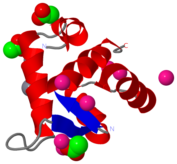
| 01 | AC1 | SOFTWARE | ASP A:33 , CYS A:35 , ACT A:109 , HOH A:224 | BINDING SITE FOR RESIDUE CD A 101 |
| 02 | AC2 | SOFTWARE | ASP A:65 , THR A:71 , GLU A:76 , HOH A:209 , HOH A:235 , HOH A:236 | BINDING SITE FOR RESIDUE CD A 102 |
| 03 | AC3 | SOFTWARE | PHE A:27 , GLU A:40 , GLU A:63 , HOH A:225 , HOH A:226 | BINDING SITE FOR RESIDUE CD A 103 |
| 04 | AC4 | SOFTWARE | GLU A:56 , CYS A:84 , ASP A:87 , SER A:89 , CD A:105 | BINDING SITE FOR RESIDUE CD A 104 |
| 05 | AC5 | SOFTWARE | GLN A:50 , CYS A:84 , ASP A:87 , ASP A:88 , SER A:89 , CD A:104 | BINDING SITE FOR RESIDUE CD A 105 |
| 06 | AC6 | SOFTWARE | CYS A:35 , GLU A:66 , ASP A:73 , ASP A:75 | BINDING SITE FOR RESIDUE CD A 106 |
| 07 | AC7 | SOFTWARE | MET A:1 , GLU A:59 , HOH A:245 , HOH A:246 , HOH A:247 , HOH A:248 | BINDING SITE FOR RESIDUE CD A 107 |
| 08 | AC8 | SOFTWARE | GLU A:15 , GLU A:19 , CA A:111 , CA A:112 , HOH A:257 | BINDING SITE FOR RESIDUE ACT A 108 |
| 09 | AC9 | SOFTWARE | MET A:1 , ASP A:33 , CYS A:35 , SER A:37 , THR A:71 , CD A:101 , HOH A:225 , HOH A:226 , HOH A:248 | BINDING SITE FOR RESIDUE ACT A 109 |
| 10 | BC1 | SOFTWARE | GLU A:10 , LEU A:12 , THR A:13 | BINDING SITE FOR RESIDUE ACT A 110 |
| 11 | BC2 | SOFTWARE | GLU A:15 , GLU A:19 , ACT A:108 , HOH A:255 | BINDING SITE FOR RESIDUE CA A 111 |
| 12 | BC3 | SOFTWARE | GLU A:10 , GLU A:19 , ACT A:108 , HOH A:220 , HOH A:257 | BINDING SITE FOR RESIDUE CA A 112 |
|
 Description