Show PDB file:   
         Plain Text   HTML   (compressed file size)
QuickSearch:   
by PDB,NDB,UniProt,PROSITE Code or Search Term(s)  
(-)Asym./Biol. Unit
(-)Asym./Biol. Unit - sites
collapse expand < >
Image Asym./Biol. Unit
Asym./Biol. Unit  (Jmol Viewer)
Image Asym./Biol. Unit - sites
Asym./Biol. Unit - sites  (Jmol Viewer)

(-) Description

Title :  CRYSTAL STRUCTURES REVEAL THE MULTI-LIGAND BINDING MECHANISM OF THE STAPHYLOCOCCUS AUREUS CLFB
 
Authors :  M. J. Yang, H. Xiang, J. W. Wang, B. Liu, Y. G. Chen, L. Liu, X. M. Deng, Y. Fe
Date :  07 May 12  (Deposition) - 08 Aug 12  (Release) - 04 Feb 15  (Revision)
Method :  X-RAY DIFFRACTION
Resolution :  1.92
Chains :  Asym./Biol. Unit :  A,Q
Keywords :  Dev-Igg Fold, Protein-Peptide Complex, Fibronogen, Cell Surface, Cell Adhesion-Blood Clotting Complex (Keyword Search: [Gene Ontology, PubMed, Web (Google))
 
Reference :  H. Xiang, Y. Feng, J. W. Wang, B. Liu, Y. G. Chen, L. Liu, X. M. Deng, M. J. Yang
Crystal Structures Reveal The Multi-Ligand Binding Mechanis Of Staphylococcus Aureus Clfb
Plos Pathog. V. 8 02751 2012
PubMed-ID: 22719251  |  Reference-DOI: 10.1371/JOURNAL.PPAT.1002751

(-) Compounds

Molecule 1 - CLUMPING FACTOR B
    ChainsA
    EngineeredYES
    Expression SystemESCHERICHIA COLI
    Expression System Taxid562
    Expression System Vector TypePLASMID
    FragmentUNP RESIDUES 197-542
    GeneCLFB, SAR2709
    Organism ScientificSTAPHYLOCOCCUS AUREUS
    Organism Taxid282458
    StrainMRSA252
    SynonymFIBRINOGEN RECEPTOR B, FIBRINOGEN-BINDING PROTEIN B
 
Molecule 2 - PEPTIDE FROM FIBRINOGEN ALPHA CHAIN
    ChainsQ
    EngineeredYES
    Organism CommonHUMAN
    Organism ScientificHOMO SAPIENS
    Organism Taxid9606
    Other DetailsTHIS SEQUENCE OCCURS NATURALLY IN HUMANS.
    SynonymFIBRINOPEPTIDE A, FIBRINOGEN ALPHA CHAIN
    SyntheticYES

 Structural Features

(-) Chains, Units

  12
Asymmetric/Biological Unit AQ

Summary Information (see also Sequences/Alignments below)
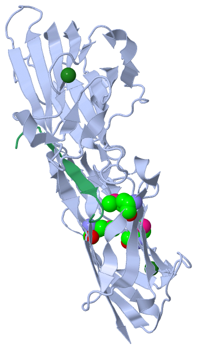
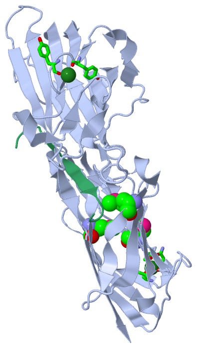
(-) Ligands, Modified Residues, Ions  (2, 5)

Asymmetric/Biological Unit (2, 5)
No.NameCountTypeFull Name
1MG2Ligand/IonMAGNESIUM ION
2MSE3Mod. Amino AcidSELENOMETHIONINE

(-) Sites  (2, 2)

Asymmetric Unit (2, 2)
No.NameEvidenceResiduesDescription
1AC1SOFTWARETYR A:468 , TYR A:470 , HOH A:707 , HOH A:714 , HOH A:791 , HOH A:792BINDING SITE FOR RESIDUE MG A 601
2AC2SOFTWAREASN A:216 , VAL A:219 , ALA A:347 , HOH A:766BINDING SITE FOR RESIDUE MG A 602

(-) SS Bonds  (0, 0)

(no "SS Bond" information available for 4F27)

(-) Cis Peptide Bonds  (0, 0)

(no "Cis Peptide Bond" information available for 4F27)

 Sequence-Structure Mapping

(-) SAPs(SNPs)/Variants  (0, 0)

(no "SAP(SNP)/Variant" information available for 4F27)

(-) PROSITE Motifs  (0, 0)

(no "PROSITE Motif" information available for 4F27)

(-) Exons   (0, 0)

(no "Exon" information available for 4F27)

(-) Sequences/Alignments

Asymmetric/Biological Unit
   Reformat: Number of residues per line =  ('0' or empty: single-line sequence representation)
  Number of residues per labelling interval =   
  UniProt sequence: complete  aligned part    
   Show mapping: SCOP domains CATH domains Pfam domains Secondary structure (by author)
SAPs(SNPs) PROSITE motifs Exons
(details for a mapped element are shown in a popup box when the mouse pointer rests over it)
Chain A from PDB  Type:PROTEIN  Length:331
                                                                                                                                                                                                                                                                                                                                                                           
               SCOP domains ------------------------------------------------------------------------------------------------------------------------------------------------------------------------------------------------------------------------------------------------------------------------------------------------------------------------------------------- SCOP domains
               CATH domains ------------------------------------------------------------------------------------------------------------------------------------------------------------------------------------------------------------------------------------------------------------------------------------------------------------------------------------------- CATH domains
               Pfam domains ------------------------------------------------------------------------------------------------------------------------------------------------------------------------------------------------------------------------------------------------------------------------------------------------------------------------------------------- Pfam domains
         Sec.struct. author ......hhh.eeeeeeee...eehhhhh..eeeeeeeee........eeeee....ee......hhhhh.eeeeeeee.....eeeeeeee....eeeeee.hhhh..eeeeeeeeeeeee........eeee..eee..eee..eeee................eeeeeeee.......eeeeeeee......eeeeeeeee....hhhhh.ee......eeeeeee.hhhhh............eee.hhhh...ee.....eeeeeeeee...eeeeeeeee.......eeeeeeeee........eeeeeeeeeeee..eeeeeee. Sec.struct. author
                 SAPs(SNPs) ------------------------------------------------------------------------------------------------------------------------------------------------------------------------------------------------------------------------------------------------------------------------------------------------------------------------------------------- SAPs(SNPs)
                    PROSITE ------------------------------------------------------------------------------------------------------------------------------------------------------------------------------------------------------------------------------------------------------------------------------------------------------------------------------------------- PROSITE
                 Transcript ------------------------------------------------------------------------------------------------------------------------------------------------------------------------------------------------------------------------------------------------------------------------------------------------------------------------------------------- Transcript
                 4f27 A 209 DAKGTNVNDKVTASDFKLEKTAFDPNQSGNTFmAANFKVTGQVKSGDYFTAKLPDSVTGNGDVDYSNSNNTmPIADIKSTNGDVVAKATYDILTKTYTFVFTDYVNDKENINGQFSLPLFTDRAKAPKSGTYDANINIADEmFDNKITYNYSSPIAGIDKPNGANISSQIIGVDTASGQNTYKQTVFVNPKQRVLGNTWVYIKGYQDKIEESSGKVSATDTKLRIFEVNDTSKLSDSYYADPNDSNLKEVTGEFKDKISYKYDNVASINFGDINKTYVVLVEGHYDNTGKNLKTQVIQENIDPATGKDYSIFGWNNENVVRYGGGSADGDS 539
                                   218       228       238  |    248       258       268       278 |     288       298       308       318       328       338       348 |     358       368       378       388       398       408       418       428       438       448       458       468       478       488       498       508       518       528       538 
                                                          241-MSE                                280-MSE                                                               350-MSE                                                                                                                                                                                         

Chain Q from PDB  Type:PROTEIN  Length:13
                                             
               SCOP domains ------------- SCOP domains
               CATH domains ------------- CATH domains
               Pfam domains ------------- Pfam domains
         Sec.struct. author ...eeeeee.... Sec.struct. author
                 SAPs(SNPs) ------------- SAPs(SNPs)
                    PROSITE ------------- PROSITE
                 Transcript ------------- Transcript
                 4f27 Q   9 ASGSSGTGSTGNQ  21
                                    18   

   Legend:   → Mismatch (orange background)
  - → Gap (green background, '-', border residues have a numbering label)
    → Modified Residue (blue background, lower-case, 'x' indicates undefined single-letter code, labelled with number + name)
  x → Chemical Group (purple background, 'x', labelled with number + name, e.g. ACE or NH2)
  extra numbering lines below/above indicate numbering irregularities and modified residue names etc., number ends below/above '|'

 Classification and Annotation

(-) SCOP Domains  (0, 0)

(no "SCOP Domain" information available for 4F27)

(-) CATH Domains  (0, 0)

(no "CATH Domain" information available for 4F27)

(-) Pfam Domains  (0, 0)

(no "Pfam Domain" information available for 4F27)

(-) Gene Ontology  (65, 66)

Asymmetric/Biological Unit(hide GO term definitions)

 Visualization

(-) Interactive Views

Asymmetric/Biological Unit
  Complete Structure
    Jena3D(integrated viewing of ligand, site, SAP, PROSITE, SCOP information)
    WebMol | AstexViewer[tm]@PDBe
(Java Applets, require no local installation except for Java; loading may be slow)
    STRAP
(Java WebStart application, automatic local installation, requires Java; full application with system access!)
    RasMol
(require local installation)
    Molscript (VRML)
(requires installation of a VRML viewer; select preferred view via VRML and generate a mono or stereo PDF format file)
 
  Ligands, Modified Residues, Ions
    MG  [ RasMol | Jena3D ]  +environment [ RasMol | Jena3D ]
    MSE  [ RasMol | Jena3D ]  +environment [ RasMol | Jena3D ]
 
  Sites
    AC1  [ RasMol ]  +environment [ RasMol ]
    AC2  [ RasMol ]  +environment [ RasMol ]
 
  Cis Peptide Bonds
(no "Cis Peptide Bonds" information available for 4f27)
 

(-) Still Images

Jmol
  protein: cartoon or spacefill or dots and stick; nucleic acid: cartoon and stick; ligands: spacefill; active site: stick
Molscript
  protein, nucleic acid: cartoon; ligands: spacefill; active site: ball and stick

 Databases and Analysis Tools

(-) Databases

Access by PDB/NDB ID
  4f27
    Family and Domain InformationProDom | SYSTERS
    General Structural InformationGlycoscienceDB | MMDB | NDB | OCA | PDB | PDBe | PDBj | PDBsum | PDBWiki | PQS | PROTEOPEDIA
    Orientation in MembranesOPM
    Protein SurfaceSURFACE
    Secondary StructureDSSP (structure derived) | HSSP (homology derived)
    Structural GenomicsGeneCensus
    Structural NeighboursCE | VAST
    Structure ClassificationCATH | Dali | SCOP
    Validation and Original DataBMRB Data View | BMRB Restraints Grid | EDS | PROCHECK | RECOORD | WHAT_CHECK
 
Access by UniProt ID/Accession number
  CLFB_STAAR | Q6GDH2
    Comparative Protein Structure ModelsModBase
    Genomic InformationEnsembl
    Protein-protein InteractionDIP
    Sequence, Family and Domain InformationInterPro | Pfam | SMART | UniProtKB/SwissProt
  FIBA_HUMAN | P02671
    Comparative Protein Structure ModelsModBase
    Genomic InformationEnsembl
    Protein-protein InteractionDIP
    Sequence, Family and Domain InformationInterPro | Pfam | SMART | UniProtKB/SwissProt
 
Access by Enzyme Classificator   (EC Number)
  (no 'Enzyme Classificator' available)
    General Enzyme InformationBRENDA | EC-PDB | Enzyme | IntEnz
    PathwayKEGG | MetaCyc
 
Access by Disease Identifier   (MIM ID)
  (no 'MIM ID' available)
    Disease InformationOMIM
 
Access by GenAge ID
  (no 'GenAge ID' available)
    Age Related InformationGenAge

(-) Analysis Tools

Access by PDB/NDB ID
    Domain InformationXDom
    Interatomic Contacts of Structural UnitsCSU
    Ligand-protein ContactsLPC
    Protein CavitiescastP
    Sequence and Secondary StructurePDBCartoon
    Structure AlignmentSTRAP(Java WebStart application, automatic local installation, requires Java; full application with system access!)
    Structure and Sequence BrowserSTING
 
Access by UniProt ID/Accession number
  CLFB_STAAR | Q6GDH2
    Protein Disorder PredictionDisEMBL | FoldIndex | GLOBPLOT (for more information see DisProt)
  FIBA_HUMAN | P02671
    Protein Disorder PredictionDisEMBL | FoldIndex | GLOBPLOT (for more information see DisProt)

 Related Entries

(-) Entries Sharing at Least One Protein Chain (UniProt ID)

UniProtKB/Swiss-Prot
        CLFB_STAAR | Q6GDH24f1z 4f20 4f24
        FIBA_HUMAN | P026711bbr 1dm4 1fph 1fza 1fzb 1fzc 1fzd 1fze 1fzf 1fzg 1lt9 1ltj 1n86 1n8e 1re3 1re4 1rf0 1rf1 1ycp 2a45 2ffd 2h43 2hlo 2hod 2hpc 2oyh 2oyi 2q9i 2xnx 2xny 2z4e 3at0 3bvh 3e1i 3ghg 3h32 3hus 5cfa

(-) Related Entries Specified in the PDB File

4f1z THE SAME PROTEIN COMPLEXED WITH CK10.
4f20 THE SAME PROTEIN COMPLEXED WITH DERMOKINE.
4f24 THE SAME PROTEIN WITHOUT LIGANDS.