|
|
|
|
 Description
|
|
Compounds
|
||||||||||||||||||||||||||||||||||||||||||||||||||||||||||
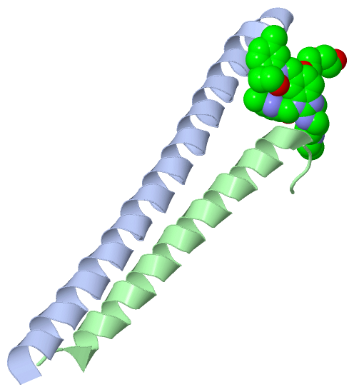
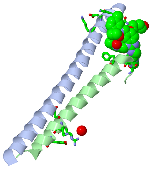
Chains, Units
Summary Information (see also Sequences/Alignments below) |
Ligands, Modified Residues, Ions (2, 2)| Asymmetric Unit (2, 2) Biological Unit 1 (2, 6) |
Sites (2, 2)
Asymmetric Unit (2, 2)
|
SS Bonds (0, 0)| (no "SS Bond" information available for 3KPE) |
Cis Peptide Bonds (1, 1)
Asymmetric Unit
|
||||||||
SAPs(SNPs)/Variants (0, 0)| (no "SAP(SNP)/Variant" information available for 3KPE) |
PROSITE Motifs (0, 0)| (no "PROSITE Motif" information available for 3KPE) |
Exons (0, 0)| (no "Exon" information available for 3KPE) |
Sequences/Alignments
Asymmetric UnitChain A from PDB Type:PROTEIN Length:49 aligned with FUS_HRSVA | P03420 from UniProtKB/Swiss-Prot Length:574 Alignment length:49 168 178 188 198 FUS_HRSVA 159 HLEGEVNKIKSALLSTNKAVVSLSNGVSVLTSKVLDLKNYIDKQLLPIV 207 SCOP domains ------------------------------------------------- SCOP domains CATH domains 3kpeA00 A:159-207 CATH domains Pfam domains ------------------------------------------------- Pfam domains SAPs(SNPs) ------------------------------------------------- SAPs(SNPs) PROSITE ------------------------------------------------- PROSITE Transcript ------------------------------------------------- Transcript 3kpe A 159 HLEGEVNKIKSALLSTNKAVVSLSNGVSVLTSKVLDLKNYIDKQLLPIV 207 168 178 188 198 Chain B from PDB Type:PROTEIN Length:35 aligned with FUS_HRSVA | P03420 from UniProtKB/Swiss-Prot Length:574 Alignment length:35 492 502 512 FUS_HRSVA 483 FPSDEFDASISQVNEKINQSLAFIRKSDELLHNVN 517 SCOP domains ----------------------------------- SCOP domains CATH domains ----------------------------------- CATH domains Pfam domains (1) Fusion_gly-3kpeB01 B:483-517 Pfam domains (1) Pfam domains (2) Fusion_gly-3kpeB02 B:483-517 Pfam domains (2) SAPs(SNPs) ----------------------------------- SAPs(SNPs) PROSITE ----------------------------------- PROSITE Transcript ----------------------------------- Transcript 3kpe B 483 FPSDEFDASISQVNEKINQSLAFIRKSDELLHNVN 517 492 502 512
|
||||||||||||||||||||
SCOP Domains (0, 0)| (no "SCOP Domain" information available for 3KPE) |
CATH Domains (1, 1)
Asymmetric Unit
 |
Pfam Domains (1, 2)
Asymmetric Unit
|
Gene Ontology (12, 12)|
Asymmetric Unit(hide GO term definitions) Chain A,B (FUS_HRSVA | P03420)
|
||||||||||||||||||||||||||||||||||||||||||||||||||||||||||||||||||||||||||||||||||||
Interactive Views
|
|||||||||||||||||||||||||||||||||||||||||||||||||||||||||||||||||||||||||||||||||||||||||||||||||||||||||||||||||||||||||||||||||||||||||||||||||||||||
Still Images
|
||||||||||||||||
Databases
|
||||||||||||||||||||||||||||||||||||||||||||||||||||||||||||||||||||||||||||||||||||||||||||||||||||||||||||||||||||||||||||||||||||||||||||||||||||||||||||||||
Analysis Tools
|
|||||||||||||||||||||||||||||||||||||||||||||||||||||||||||||
Entries Sharing at Least One Protein Chain (UniProt ID)
Related Entries Specified in the PDB File
|
|