|
|
|
|
 Description
|
|
Compounds
|
||||||||||||||||||||||||||||||||||||||||||||
Chains, Units
Summary Information (see also Sequences/Alignments below) |
Ligands, Modified Residues, Ions (1, 1)
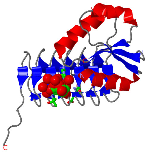
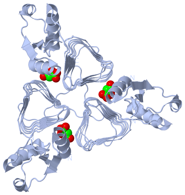
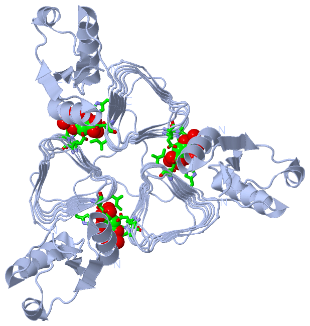
Asymmetric Unit (1, 1)
|
Sites (1, 1)
Asymmetric Unit (1, 1)
|
SS Bonds (0, 0)| (no "SS Bond" information available for 3BSW) |
Cis Peptide Bonds (0, 0)| (no "Cis Peptide Bond" information available for 3BSW) |
SAPs(SNPs)/Variants (0, 0)| (no "SAP(SNP)/Variant" information available for 3BSW) |
PROSITE Motifs (0, 0)| (no "PROSITE Motif" information available for 3BSW) |
Exons (0, 0)| (no "Exon" information available for 3BSW) |
Sequences/Alignments
Asymmetric UnitChain A from PDB Type:PROTEIN Length:191 aligned with PGLD_CAMJE | Q0P9D1 from UniProtKB/Swiss-Prot Length:195 Alignment length:193 12 22 32 42 52 62 72 82 92 102 112 122 132 142 152 162 172 182 192 PGLD_CAMJE 3 RTEKIYIYGASGHGLVCEDVAKNMGYKECIFLDDFKGMKFESTLPKYDFFIAIGNNEIRKKIYQKISENGFKIVNLIHKSALISPSAIVEENAGILIMPYVVINAKAKIEKGVILNTSSVIEHECVIGEFSHVSVGAKCAGNVKIGKNCFLGINSCVLPNLSLADDSILGGGATLVKNQDEKGVFVGVPAKRM 195 SCOP domains d3bswa_ A : Acetyltransferase PglD SCOP domains CATH domains 3bswA01 A :3-76 [code=3.40.50.20, no name defined] 3bswA02 A:77-188 Hexapeptide repeat proteins ------- CATH domains Pfam domains ------------------------------------------------------------------------------------------------------------------------------------------------------------------------------------------------- Pfam domains SAPs(SNPs) ------------------------------------------------------------------------------------------------------------------------------------------------------------------------------------------------- SAPs(SNPs) PROSITE ------------------------------------------------------------------------------------------------------------------------------------------------------------------------------------------------- PROSITE Transcript ------------------------------------------------------------------------------------------------------------------------------------------------------------------------------------------------- Transcript 3bsw A 3 RTEKIYIYG--GHGLVCEDVAKNMGYKECIFLDDFKGMKFESTLPKYDFFIAIGNNEIRKKIYQKISENGFKIVNLIHKSALISPSAIVEENAGILIMPYVVINAKAKIEKGVILNTSSVIEHECVIGEFSHVSVGAKCAGNVKIGKNCFLGINSCVLPNLSLADDSILGGGATLVKNQDEKGVFVGVPAKRM 195 |- | 22 32 42 52 62 72 82 92 102 112 122 132 142 152 162 172 182 192 11 14
|
||||||||||||||||||||
SCOP Domains (1, 1)
Asymmetric Unit
 |
CATH Domains (2, 2)
Asymmetric Unit
|
Pfam Domains (0, 0)| (no "Pfam Domain" information available for 3BSW) |
Gene Ontology (5, 5)|
Asymmetric Unit(hide GO term definitions) Chain A (PGLD_CAMJE | Q0P9D1)
|
||||||||||||||||||||||||||||||||||||||||||
Interactive Views
|
||||||||||||||||||||||||||||||||||||||||||||||||||||||||||||||||||||||||||||||||||||||||||||||||||||||||||||||||||||||||||||||||||||||||
Still Images
|
||||||||||||||||
Databases
|
||||||||||||||||||||||||||||||||||||||||||||||||||||||||||||||||||||||||||||||||||||||||||||||||||||||||||||||||||||||||||||||||||||||||||||||||||||||||||||||||
Analysis Tools
|
|||||||||||||||||||||||||||||||||||||||||||||||||||||||||||||
Entries Sharing at Least One Protein Chain (UniProt ID)
Related Entries Specified in the PDB File
|
|