|
|
|
|
 Description
|
|
Compounds
|
||||||||||||||||||||||||||||||||||||||||||||
Chains, Units
Summary Information (see also Sequences/Alignments below) |
Ligands, Modified Residues, Ions (1, 1)
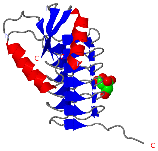
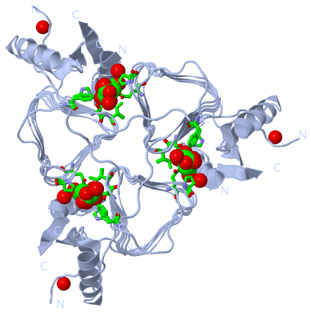
Asymmetric Unit (1, 1)
|
Sites (1, 1)
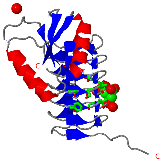
Asymmetric Unit (1, 1)
|
SS Bonds (0, 0)| (no "SS Bond" information available for 3BFP) |
Cis Peptide Bonds (0, 0)| (no "Cis Peptide Bond" information available for 3BFP) |
SAPs(SNPs)/Variants (0, 0)| (no "SAP(SNP)/Variant" information available for 3BFP) |
PROSITE Motifs (0, 0)| (no "PROSITE Motif" information available for 3BFP) |
Exons (0, 0)| (no "Exon" information available for 3BFP) |
Sequences/Alignments
Asymmetric UnitChain A from PDB Type:PROTEIN Length:184 aligned with PGLD_CAMJE | Q0P9D1 from UniProtKB/Swiss-Prot Length:195 Alignment length:194 11 21 31 41 51 61 71 81 91 101 111 121 131 141 151 161 171 181 191 PGLD_CAMJE 2 ARTEKIYIYGASGHGLVCEDVAKNMGYKECIFLDDFKGMKFESTLPKYDFFIAIGNNEIRKKIYQKISENGFKIVNLIHKSALISPSAIVEENAGILIMPYVVINAKAKIEKGVILNTSSVIEHECVIGEFSHVSVGAKCAGNVKIGKNCFLGINSCVLPNLSLADDSILGGGATLVKNQDEKGVFVGVPAKRM 195 SCOP domains d3bfpa_ A: Acetyltransferase Pgl D SCOP domains CATH domains 3bfpA01 A: 2-76 [code=3.40.50.20 , no name defined] 3bfpA02 A:77-195 Hexapeptide repeat proteins CATH domains Pfam domains -------------------------------------------------------------------------------------------------------------------------------------------------------------------------------------------------- Pfam domains SAPs(SNPs) -------------------------------------------------------------------------------------------------------------------------------------------------------------------------------------------------- SAPs(SNPs) PROSITE -------------------------------------------------------------------------------------------------------------------------------------------------------------------------------------------------- PROSITE Transcript -------------------------------------------------------------------------------------------------------------------------------------------------------------------------------------------------- Transcript 3bfp A 2 ARTEKIYIYG--GHGLVCEDVAKNMGYKECIFLD--------STLPKYDFFIAIGNNEIRKKIYQKISENGFKIVNLIHKSALISPSAIVEENAGILIMPYVVINAKAKIEKGVILNTSSVIEHECVIGEFSHVSVGAKCAGNVKIGKNCFLGINSCVLPNLSLADDSILGGGATLVKNQDEKGVFVGVPAKRM 195 11 | 21 31 | - | 51 61 71 81 91 101 111 121 131 141 151 161 171 181 191 11 14 35 44
|
||||||||||||||||||||
SCOP Domains (1, 1)
Asymmetric Unit
 |
CATH Domains (2, 2)
Asymmetric Unit
|
Pfam Domains (0, 0)| (no "Pfam Domain" information available for 3BFP) |
Gene Ontology (5, 5)|
Asymmetric Unit(hide GO term definitions) Chain A (PGLD_CAMJE | Q0P9D1)
|
||||||||||||||||||||||||||||||||||||||||||
Interactive Views
|
||||||||||||||||||||||||||||||||||||||||||||||||||||||||||||||||||||||||||||||||||||||||||||||||||||||||||||||||||||||||||||||||||||||||
Still Images
|
||||||||||||||||
Databases
|
||||||||||||||||||||||||||||||||||||||||||||||||||||||||||||||||||||||||||||||||||||||||||||||||||||||||||||||||||||||||||||||||||||||||||||||||||||||||||||||||
Analysis Tools
|
|||||||||||||||||||||||||||||||||||||||||||||||||||||||||||||
Entries Sharing at Least One Protein Chain (UniProt ID)
Related Entries Specified in the PDB File
|
|