Show PDB file:   
         Plain Text   HTML   (compressed file size)
QuickSearch:   
by PDB,NDB,UniProt,PROSITE Code or Search Term(s)  
(-)Asymmetric Unit
(-)Asym. Unit - sites
(-)Biological Unit 1
(-)Biol. Unit 1 - sites
(-)Biological Unit 2
collapse expand < >
Image Asymmetric Unit
Asymmetric Unit  (Jmol Viewer)
Image Asym. Unit - sites
Asym. Unit - sites  (Jmol Viewer)
Image Biological Unit 1
Biological Unit 1  (Jmol Viewer)
Image Biol. Unit 1 - sites
Biol. Unit 1 - sites  (Jmol Viewer)
Image Biological Unit 2
Biological Unit 2  (Jmol Viewer)

(-) Description

Title :  CRYSTAL STRUCTURE OF P. AERUGINOAS AZURIN CONTAINING A TYR-HIS HYDROGEN BONDED PAIR
 
Authors :  J. J. Warren, J. R. Winkler, H. B. Gray
Date :  30 Sep 11  (Deposition) - 28 Dec 11  (Release) - 13 Jun 12  (Revision)
Method :  X-RAY DIFFRACTION
Resolution :  1.18
Chains :  Asym. Unit :  A,B
Biol. Unit 1:  A  (1x)
Biol. Unit 2:  B  (1x)
Keywords :  Azurin, Cupredoxin, Electron Transport (Keyword Search: [Gene Ontology, PubMed, Web (Google))
 
Reference :  J. J. Warren, J. R. Winkler, H. B. Gray
Redox Properties Of Tyrosine And Related Molecules.
Febs Lett. V. 586 596 2012
PubMed-ID: 22210190  |  Reference-DOI: 10.1016/J.FEBSLET.2011.12.014

(-) Compounds

Molecule 1 - AZURIN
    ChainsA, B
    EngineeredYES
    Expression SystemESCHERICHIA COLI
    Expression System PlasmidPET3A
    Expression System StrainBL21DE3
    Expression System Taxid469008
    Expression System Vector TypePLASMID
    FragmentUNP RESIDUES 22-148
    GeneAZU, PA4922
    MutationYES
    Organism ScientificPSEUDOMONAS AERUGINOSA
    Organism Taxid287

 Structural Features

(-) Chains, Units

  12
Asymmetric Unit AB
Biological Unit 1 (1x)A 
Biological Unit 2 (1x) B

Summary Information (see also Sequences/Alignments below)

(-) Ligands, Modified Residues, Ions  (2, 4)

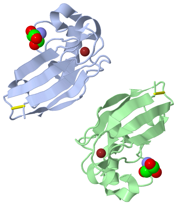
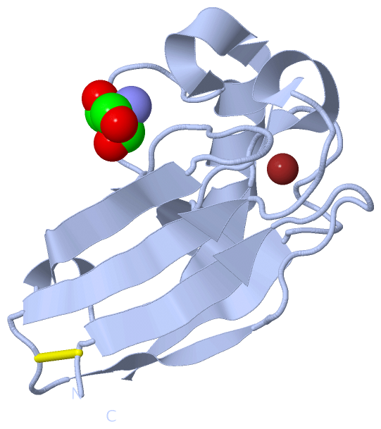
Asymmetric Unit (2, 4)
No.NameCountTypeFull Name
1CU2Ligand/IonCOPPER (II) ION
2TRS2Ligand/Ion2-AMINO-2-HYDROXYMETHYL-PROPANE-1,3-DIOL
Biological Unit 1 (1, 1)
No.NameCountTypeFull Name
1CU-1Ligand/IonCOPPER (II) ION
2TRS1Ligand/Ion2-AMINO-2-HYDROXYMETHYL-PROPANE-1,3-DIOL
Biological Unit 2 (1, 1)
No.NameCountTypeFull Name
1CU-1Ligand/IonCOPPER (II) ION
2TRS1Ligand/Ion2-AMINO-2-HYDROXYMETHYL-PROPANE-1,3-DIOL

(-) Sites  (4, 4)

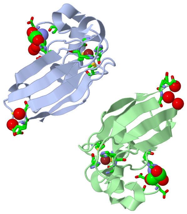
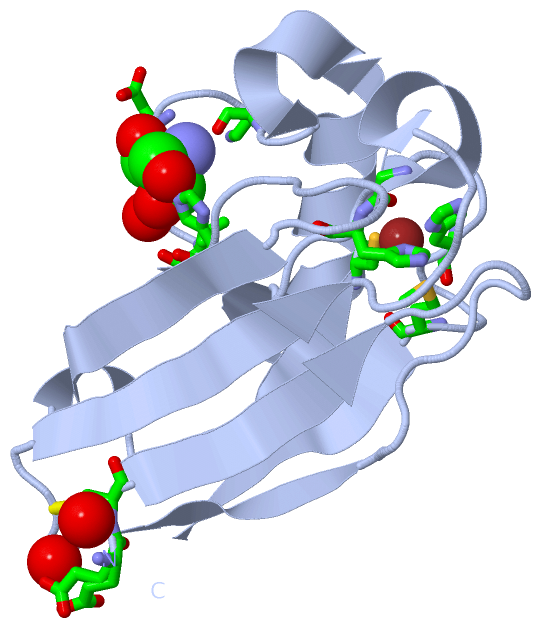
Asymmetric Unit (4, 4)
No.NameEvidenceResiduesDescription
1AC1SOFTWAREGLY A:45 , HIS A:46 , CYS A:112 , HIS A:117 , MET A:121BINDING SITE FOR RESIDUE CU A 1
2AC2SOFTWAREGLU A:2 , CYS A:3 , LYS A:74 , ASP A:76 , VAL A:80 , HIS A:83 , HOH A:144 , HOH A:151 , HOH A:194BINDING SITE FOR RESIDUE TRS A 129
3AC3SOFTWAREGLY B:45 , HIS B:46 , CYS B:112 , HIS B:117 , MET B:121BINDING SITE FOR RESIDUE CU B 129
4AC4SOFTWAREGLU B:2 , CYS B:3 , LYS B:74 , ASP B:76 , ASP B:77 , HIS B:83 , HOH B:193 , HOH B:200 , HOH B:248BINDING SITE FOR RESIDUE TRS B 1

(-) SS Bonds  (2, 2)

Asymmetric Unit
No.Residues
1A:3 -A:26
2B:3 -B:26

(-) Cis Peptide Bonds  (0, 0)

(no "Cis Peptide Bond" information available for 3U25)

 Sequence-Structure Mapping

(-) SAPs(SNPs)/Variants  (0, 0)

(no "SAP(SNP)/Variant" information available for 3U25)

(-) PROSITE Motifs  (1, 2)

Asymmetric Unit (1, 2)
 PROSITEUniProtKBPDB
No.IDACDescriptionIDLocationCountLocation
1COPPER_BLUEPS00196 Type-1 copper (blue) proteins signature.AZUR_PSEAE125-141
 
  2A:105-121
B:105-121
Biological Unit 1 (1, 1)
 PROSITEUniProtKBPDB
No.IDACDescriptionIDLocationCountLocation
1COPPER_BLUEPS00196 Type-1 copper (blue) proteins signature.AZUR_PSEAE125-141
 
  1A:105-121
-
Biological Unit 2 (1, 1)
 PROSITEUniProtKBPDB
No.IDACDescriptionIDLocationCountLocation
1COPPER_BLUEPS00196 Type-1 copper (blue) proteins signature.AZUR_PSEAE125-141
 
  1-
B:105-121

(-) Exons   (0, 0)

(no "Exon" information available for 3U25)

(-) Sequences/Alignments

Asymmetric Unit
   Reformat: Number of residues per line =  ('0' or empty: single-line sequence representation)
  Number of residues per labelling interval =   
  UniProt sequence: complete  aligned part    
   Show mapping: SCOP domains CATH domains Pfam domains Secondary structure (by author)
SAPs(SNPs) PROSITE motifs Exons
(details for a mapped element are shown in a popup box when the mouse pointer rests over it)
Chain A from PDB  Type:PROTEIN  Length:127
 aligned with AZUR_PSEAE | P00282 from UniProtKB/Swiss-Prot  Length:148

    Alignment length:127
                                    31        41        51        61        71        81        91       101       111       121       131       141       
           AZUR_PSEAE    22 ECSVDIQGNDQMQFNTNAITVDKSCKQFTVNLSHPGNLPKNVMGHNWVLSTAADMQGVVTDGMASGLDKDYLKPDDSRVIAHTKLIGSGEKDSVTFDVSKLKEGEQYMFFCTFPGHSALMKGTLTLK 148
               SCOP domains d3u25a_ A: Azurin                                                                                                               SCOP domains
               CATH domains ------------------------------------------------------------------------------------------------------------------------------- CATH domains
               Pfam domains ------------------------------------------------------------------------------------------------------------------------------- Pfam domains
         Sec.struct. author ..eeeee..........eeeee....eeeeeee.....hhhhhh...eeee..hhhhhhhhhhh.hhhhh..........ee........eeeeeee.hhh.....eeee..........eeeeeee Sec.struct. author
                 SAPs(SNPs) ------------------------------------------------------------------------------------------------------------------------------- SAPs(SNPs)
                    PROSITE -------------------------------------------------------------------------------------------------------COPPER_BLUE      ------- PROSITE
                 Transcript ------------------------------------------------------------------------------------------------------------------------------- Transcript
                 3u25 A   2 ECSVDIQGNDQMQFNTNAHTVDKSCKQFTVNLSHPGNLPKNVMGHNYVLSTAADMQGVVTDGMASGLDKDFLKPDDSRVIAHTKLIGSGEKDSVTFDVSKLKEGEQFMFFCTFPGHSALMKGTLTLK 128
                                    11        21        31        41        51        61        71        81        91       101       111       121       

Chain B from PDB  Type:PROTEIN  Length:127
 aligned with AZUR_PSEAE | P00282 from UniProtKB/Swiss-Prot  Length:148

    Alignment length:127
                                    31        41        51        61        71        81        91       101       111       121       131       141       
           AZUR_PSEAE    22 ECSVDIQGNDQMQFNTNAITVDKSCKQFTVNLSHPGNLPKNVMGHNWVLSTAADMQGVVTDGMASGLDKDYLKPDDSRVIAHTKLIGSGEKDSVTFDVSKLKEGEQYMFFCTFPGHSALMKGTLTLK 148
               SCOP domains d3u25b_ B: Azurin                                                                                                               SCOP domains
               CATH domains ------------------------------------------------------------------------------------------------------------------------------- CATH domains
               Pfam domains ------------------------------------------------------------------------------------------------------------------------------- Pfam domains
         Sec.struct. author ..eeeee..........eeeee....eeeeeee.....hhhhhh...eeee..hhhhhhhhhhhhhhhhh..........ee........eeeeeee.hhh.....eeee..........eeeeeee Sec.struct. author
                 SAPs(SNPs) ------------------------------------------------------------------------------------------------------------------------------- SAPs(SNPs)
                    PROSITE -------------------------------------------------------------------------------------------------------COPPER_BLUE      ------- PROSITE
                 Transcript ------------------------------------------------------------------------------------------------------------------------------- Transcript
                 3u25 B   2 ECSVDIQGNDQMQFNTNAHTVDKSCKQFTVNLSHPGNLPKNVMGHNYVLSTAADMQGVVTDGMASGLDKDFLKPDDSRVIAHTKLIGSGEKDSVTFDVSKLKEGEQFMFFCTFPGHSALMKGTLTLK 128
                                    11        21        31        41        51        61        71        81        91       101       111       121       

   Legend:   → Mismatch (orange background)
  - → Gap (green background, '-', border residues have a numbering label)
    → Modified Residue (blue background, lower-case, 'x' indicates undefined single-letter code, labelled with number + name)
  x → Chemical Group (purple background, 'x', labelled with number + name, e.g. ACE or NH2)
  extra numbering lines below/above indicate numbering irregularities and modified residue names etc., number ends below/above '|'

 Classification and Annotation

(-) SCOP Domains  (1, 2)

Asymmetric Unit

(-) CATH Domains  (0, 0)

(no "CATH Domain" information available for 3U25)

(-) Pfam Domains  (0, 0)

(no "Pfam Domain" information available for 3U25)

(-) Gene Ontology  (7, 7)

Asymmetric Unit(hide GO term definitions)
Chain A,B   (AZUR_PSEAE | P00282)
molecular function
    GO:0005507    copper ion binding    Interacting selectively and non-covalently with copper (Cu) ions.
    GO:0009055    electron carrier activity    Any molecular entity that serves as an electron acceptor and electron donor in an electron transport chain. An electron transport chain is a process in which a series of electron carriers operate together to transfer electrons from donors to any of several different terminal electron acceptors to generate a transmembrane electrochemical gradient.
    GO:0046872    metal ion binding    Interacting selectively and non-covalently with any metal ion.
    GO:0046914    transition metal ion binding    Interacting selectively and non-covalently with a transition metal ions; a transition metal is an element whose atom has an incomplete d-subshell of extranuclear electrons, or which gives rise to a cation or cations with an incomplete d-subshell. Transition metals often have more than one valency state. Biologically relevant transition metals include vanadium, manganese, iron, copper, cobalt, nickel, molybdenum and silver.
    GO:0008270    zinc ion binding    Interacting selectively and non-covalently with zinc (Zn) ions.
biological process
    GO:0055114    oxidation-reduction process    A metabolic process that results in the removal or addition of one or more electrons to or from a substance, with or without the concomitant removal or addition of a proton or protons.
cellular component
    GO:0042597    periplasmic space    The region between the inner (cytoplasmic) and outer membrane (Gram-negative Bacteria) or cytoplasmic membrane and cell wall (Fungi and Gram-positive Bacteria).

 Visualization

(-) Interactive Views

Asymmetric Unit
  Complete Structure
    Jena3D(integrated viewing of ligand, site, SAP, PROSITE, SCOP information)
    WebMol | AstexViewer[tm]@PDBe
(Java Applets, require no local installation except for Java; loading may be slow)
    STRAP
(Java WebStart application, automatic local installation, requires Java; full application with system access!)
    RasMol
(require local installation)
    Molscript (VRML)
(requires installation of a VRML viewer; select preferred view via VRML and generate a mono or stereo PDF format file)
 
  Ligands, Modified Residues, Ions
    CU  [ RasMol | Jena3D ]  +environment [ RasMol | Jena3D ]
    TRS  [ RasMol | Jena3D ]  +environment [ RasMol | Jena3D ]
 
  Sites
    AC1  [ RasMol ]  +environment [ RasMol ]
    AC2  [ RasMol ]  +environment [ RasMol ]
    AC3  [ RasMol ]  +environment [ RasMol ]
    AC4  [ RasMol ]  +environment [ RasMol ]
 
  Cis Peptide Bonds
(no "Cis Peptide Bonds" information available for 3u25)
 
Biological Units
  Complete Structure
    Biological Unit 1  [ Jena3D ]
    Biological Unit 2  [ Jena3D ]

(-) Still Images

Jmol
  protein: cartoon or spacefill or dots and stick; nucleic acid: cartoon and stick; ligands: spacefill; active site: stick
Molscript
  protein, nucleic acid: cartoon; ligands: spacefill; active site: ball and stick

 Databases and Analysis Tools

(-) Databases

Access by PDB/NDB ID
  3u25
    Family and Domain InformationProDom | SYSTERS
    General Structural InformationGlycoscienceDB | MMDB | NDB | OCA | PDB | PDBe | PDBj | PDBsum | PDBWiki | PQS | PROTEOPEDIA
    Orientation in MembranesOPM
    Protein SurfaceSURFACE
    Secondary StructureDSSP (structure derived) | HSSP (homology derived)
    Structural GenomicsGeneCensus
    Structural NeighboursCE | VAST
    Structure ClassificationCATH | Dali | SCOP
    Validation and Original DataBMRB Data View | BMRB Restraints Grid | EDS | PROCHECK | RECOORD | WHAT_CHECK
 
Access by UniProt ID/Accession number
  AZUR_PSEAE | P00282
    Comparative Protein Structure ModelsModBase
    Genomic InformationEnsembl
    Protein-protein InteractionDIP
    Sequence, Family and Domain InformationInterPro | Pfam | SMART | UniProtKB/SwissProt
 
Access by Enzyme Classificator   (EC Number)
  (no 'Enzyme Classificator' available)
    General Enzyme InformationBRENDA | EC-PDB | Enzyme | IntEnz
    PathwayKEGG | MetaCyc
 
Access by Disease Identifier   (MIM ID)
  (no 'MIM ID' available)
    Disease InformationOMIM
 
Access by GenAge ID
  (no 'GenAge ID' available)
    Age Related InformationGenAge

(-) Analysis Tools

Access by PDB/NDB ID
    Domain InformationXDom
    Interatomic Contacts of Structural UnitsCSU
    Ligand-protein ContactsLPC
    Protein CavitiescastP
    Sequence and Secondary StructurePDBCartoon
    Structure AlignmentSTRAP(Java WebStart application, automatic local installation, requires Java; full application with system access!)
    Structure and Sequence BrowserSTING
 
Access by UniProt ID/Accession number
  AZUR_PSEAE | P00282
    Protein Disorder PredictionDisEMBL | FoldIndex | GLOBPLOT (for more information see DisProt)

 Related Entries

(-) Entries Sharing at Least One Protein Chain (UniProt ID)

UniProtKB/Swiss-Prot
        AZUR_PSEAE | P002821ag0 1azn 1azr 1azu 1bex 1cc3 1e5y 1e5z 1e65 1e67 1etj 1ezl 1gr7 1i53 1ils 1ilu 1jvl 1jvo 1jze 1jzf 1jzg 1jzh 1jzi 1jzj 1nzr 1r1c 1vlx 1xb3 1xb6 1xb8 2azu 2fnw 2ft6 2ft7 2ft8 2fta 2ghz 2gi0 2hx7 2hx8 2hx9 2hxa 2i7o 2i7s 2idf 2iwe 2oj1 2tsa 2tsb 2xv0 2xv2 2xv3 3azu 3fpy 3fq1 3fq2 3fqy 3fs9 3fsa 3fsv 3fsw 3fsz 3ft0 3ibo 3in0 3in2 3jt2 3jtb 3n2j 3np3 3np4 3oqr 3uge 4azu 4bww 4hhg 4hhw 4hip 4hz1 4jkn 4k9j 4ko5 4ko6 4ko7 4ko9 4kob 4koc 4mfh 4qkt 4qlw 4wkx 5azu 5i26 5i28

(-) Related Entries Specified in the PDB File

(no "Related Entries Specified in the PDB File" available for 3U25)