|
|
|
|
 Description
|
|
Compounds
|
||||||||||||||||||||||||||||||||||||||||||||||||||||||||||||||||||||||||||||||||||||||||||||||||||||||||||||||
Chains, Units
Summary Information (see also Sequences/Alignments below) |
Ligands, Modified Residues, Ions (1, 1)
Asymmetric Unit (1, 1)
|
Sites (1, 1)
Asymmetric Unit (1, 1)
|
SS Bonds (0, 0)| (no "SS Bond" information available for 3BZX) |
Cis Peptide Bonds (0, 0)| (no "Cis Peptide Bond" information available for 3BZX) |
SAPs(SNPs)/Variants (0, 0)| (no "SAP(SNP)/Variant" information available for 3BZX) |
PROSITE Motifs (0, 0)| (no "PROSITE Motif" information available for 3BZX) |
Exons (0, 0)| (no "Exon" information available for 3BZX) |
Sequences/Alignments
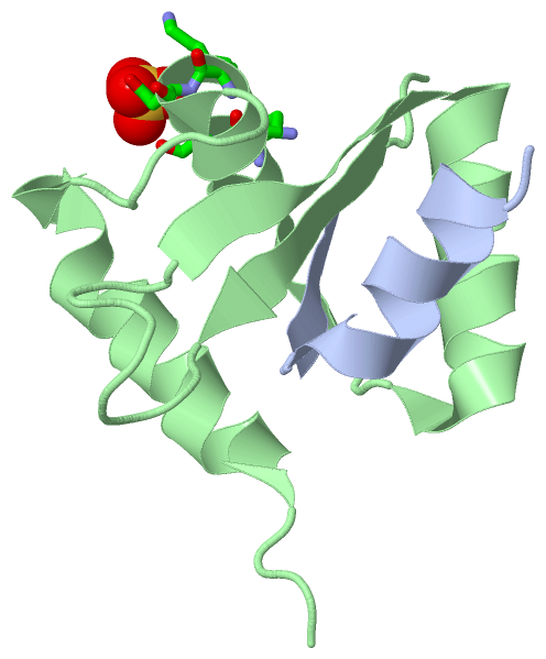
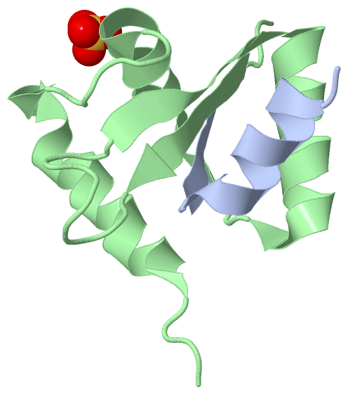
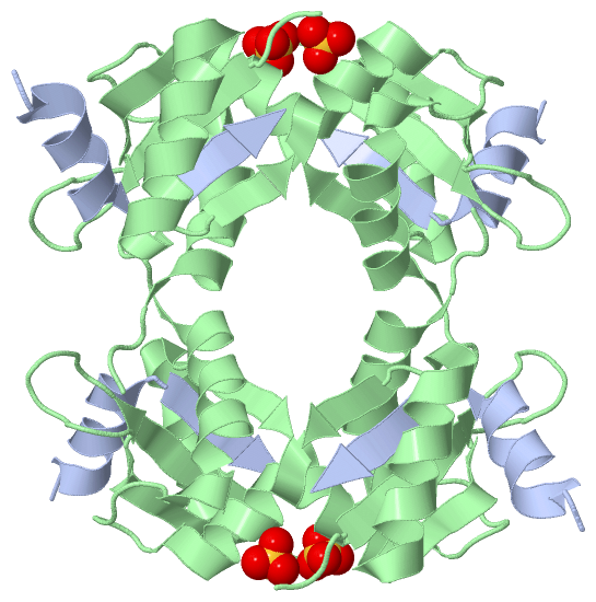
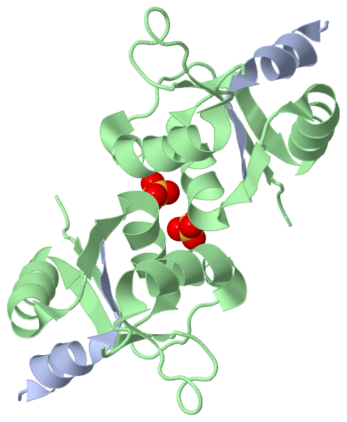
Asymmetric UnitChain A from PDB Type:PROTEIN Length:17 aligned with Q9AJ26_ECOLX | Q9AJ26 from UniProtKB/TrEMBL Length:345 Alignment length:17 255 Q9AJ26_ECOLX 246 SGSLANNIKKSTVIVKN 262 SCOP domains d3bzx.1 SCOP domains CATH domains ----------------- CATH domains Pfam domains ----------------- Pfam domains SAPs(SNPs) ----------------- SAPs(SNPs) PROSITE ----------------- PROSITE Transcript ----------------- Transcript 3bzx A 246 SGSLANNIKKSTVIVKN 262 255 Chain B from PDB Type:PROTEIN Length:83 aligned with Q9AJ26_ECOLX | Q9AJ26 from UniProtKB/TrEMBL Length:345 Alignment length:83 272 282 292 302 312 322 332 342 Q9AJ26_ECOLX 263 PTHIAICLYYKLGETPLPLVIETGKDAKALQIIKLAELYDIPVIEDIPLARSLYKNIHKGQYITEDFFEPVAQLIRIAIDLDY 345 SCOP domains d3bzx.1 A:246-262,B:263-345 Type III secretion proteins EscU SCOP domains CATH domains 3bzxB00 B:263-345 secretion proteins EscU CATH domains Pfam domains ----------------------------------------------------------------------------------- Pfam domains SAPs(SNPs) ----------------------------------------------------------------------------------- SAPs(SNPs) PROSITE ----------------------------------------------------------------------------------- PROSITE Transcript ----------------------------------------------------------------------------------- Transcript 3bzx B 263 PTAIAICLYYKLGETPLPLVIETGKDAKALQIIKLAELYDIPVIEDIPLARSLYKNIHKGQYITEDFFEPVAQLIRIAIDLDY 345 272 282 292 302 312 322 332 342
|
||||||||||||||||||||
SCOP Domains (1, 1)
Asymmetric Unit

|
CATH Domains (1, 1)
Asymmetric Unit
|
Pfam Domains (0, 0)| (no "Pfam Domain" information available for 3BZX) |
Gene Ontology (6, 6)|
Asymmetric Unit(hide GO term definitions) Chain A,B (Q9AJ26_ECOLX | Q9AJ26)
|
||||||||||||||||||||||||||||||||||||||||||||||||||||||
Interactive Views
|
|||||||||||||||||||||||||||||||||||||||||||||||||||||||||||||||||||||||||||||||||||||||||||||||||||||||||||||||||||||||||||||||||||||||||||||||||||||||
Still Images
|
||||||||||||||||
Databases
|
||||||||||||||||||||||||||||||||||||||||||||||||||||||||||||||||||||||||||||||||||||||||||||||||||||||||||||||||||||||||||||||||||||||||||||||||||||||||||||||||
Analysis Tools
|
|||||||||||||||||||||||||||||||||||||||||||||||||||||||||||||
Entries Sharing at Least One Protein Chain (UniProt ID)
Related Entries Specified in the PDB File
|
|