| molecular function |
|---|
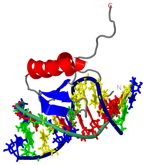
| | GO:0003677 | | DNA binding | | Any molecular function by which a gene product interacts selectively and non-covalently with DNA (deoxyribonucleic acid). |
| | GO:0016787 | | hydrolase activity | | Catalysis of the hydrolysis of various bonds, e.g. C-O, C-N, C-C, phosphoric anhydride bonds, etc. Hydrolase is the systematic name for any enzyme of EC class 3. |
| | GO:0008907 | | integrase activity | | Catalysis of the integration of one segment of DNA into another. |
| | GO:0003676 | | nucleic acid binding | | Interacting selectively and non-covalently with any nucleic acid. |
| | GO:0016740 | | transferase activity | | Catalysis of the transfer of a group, e.g. a methyl group, glycosyl group, acyl group, phosphorus-containing, or other groups, from one compound (generally regarded as the donor) to another compound (generally regarded as the acceptor). Transferase is the systematic name for any enzyme of EC class 2. |
| biological process |
|---|
| | GO:0015074 | | DNA integration | | The process in which a segment of DNA is incorporated into another, usually larger, DNA molecule such as a chromosome. |
| | GO:0006310 | | DNA recombination | | Any process in which a new genotype is formed by reassortment of genes resulting in gene combinations different from those that were present in the parents. In eukaryotes genetic recombination can occur by chromosome assortment, intrachromosomal recombination, or nonreciprocal interchromosomal recombination. Intrachromosomal recombination occurs by crossing over. In bacteria it may occur by genetic transformation, conjugation, transduction, or F-duction. |
| | GO:0075713 | | establishment of integrated proviral latency | | A process by which the virus integrates into the host genome and establishes as a stable provirus or prophage. |
| | GO:0046718 | | viral entry into host cell | | The process that occurs after viral attachment by which a virus, or viral nucleic acid, breaches the plasma membrane or cell envelope and enters the host cell. The process ends when the viral nucleic acid is released into the host cell cytoplasm. |
 Description