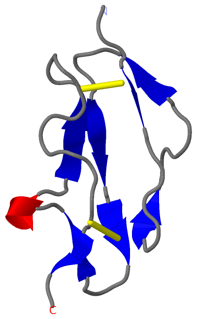
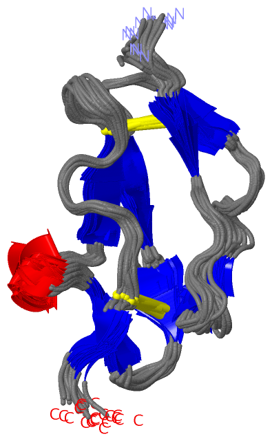
Show PDB file:   
         Plain Text   HTML   (compressed file size)
QuickSearch:   
by PDB,NDB,UniProt,PROSITE Code or Search Term(s)  
(-)NMR Structure - model 1
(-)NMR Structure - all models
collapse expand < >
Image NMR Structure - model 1
NMR Structure - model 1  (Jmol Viewer)
Image NMR Structure - all models
NMR Structure - all models  (Jmol Viewer)

(-) Description

Title :  STRUCTURE OF CCP MODULE 7 OF COMPLEMENT FACTOR H - THE AMD AT RISK VARIENT (402H)
 
Authors :  A. P. Herbert, J. A. Deakin, C. Q. Schmidt, B. S. Blaum, C. Egan, V. P. Ferreira, M. K. Pangburn, M. Lyon, D. Uhrin, P. N. Barlow
Date :  16 Feb 07  (Deposition) - 20 Mar 07  (Release) - 24 Feb 09  (Revision)
Method :  SOLUTION NMR
Resolution :  NOT APPLICABLE
Chains :  NMR Structure  :  A  (20x)
Keywords :  Age Related Macular Degeneration, Age-Related Macular Degeneration, Disease Mutation, Glycosaminoglycan, Alternative Splicing, Complement Alternate Pathway, Glycoprotein, Innate Immunity, Immune Response, Sushi, Factor H, Complement, Polymorphism (Keyword Search: [Gene Ontology, PubMed, Web (Google))
 
Reference :  A. P. Herbert, J. A. Deakin, C. Q. Schmidt, B. S. Blaum, C. Egan, V. P. Ferreira, M. K. Pangburn, M. Lyon, D. Uhrin, P. N. Barlow
Structure Shows That A Glycosaminoglycan And Protein Recognition Site In Factor H Is Perturbed By Age-Related Macular Degeneration-Linked Single Nucleotide Polymorphism.
J. Biol. Chem. V. 282 18960 2007
PubMed-ID: 17360715  |  Reference-DOI: 10.1074/JBC.M609636200

(-) Compounds

Molecule 1 - COMPLEMENT FACTOR H
    ChainsA
    EngineeredYES
    Expression SystemPICHIA PASTORIS
    Expression System PlasmidPPICZALPHAB
    Expression System StrainKM71H
    Expression System Taxid4922
    FragmentCCP MODULE 7, RESIDUES 386-446
    Organism CommonHUMAN
    Organism ScientificHOMO SAPIENS
    Organism Taxid9606
    SynonymH FACTOR 1

 Structural Features

(-) Chains, Units

  
NMR Structure (20x)

Summary Information (see also Sequences/Alignments below)

(-) Ligands, Modified Residues, Ions  (0, 0)

(no "Ligand,Modified Residues,Ions" information available for 2JGW)

(-) Sites  (0, 0)

(no "Site" information available for 2JGW)

(-) SS Bonds  (2, 2)

NMR Structure
No.Residues
1A:389 -A:431
2A:416 -A:442

(-) Cis Peptide Bonds  (0, 0)

(no "Cis Peptide Bond" information available for 2JGW)

 Sequence-Structure Mapping

(-) SAPs(SNPs)/Variants  (3, 3)

NMR Structure (3, 3)
  dbSNPPDB
No.SourceVariant IDVariantUniProt IDStatusIDChainVariant
1UniProtVAR_031980Q400KCFAH_HUMANDisease (AHUS1)  ---AQ400K
2UniProtVAR_001979Y402HCFAH_HUMANPolymorphism1061170AH402H
3UniProtVAR_031981C431SCFAH_HUMANDisease (CFHD)  ---AC431S

  SNP/SAP Summary Statistics (UniProtKB/Swiss-Prot)

(-) PROSITE Motifs  (1, 2)

NMR Structure (1, 2)
 PROSITEUniProtKBPDB
No.IDACDescriptionIDLocationCountLocation
1SUSHIPS50923 Sushi/CCP/SCR domain profile.CFAH_HUMAN19-82
83-143
144-207
208-264
265-322
387-444
446-507
515-566
567-625
628-686
689-746
751-805
809-866
868-928
929-986
987-1045
1046-1104
1107-1165
1194-1230
  2-
-
-
-
-
A:387-444
A:446-446
-
-
-
-
-
-
-
-
-
-
-
-

(-) Exons   (3, 3)

NMR Structure (3, 3)
 ENSEMBLUniProtKBPDB
No.Transcript IDExonExon IDGenome LocationLengthIDLocationLengthCountLocationLength
1.1aENST000003674291aENSE00001444484chr1:196621008-196621305298CFAH_HUMAN1-20200--
1.2aENST000003674292aENSE00001134426chr1:196642108-196642293186CFAH_HUMAN20-82630--
1.3ENST000003674293ENSE00000791013chr1:196642987-196643092106CFAH_HUMAN82-117360--
1.4ENST000003674294ENSE00000791014chr1:196645119-19664519577CFAH_HUMAN117-143270--
1.5bENST000003674295bENSE00000469039chr1:196646606-196646797192CFAH_HUMAN143-207650--
1.5cENST000003674295cENSE00000791016chr1:196648753-196648923171CFAH_HUMAN207-264580--
1.6ENST000003674296ENSE00000469041chr1:196654194-196654367174CFAH_HUMAN264-322590--
1.7ENST000003674297ENSE00001769047chr1:196658550-196658744195CFAH_HUMAN322-387661A:386-3872
1.8aENST000003674298aENSE00001597868chr1:196659193-196659369177CFAH_HUMAN387-446601A:387-44660
1.12ENST0000036742912ENSE00000469044chr1:196682865-196683047183CFAH_HUMAN446-507621A:446-4461
1.13ENST0000036742913ENSE00000469045chr1:196684723-196684899177CFAH_HUMAN507-566600--
1.14ENST0000036742914ENSE00000791022chr1:196694251-196694427177CFAH_HUMAN566-625600--
1.15ENST0000036742915ENSE00000791023chr1:196695600-196695782183CFAH_HUMAN625-686620--
1.16ENST0000036742916ENSE00000469048chr1:196695891-196696070180CFAH_HUMAN686-746610--
1.17ENST0000036742917ENSE00000791025chr1:196697476-196697652177CFAH_HUMAN746-805600--
1.18aENST0000036742918aENSE00000791026chr1:196705954-196706136183CFAH_HUMAN805-866620--
1.19ENST0000036742919ENSE00000791027chr1:196706605-196706790186CFAH_HUMAN866-928630--
1.20aENST0000036742920aENSE00000791028chr1:196709749-196709922174CFAH_HUMAN928-986590--
1.20dENST0000036742920dENSE00000469053chr1:196711005-196711181177CFAH_HUMAN986-1045600--
1.21ENST0000036742921ENSE00001622338chr1:196712582-196712758177CFAH_HUMAN1045-1104600--
1.22ENST0000036742922ENSE00001719380chr1:196714947-196715129183CFAH_HUMAN1104-1165620--
1.23ENST0000036742923ENSE00001931000chr1:196716241-196716634394CFAH_HUMAN1165-1231670--

(-) Sequences/Alignments

NMR Structure
   Reformat: Number of residues per line =  ('0' or empty: single-line sequence representation)
  Number of residues per labelling interval =   
  UniProt sequence: complete  aligned part    
   Show mapping: SCOP domains CATH domains Pfam domains Secondary structure (by author)
SAPs(SNPs) PROSITE motifs Exons
(details for a mapped element are shown in a popup box when the mouse pointer rests over it)
Chain A from PDB  Type:PROTEIN  Length:61
 aligned with CFAH_HUMAN | P08603 from UniProtKB/Swiss-Prot  Length:1231

    Alignment length:61
                                   395       405       415       425       435       445 
           CFAH_HUMAN   386 LRKCYFPYLENGYNQNYGRKFVQGKSIDVACHPGYALPKAQTTVTCMENGWSPTPRCIRVK 446
               SCOP domains ------------------------------------------------------------- SCOP domains
               CATH domains ------------------------------------------------------------- CATH domains
           Pfam domains (1) Sushi-2jgwA01 A:386-442                                  ---- Pfam domains (1)
           Pfam domains (2) Sushi-2jgwA02 A:386-442                                  ---- Pfam domains (2)
         Sec.struct. author ...ee......ee......ee....eee.ee...ee.hhh..eee............ee.. Sec.struct. author
                 SAPs(SNPs) --------------K-H----------------------------S--------------- SAPs(SNPs)
                    PROSITE -SUSHI  PDB: A:387-444 UniProt: 387-444                    -S PROSITE
           Transcript 1 (1) 1.----------------------------------------------------------1 Transcript 1 (1)
           Transcript 1 (2) -Exon 1.8a  PDB: A:387-446 UniProt: 387-446                   Transcript 1 (2)
                 2jgw A 386 LRKCYFPYLENGYNQNHGRKFVQGKSIDVACHPGYALPKAQTTVTCMENGWSPTPRCIRVK 446
                                   395       405       415       425       435       445 

   Legend:   → Mismatch (orange background)
  - → Gap (green background, '-', border residues have a numbering label)
    → Modified Residue (blue background, lower-case, 'x' indicates undefined single-letter code, labelled with number + name)
  x → Chemical Group (purple background, 'x', labelled with number + name, e.g. ACE or NH2)
  extra numbering lines below/above indicate numbering irregularities and modified residue names etc., number ends below/above '|'

 Classification and Annotation

(-) SCOP Domains  (0, 0)

(no "SCOP Domain" information available for 2JGW)

(-) CATH Domains  (0, 0)

(no "CATH Domain" information available for 2JGW)

(-) Pfam Domains  (1, 2)

NMR Structure
(-)
Family: Sushi (50)
1aSushi-2jgwA01A:386-442
1bSushi-2jgwA02A:386-442

(-) Gene Ontology  (12, 12)

NMR Structure(hide GO term definitions)
Chain A   (CFAH_HUMAN | P08603)
molecular function
    GO:0043395    heparan sulfate proteoglycan binding    Interacting selectively and non-covalently with a heparan sulfate proteoglycan, any proteoglycan containing heparan sulfate as the glycosaminoglycan carbohydrate unit.
    GO:0008201    heparin binding    Interacting selectively and non-covalently with heparin, any member of a group of glycosaminoglycans found mainly as an intracellular component of mast cells and which consist predominantly of alternating alpha-(1->4)-linked D-galactose and N-acetyl-D-glucosamine-6-sulfate residues.
    GO:0005515    protein binding    Interacting selectively and non-covalently with any protein or protein complex (a complex of two or more proteins that may include other nonprotein molecules).
biological process
    GO:0006956    complement activation    Any process involved in the activation of any of the steps of the complement cascade, which allows for the direct killing of microbes, the disposal of immune complexes, and the regulation of other immune processes; the initial steps of complement activation involve one of three pathways, the classical pathway, the alternative pathway, and the lectin pathway, all of which lead to the terminal complement pathway.
    GO:0006957    complement activation, alternative pathway    Any process involved in the activation of any of the steps of the alternative pathway of the complement cascade which allows for the direct killing of microbes and the regulation of other immune processes.
    GO:0002376    immune system process    Any process involved in the development or functioning of the immune system, an organismal system for calibrated responses to potential internal or invasive threats.
    GO:0045087    innate immune response    Innate immune responses are defense responses mediated by germline encoded components that directly recognize components of potential pathogens.
    GO:0030449    regulation of complement activation    Any process that modulates the frequency, rate or extent of complement activation.
cellular component
    GO:0072562    blood microparticle    A phospholipid microvesicle that is derived from any of several cell types, such as platelets, blood cells, endothelial cells, or others, and contains membrane receptors as well as other proteins characteristic of the parental cell. Microparticles are heterogeneous in size, and are characterized as microvesicles free of nucleic acids.
    GO:0070062    extracellular exosome    A vesicle that is released into the extracellular region by fusion of the limiting endosomal membrane of a multivesicular body with the plasma membrane. Extracellular exosomes, also simply called exosomes, have a diameter of about 40-100 nm.
    GO:0005576    extracellular region    The space external to the outermost structure of a cell. For cells without external protective or external encapsulating structures this refers to space outside of the plasma membrane. This term covers the host cell environment outside an intracellular parasite.
    GO:0005615    extracellular space    That part of a multicellular organism outside the cells proper, usually taken to be outside the plasma membranes, and occupied by fluid.

 Visualization

(-) Interactive Views

NMR Structure
  Complete Structure
    Jena3D(integrated viewing of ligand, site, SAP, PROSITE, SCOP information)
    WebMol | AstexViewer[tm]@PDBe
(Java Applets, require no local installation except for Java; loading may be slow)
    STRAP
(Java WebStart application, automatic local installation, requires Java; full application with system access!)
    RasMol
(require local installation)
    Molscript (VRML)
(requires installation of a VRML viewer; select preferred view via VRML and generate a mono or stereo PDF format file)
 
  Ligands, Modified Residues, Ions
(no "Ligands, Modified Residues, Ions" information available for 2jgw)
 
  Sites
(no "Sites" information available for 2jgw)
 
  Cis Peptide Bonds
(no "Cis Peptide Bonds" information available for 2jgw)
 

(-) Still Images

Jmol
  protein: cartoon or spacefill or dots and stick; nucleic acid: cartoon and stick; ligands: spacefill; active site: stick
Molscript
  protein, nucleic acid: cartoon; ligands: spacefill; active site: ball and stick

 Databases and Analysis Tools

(-) Databases

Access by PDB/NDB ID
  2jgw
    Family and Domain InformationProDom | SYSTERS
    General Structural InformationGlycoscienceDB | MMDB | NDB | OCA | PDB | PDBe | PDBj | PDBsum | PDBWiki | PQS | PROTEOPEDIA
    Orientation in MembranesOPM
    Protein SurfaceSURFACE
    Secondary StructureDSSP (structure derived) | HSSP (homology derived)
    Structural GenomicsGeneCensus
    Structural NeighboursCE | VAST
    Structure ClassificationCATH | Dali | SCOP
    Validation and Original DataBMRB Data View | BMRB Restraints Grid | EDS | PROCHECK | RECOORD | WHAT_CHECK
 
Access by UniProt ID/Accession number
  CFAH_HUMAN | P08603
    Comparative Protein Structure ModelsModBase
    Genomic InformationEnsembl
    Protein-protein InteractionDIP
    Sequence, Family and Domain InformationInterPro | Pfam | SMART | UniProtKB/SwissProt
 
Access by Enzyme Classificator   (EC Number)
  (no 'Enzyme Classificator' available)
    General Enzyme InformationBRENDA | EC-PDB | Enzyme | IntEnz
    PathwayKEGG | MetaCyc
 
Access by Disease Identifier   (MIM ID)
  235400
    Disease InformationOMIM
  609814
    Disease InformationOMIM
 
Access by GenAge ID
  (no 'GenAge ID' available)
    Age Related InformationGenAge

(-) Analysis Tools

Access by PDB/NDB ID
    Domain InformationXDom
    Interatomic Contacts of Structural UnitsCSU
    Ligand-protein ContactsLPC
    Protein CavitiescastP
    Sequence and Secondary StructurePDBCartoon
    Structure AlignmentSTRAP(Java WebStart application, automatic local installation, requires Java; full application with system access!)
    Structure and Sequence BrowserSTING
 
Access by UniProt ID/Accession number
  CFAH_HUMAN | P08603
    Protein Disorder PredictionDisEMBL | FoldIndex | GLOBPLOT (for more information see DisProt)

 Related Entries

(-) Entries Sharing at Least One Protein Chain (UniProt ID)

UniProtKB/Swiss-Prot
        CFAH_HUMAN | P086031fhc 1haq 1hcc 1hfh 1hfi 1kov 2bzm 2g7i 2ic4 2jgx 2kms 2qfg 2qfh 2rlp 2rlq 2uwn 2v8e 2w80 2w81 2wii 2xqw 3gau 3gav 3gaw 3kxv 3kzj 3oxu 3r62 3rj3 3sw0 4ayd 4aye 4ayi 4aym 4b2r 4b2s 4j38 4k12 4ont 4zh1 5o32 5o35

(-) Related Entries Specified in the PDB File

1fhc C3D AND HEPARIN BINDING COMPLEMENT FACTOR H DOMAINS SCR19-20
1haq FOUR MODELS OF HUMAN FACTOR H DETERMINED BY SOLUTION SCATTERING CURVE-FITTING AND HOMOLOGY MODELLING
1hcc
1hfh FACTOR H, 15TH AND 16TH C-MODULE PAIR ( NMR, MINIMIZED AVERAGED STRUCTURE)
1hfi FACTOR H, 15TH C-MODULE PAIR (NMR, MINIMIZED AVERAGED STRUCTURE)
1kov HOMOLOGY MODEL OF HUMAN FACTOR H SCRS 6 AND 7
2bzm SOLUTION STRUCTURE OF THE PRIMARY HOST RECOGNITION REGION OF COMPLEMENT FACTOR H
2g7i STRUCTURE OF HUMAN COMPLEMENT FACTOR H CARBOXYL TERMINALDOMAINS 19-20: A BASIS FOR ATYPICAL HEMOLYTIC UREMICSYNDROME
2jgx STRUCTURE OF CCP MODULE 7 OF COMPLEMENT FACTOR H - THE AMD NOT AT RISK VARIENT (402Y)