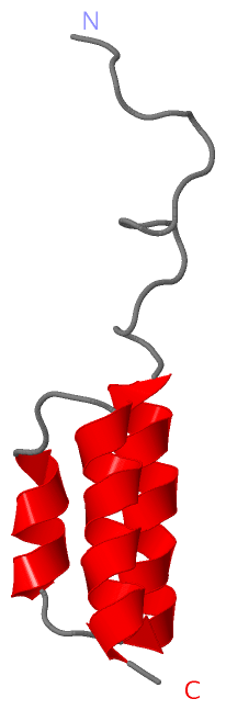
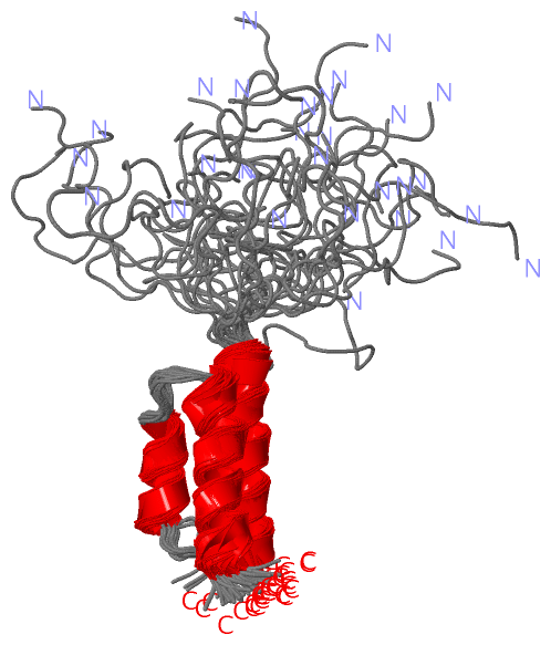
Show PDB file:   
         Plain Text   HTML   (compressed file size)
QuickSearch:   
by PDB,NDB,UniProt,PROSITE Code or Search Term(s)  
(-)NMR Structure - model 1
(-)NMR Structure - all models
collapse expand < >
Image NMR Structure - model 1
NMR Structure - model 1  (Jmol Viewer)
Image NMR Structure - all models
NMR Structure - all models  (Jmol Viewer)

(-) Description

Title :  SOLUTION STRUCTURE OF THE ALBUMIN BINDING DOMAIN OF STREPTOCOCCAL PROTEIN G
 
Authors :  M. U. Johansson, I. M. Frick, H. Nilsson, P. J. Kraulis, S. Hober, P. Jonasson, P. A. Nygren, M. Uhlen, L. Bjorck, T. Drakenberg, S. Forse M. Wikstrom
Date :  02 Aug 01  (Deposition) - 09 Aug 01  (Release) - 08 Apr 15  (Revision)
Method :  SOLUTION NMR
Resolution :  NOT APPLICABLE
Chains :  NMR Structure  :  A  (30x)
NMR Structure *:  A  (1x)
Keywords :  Immunoglobulin-Binding Protein, Bacterial Surface Protein, Albumin Binding, Protein G (Keyword Search: [Gene Ontology, PubMed, Web (Google))
 
Reference :  M. Johansson, I. Frick, H. Nilsson, P. Kraulis, S. Hober, P. Jonasson, M. Linhult, P. Nygren, M. Uhlen, L. Bjorck, T. Drakenberg, S. Forsen, M. Wikstrom
Structure, Specificity, And Mode Of Interaction For Bacterial Albumin-Binding Modules
J. Biol. Chem. V. 277 8114 2002
PubMed-ID: 11751858  |  Reference-DOI: 10.1074/JBC.M109943200

(-) Compounds

Molecule 1 - IMMUNOGLOBULIN G BINDING PROTEIN G
    ChainsA
    EngineeredYES
    Expression SystemESCHERICHIA COLI
    Expression System Taxid562
    FragmentALBUMIN-BINDING DOMAIN RESIDUES 254-299
    Organism CommonGROUP G STREPTOCOCCI
    Organism ScientificSTREPTOCOCCUS SP.
    Organism Taxid1306
    SynonymABD, ALBUMIN-BINDING DOMAIN OF PROTEIN G, IGG BINDING PROTEIN G

 Structural Features

(-) Chains, Units

  1
NMR Structure (30x)A
NMR Structure * (1x)A

Summary Information (see also Sequences/Alignments below)

(-) Ligands, Modified Residues, Ions  (0, 0)

(no "Ligand,Modified Residues,Ions" information available for 1GJS)

(-) Sites  (0, 0)

(no "Site" information available for 1GJS)

(-) SS Bonds  (0, 0)

(no "SS Bond" information available for 1GJS)

(-) Cis Peptide Bonds  (0, 0)

(no "Cis Peptide Bond" information available for 1GJS)

 Sequence-Structure Mapping

(-) SAPs(SNPs)/Variants  (0, 0)

(no "SAP(SNP)/Variant" information available for 1GJS)

(-) PROSITE Motifs  (0, 0)

(no "PROSITE Motif" information available for 1GJS)

(-) Exons   (0, 0)

(no "Exon" information available for 1GJS)

(-) Sequences/Alignments

NMR Structure
   Reformat: Number of residues per line =  ('0' or empty: single-line sequence representation)
  Number of residues per labelling interval =   
  UniProt sequence: complete  aligned part    
   Show mapping: SCOP domains CATH domains Pfam domains Secondary structure (by author)
SAPs(SNPs) PROSITE motifs Exons
(details for a mapped element are shown in a popup box when the mouse pointer rests over it)
Chain A from PDB  Type:PROTEIN  Length:65
 aligned with SPG2_STRSG | P19909 from UniProtKB/Swiss-Prot  Length:593

    Alignment length:85
                                   224       234       244       254       264       274       284       294     
           SPG2_STRSG   215 LQAQVVESAKKARISEATDGLSDFLKSQTPAEDTVKSIELAEAKVLANRELDKYGVSDYYKNLINNAKTVEGVKALIDEILAALP 299
               SCOP domains d1gjsa_ A: I   gG bind                 ing protein G                                  SCOP domains
               CATH domains 1gjsA00 A:1-   65 Albu                 min-binding domain                             CATH domains
               Pfam domains ------------------------------------------------------------------------------------- Pfam domains
         Sec.struct. author ............---......h-----------------hhhhhhhhhhhhhhh...hhhhhhhh....hhhhhhhhhhhhhh.. Sec.struct. author
                 SAPs(SNPs) ------------------------------------------------------------------------------------- SAPs(SNPs)
                    PROSITE ------------------------------------------------------------------------------------- PROSITE
                 Transcript ------------------------------------------------------------------------------------- Transcript
                 1gjs A   1 MKAIFVLNAQHD---EAVDANS-----------------LAEAKVLANRELDKYGVSDYYKNLINNAKTVEGVKALIDEILAALP  65
                                    10 |   |  17 |       -        20        30        40        50        60     
                                      12  13    19                20                                             

   Legend:   → Mismatch (orange background)
  - → Gap (green background, '-', border residues have a numbering label)
    → Modified Residue (blue background, lower-case, 'x' indicates undefined single-letter code, labelled with number + name)
  x → Chemical Group (purple background, 'x', labelled with number + name, e.g. ACE or NH2)
  extra numbering lines below/above indicate numbering irregularities and modified residue names etc., number ends below/above '|'

 Classification and Annotation

(-) SCOP Domains  (1, 1)

NMR Structure

(-) CATH Domains  (1, 1)

NMR Structure

(-) Pfam Domains  (0, 0)

(no "Pfam Domain" information available for 1GJS)

(-) Gene Ontology  (5, 5)

NMR Structure(hide GO term definitions)
Chain A   (SPG2_STRSG | P19909)
molecular function
    GO:0019864    IgG binding    Interacting selectively and non-covalently with an immunoglobulin of an IgG isotype.
biological process
    GO:0009405    pathogenesis    The set of specific processes that generate the ability of an organism to induce an abnormal, generally detrimental state in another organism.
cellular component
    GO:0005618    cell wall    The rigid or semi-rigid envelope lying outside the cell membrane of plant, fungal, most prokaryotic cells and some protozoan parasites, maintaining their shape and protecting them from osmotic lysis. In plants it is made of cellulose and, often, lignin; in fungi it is composed largely of polysaccharides; in bacteria it is composed of peptidoglycan; in protozoan parasites such as Giardia species, it's made of carbohydrates and proteins.
    GO:0005576    extracellular region    The space external to the outermost structure of a cell. For cells without external protective or external encapsulating structures this refers to space outside of the plasma membrane. This term covers the host cell environment outside an intracellular parasite.
    GO:0016020    membrane    A lipid bilayer along with all the proteins and protein complexes embedded in it an attached to it.

 Visualization

(-) Interactive Views

NMR Structure
  Complete Structure
    Jena3D(integrated viewing of ligand, site, SAP, PROSITE, SCOP information)
    WebMol | AstexViewer[tm]@PDBe
(Java Applets, require no local installation except for Java; loading may be slow)
    STRAP
(Java WebStart application, automatic local installation, requires Java; full application with system access!)
    RasMol
(require local installation)
    Molscript (VRML)
(requires installation of a VRML viewer; select preferred view via VRML and generate a mono or stereo PDF format file)
 
  Ligands, Modified Residues, Ions
(no "Ligands, Modified Residues, Ions" information available for 1gjs)
 
  Sites
(no "Sites" information available for 1gjs)
 
  Cis Peptide Bonds
(no "Cis Peptide Bonds" information available for 1gjs)
 

(-) Still Images

Jmol
  protein: cartoon or spacefill or dots and stick; nucleic acid: cartoon and stick; ligands: spacefill; active site: stick
Molscript
  protein, nucleic acid: cartoon; ligands: spacefill; active site: ball and stick

 Databases and Analysis Tools

(-) Databases

Access by PDB/NDB ID
  1gjs
    Family and Domain InformationProDom | SYSTERS
    General Structural InformationGlycoscienceDB | MMDB | NDB | OCA | PDB | PDBe | PDBj | PDBsum | PDBWiki | PQS | PROTEOPEDIA
    Orientation in MembranesOPM
    Protein SurfaceSURFACE
    Secondary StructureDSSP (structure derived) | HSSP (homology derived)
    Structural GenomicsGeneCensus
    Structural NeighboursCE | VAST
    Structure ClassificationCATH | Dali | SCOP
    Validation and Original DataBMRB Data View | BMRB Restraints Grid | EDS | PROCHECK | RECOORD | WHAT_CHECK
 
Access by UniProt ID/Accession number
  SPG2_STRSG | P19909
    Comparative Protein Structure ModelsModBase
    Genomic InformationEnsembl
    Protein-protein InteractionDIP
    Sequence, Family and Domain InformationInterPro | Pfam | SMART | UniProtKB/SwissProt
 
Access by Enzyme Classificator   (EC Number)
  (no 'Enzyme Classificator' available)
    General Enzyme InformationBRENDA | EC-PDB | Enzyme | IntEnz
    PathwayKEGG | MetaCyc
 
Access by Disease Identifier   (MIM ID)
  (no 'MIM ID' available)
    Disease InformationOMIM
 
Access by GenAge ID
  (no 'GenAge ID' available)
    Age Related InformationGenAge

(-) Analysis Tools

Access by PDB/NDB ID
    Domain InformationXDom
    Interatomic Contacts of Structural UnitsCSU
    Ligand-protein ContactsLPC
    Protein CavitiescastP
    Sequence and Secondary StructurePDBCartoon
    Structure AlignmentSTRAP(Java WebStart application, automatic local installation, requires Java; full application with system access!)
    Structure and Sequence BrowserSTING
 
Access by UniProt ID/Accession number
  SPG2_STRSG | P19909
    Protein Disorder PredictionDisEMBL | FoldIndex | GLOBPLOT (for more information see DisProt)

 Related Entries

(-) Entries Sharing at Least One Protein Chain (UniProt ID)

UniProtKB/Swiss-Prot
        SPG2_STRSG | P199091fcc 1fcl 1fd6 1gb4 1gjt 1ibx 1p7e 1p7f 1qkz 1uwx 1zxh 2gi9 2i2y 2i38 2igg 2jsv 2ju6 2khu 2khw 2kn4 2kq4 2kwd 2lum 2n9k 2n9l 2oed 2on8 2onq 2plp 2qmt 3fil 3ui3 3v3x 4oza 4ozb 4ozc 4wh4 5bmg 5bmh 5bmi 5hfy 5hg2 5hi1

(-) Related Entries Specified in the PDB File

1fcc
1fcl DELTA1.5: A COMPUTATIONALLY DESIGNED CORE VARIANT OF THE B1DOMAIN OF STREPTOCOCCAL PROTEIN G
1fd6 DELTA0: A COMPUTATIONALLY DESIGNED CORE VARIANT OF THE B1DOMAIN OF STREPTOCOCCAL PROTEIN G
1gb4 HYPERTHERMOPHILIC VARIANT OF THE B1 DOMAIN FROMSTREPTOCOCCAL PROTEIN G, NMR, 47 STRUCTURES
1gjt SOLUTION STRUCTURE OF THE ALBUMIN BINDING DOMAIN OF STREPTOCOCCAL PROTEIN G
1ibx NMR STRUCTURE OF DFF40 AND DFF45 N- TERMINAL DOMAIN COMPLEX
1p7e GB3 SOLUTION STRUCTURE OBTAINED BY REFINEMENT OF X-RAYSTRUCTURE WITH DIPOLAR COUPLINGS
1p7f GB3 SOLUTION STRUCTURE OBTAINED BY REFINEMENT OF X-RAYSTRUCTURE WITH DIPOLAR COUPLINGS
2igg