|
|
|
|
 Description
|
|
Compounds
|
||||||||||||||||||||||||||||||||||||||||||||||||||||
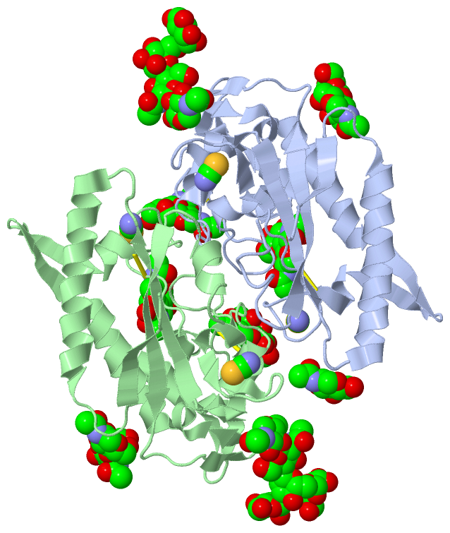
Chains, Units
Summary Information (see also Sequences/Alignments below) |
Ligands, Modified Residues, Ions (4, 27)
Asymmetric Unit (4, 27)
|
Sites (15, 15)
Asymmetric Unit (15, 15)
|
SS Bonds (4, 4)
Asymmetric Unit
|
||||||||||||||||||||
Cis Peptide Bonds (2, 2)
Asymmetric Unit
|
||||||||||||
SAPs(SNPs)/Variants (0, 0)| (no "SAP(SNP)/Variant" information available for 5HNS) |
PROSITE Motifs (0, 0)| (no "PROSITE Motif" information available for 5HNS) |
Exons (0, 0)| (no "Exon" information available for 5HNS) |
Sequences/Alignments
Asymmetric Unit
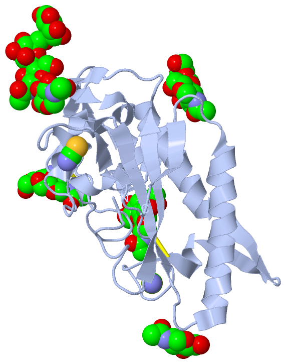
Chain A from PDB Type:PROTEIN Length:215
SCOP domains ----------------------------------------------------------------------------------------------------------------------------------------------------------------------------------------------------------------------- SCOP domains
CATH domains ----------------------------------------------------------------------------------------------------------------------------------------------------------------------------------------------------------------------- CATH domains
Pfam domains ----------------------------------------------------------------------------------------------------------------------------------------------------------------------------------------------------------------------- Pfam domains
SAPs(SNPs) ----------------------------------------------------------------------------------------------------------------------------------------------------------------------------------------------------------------------- SAPs(SNPs)
PROSITE ----------------------------------------------------------------------------------------------------------------------------------------------------------------------------------------------------------------------- PROSITE
Transcript ----------------------------------------------------------------------------------------------------------------------------------------------------------------------------------------------------------------------- Transcript
5hns A 392 KEYFDQHFGPFFRTEQLIIRAPLTDKHIYQPYPSGADVPFGPPLDIQILHQVLDLQIAIENITASYDNETVTLQDICLAPLSPYNTNCTILSVLNYFQNSHSVLDHKKGDDFFVYADYHTHFLYCVRAPASLNDTSLLHDPCLGTFGGPVFPWLVLGGYDDQNYNNATALVITFPVNNYYNDTEKLQRAQAWEKEFINFVKNYKNPNLTISFTAE 606
401 411 421 431 441 451 461 471 481 491 501 511 521 531 541 551 561 571 581 591 601
Chain B from PDB Type:PROTEIN Length:216
SCOP domains ------------------------------------------------------------------------------------------------------------------------------------------------------------------------------------------------------------------------ SCOP domains
CATH domains ------------------------------------------------------------------------------------------------------------------------------------------------------------------------------------------------------------------------ CATH domains
Pfam domains ------------------------------------------------------------------------------------------------------------------------------------------------------------------------------------------------------------------------ Pfam domains
SAPs(SNPs) ------------------------------------------------------------------------------------------------------------------------------------------------------------------------------------------------------------------------ SAPs(SNPs)
PROSITE ------------------------------------------------------------------------------------------------------------------------------------------------------------------------------------------------------------------------ PROSITE
Transcript ------------------------------------------------------------------------------------------------------------------------------------------------------------------------------------------------------------------------ Transcript
5hns B 391 EKEYFDQHFGPFFRTEQLIIRAPLTDKHIYQPYPSGADVPFGPPLDIQILHQVLDLQIAIENITASYDNETVTLQDICLAPLSPYNTNCTILSVLNYFQNSHSVLDHKKGDDFFVYADYHTHFLYCVRAPASLNDTSLLHDPCLGTFGGPVFPWLVLGGYDDQNYNNATALVITFPVNNYYNDTEKLQRAQAWEKEFINFVKNYKNPNLTISFTAE 606
400 410 420 430 440 450 460 470 480 490 500 510 520 530 540 550 560 570 580 590 600
|
||||||||||||||||||||
SCOP Domains (0, 0)| (no "SCOP Domain" information available for 5HNS) |
CATH Domains (0, 0)| (no "CATH Domain" information available for 5HNS) |
Pfam Domains (0, 0)| (no "Pfam Domain" information available for 5HNS) |
Gene Ontology (47, 47)|
Asymmetric Unit(hide GO term definitions) |
Interactive Views
|
||||||||||||||||||||||||||||||||||||||||||||||||||||||||||||||||||||||||||||||||||||||||||||||||||||||||||||||||||||||||||||||||||||||||||||||||||||||||||||||||||||||||||||||||||||||||||||||||||||||||||||||||||||||||||||||||||||||||||||||||||||||||||||||||||||||||||||
Still Images
|
||||||||||||||||
Databases
|
||||||||||||||||||||||||||||||||||||||||||||||||||||||||||||||||||||||||||||||||||||||||||||||||||||||||||||||||||||||||||||||||||||||||||||||||||||||||||||||||
Analysis Tools
|
|||||||||||||||||||||||||||||||||||||||||||||||||||||||||||||
Entries Sharing at Least One Protein Chain (UniProt ID)
Related Entries Specified in the PDB File
|
|