Show PDB file:   
         Plain Text   HTML   (compressed file size)
QuickSearch:   
by PDB,NDB,UniProt,PROSITE Code or Search Term(s)  
(-)Asymmetric Unit
(-)Asym. Unit - sites
(-)Biological Unit 1
(-)Biol. Unit 1 - sites
collapse expand < >
Image Asymmetric Unit
Asymmetric Unit  (Jmol Viewer)
Image Asym. Unit - sites
Asym. Unit - sites  (Jmol Viewer)
Image Biological Unit 1
Biological Unit 1  (Jmol Viewer)
Image Biol. Unit 1 - sites
Biol. Unit 1 - sites  (Jmol Viewer)

(-) Description

Title :  CRYSTAL STRUCTURE OF THE NMDA RECEPTOR GLUN1 LIGAND BINDING DOMAIN APO STATE
 
Authors :  A. J. Berger, A. Y. Lau, M. L. Mayer
Date :  24 Apr 13  (Deposition) - 31 Jul 13  (Release) - 06 Nov 13  (Revision)
Method :  X-RAY DIFFRACTION
Resolution :  1.89
Chains :  Asym. Unit :  A
Biol. Unit 1:  A  (2x)
Keywords :  Membrane Protein (Keyword Search: [Gene Ontology, PubMed, Web (Google))
 
Reference :  Y. Yao, J. Belcher, A. J. Berger, M. L. Mayer, A. Y. Lau
Conformational Analysis Of Nmda Receptor Glun1, Glun2, And Glun3 Ligand-Binding Domains Reveals Subtype-Specific Characteristics.
Structure V. 21 1788 2013
PubMed-ID: 23972471  |  Reference-DOI: 10.1016/J.STR.2013.07.011

(-) Compounds

Molecule 1 - GLUTAMATE RECEPTOR IONOTROPIC, NMDA 1
    ChainsA
    EngineeredYES
    Expression SystemESCHERICHIA COLI
    Expression System PlasmidPET22 MODIFIED
    Expression System StrainORIGAMI B(DE3)
    Expression System Taxid562
    Expression System Vector TypePLASMID
    FragmentLIGAND BINDING DOMAIN (UNP RESIDUES 394-544, 663-800)
    GeneGRIN1, NMDAR1
    Organism CommonBROWN RAT,RAT,RATS
    Organism ScientificRATTUS NORVEGICUS
    Organism Taxid10116
    SynonymGLUN1, GLUTAMATE [NMDA] RECEPTOR SUBUNIT ZETA-1, N-METHYL-D- ASPARTATE RECEPTOR SUBUNIT NR1, NMD-R1

 Structural Features

(-) Chains, Units

  1
Asymmetric Unit A
Biological Unit 1 (2x)A

Summary Information (see also Sequences/Alignments below)

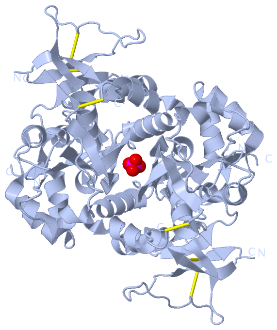
(-) Ligands, Modified Residues, Ions  (1, 1)

Asymmetric Unit (1, 1)
No.NameCountTypeFull Name
1PO41Ligand/IonPHOSPHATE ION
Biological Unit 1 (1, 2)
No.NameCountTypeFull Name
1PO42Ligand/IonPHOSPHATE ION

(-) Sites  (1, 1)

Asymmetric Unit (1, 1)
No.NameEvidenceResiduesDescription
1AC1SOFTWARELYS A:139 , LYS A:261 , HOH A:412BINDING SITE FOR RESIDUE PO4 A 301

(-) SS Bonds  (3, 3)

Asymmetric Unit
No.Residues
1A:28 -A:62
2A:44 -A:63
3A:236 -A:290

(-) Cis Peptide Bonds  (1, 1)

Asymmetric Unit
No.Residues
1Glu A:14 -Pro A:15

 Sequence-Structure Mapping

(-) SAPs(SNPs)/Variants  (0, 0)

(no "SAP(SNP)/Variant" information available for 4KCC)

(-) PROSITE Motifs  (0, 0)

(no "PROSITE Motif" information available for 4KCC)

(-) Exons   (0, 0)

(no "Exon" information available for 4KCC)

(-) Sequences/Alignments

Asymmetric Unit
   Reformat: Number of residues per line =  ('0' or empty: single-line sequence representation)
  Number of residues per labelling interval =   
  UniProt sequence: complete  aligned part    
   Show mapping: SCOP domains CATH domains Pfam domains Secondary structure (by author)
SAPs(SNPs) PROSITE motifs Exons
(details for a mapped element are shown in a popup box when the mouse pointer rests over it)
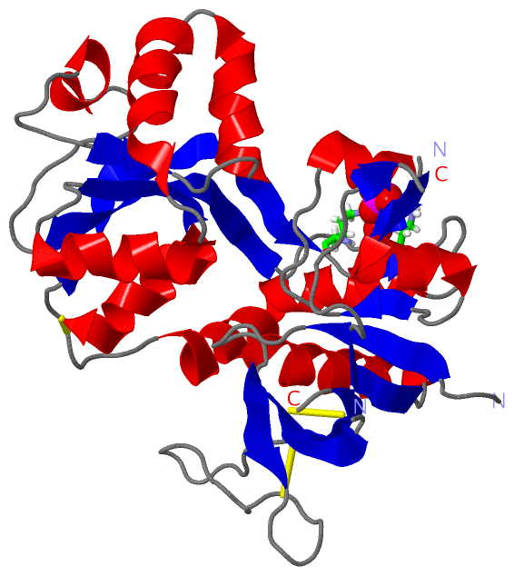
Chain A from PDB  Type:PROTEIN  Length:275
                                                                                                                                                                                                                                                                                                                   
               SCOP domains d4kcca_ A: N-methyl-D-aspartate receptor subunit 1                                                                                                                                                                                                                                  SCOP domains
               CATH domains ----------------------------------------------------------------------------------------------------------------------------------------------------------------------------------------------------------------------------------------------------------------------------------- CATH domains
               Pfam domains ----------------------------------------------------------------------------------------------------------------------------------------------------------------------------------------------------------------------------------------------------------------------------------- Pfam domains
         Sec.struct. author ...eeeee.......eeee....................eeeee....eeeeeehhhhhhhhhhhhhhh..eeeee........ee...eehhhhhhhhh....ee......hhhhhh..ee....eeeeeeeeee.........hhhhhh......ee....hhhhhhhhhh..hhhhhhhhhhhh..hhhhhhhhhhh....eeeeehhhhhhhhhhh..eeeeeeeeeeeee..ee.....hhhhhhhhhhhhhhhhhhhhhhhhhh..... Sec.struct. author
                 SAPs(SNPs) ----------------------------------------------------------------------------------------------------------------------------------------------------------------------------------------------------------------------------------------------------------------------------------- SAPs(SNPs)
                    PROSITE ----------------------------------------------------------------------------------------------------------------------------------------------------------------------------------------------------------------------------------------------------------------------------------- PROSITE
                 Transcript ----------------------------------------------------------------------------------------------------------------------------------------------------------------------------------------------------------------------------------------------------------------------------------- Transcript
                 4kcc A   3 STRLKIVTIHQEPFVYVKPTMSDGTCKEEFTVNGDPVKKVICTGPNHTVPQCCYGFCIDLLIKLARTMNFTYEVHLVADGKFGTQERVKEWNGMMGELLSGQADMIVAPLTINNERAQYIEFSKPFKYQGLTILVKKGTRITGINDPRLRNPSDKFIYATVKQSSVDIYFRRQVELSTMYRHMEKHNYESAAEAIQAVRDNKLHAFIWDSAVLEFEASQKCDLVTTGELFFRSGFGIGMRKDSPWKQNVSLSILKSHENGFMEDLDKTWVRYQEC 290
                                    12        22        32        42     || 60        70        80        90       105       115       125       135       145       155       165       175       185       195       205       215       225       235       245       255       265       275       285     
                                                                        48|                                       98|                                                                                                                                                                                          
                                                                         57                                       104                                                                                                                                                                                          

   Legend:   → Mismatch (orange background)
  - → Gap (green background, '-', border residues have a numbering label)
    → Modified Residue (blue background, lower-case, 'x' indicates undefined single-letter code, labelled with number + name)
  x → Chemical Group (purple background, 'x', labelled with number + name, e.g. ACE or NH2)
  extra numbering lines below/above indicate numbering irregularities and modified residue names etc., number ends below/above '|'

 Classification and Annotation

(-) SCOP Domains  (1, 1)

Asymmetric Unit

(-) CATH Domains  (0, 0)

(no "CATH Domain" information available for 4KCC)

(-) Pfam Domains  (0, 0)

(no "Pfam Domain" information available for 4KCC)

(-) Gene Ontology  (45, 45)

Asymmetric Unit(hide GO term definitions)

 Visualization

(-) Interactive Views

Asymmetric Unit
  Complete Structure
    Jena3D(integrated viewing of ligand, site, SAP, PROSITE, SCOP information)
    WebMol | AstexViewer[tm]@PDBe
(Java Applets, require no local installation except for Java; loading may be slow)
    STRAP
(Java WebStart application, automatic local installation, requires Java; full application with system access!)
    RasMol
(require local installation)
    Molscript (VRML)
(requires installation of a VRML viewer; select preferred view via VRML and generate a mono or stereo PDF format file)
 
  Ligands, Modified Residues, Ions
    PO4  [ RasMol | Jena3D ]  +environment [ RasMol | Jena3D ]
 
  Sites
    AC1  [ RasMol ]  +environment [ RasMol ]
 
  Cis Peptide Bonds
    Glu A:14 - Pro A:15   [ RasMol ]  
 
Biological Unit
  Complete Structure
    Biological Unit 1  [ Jena3D ]

(-) Still Images

Jmol
  protein: cartoon or spacefill or dots and stick; nucleic acid: cartoon and stick; ligands: spacefill; active site: stick
Molscript
  protein, nucleic acid: cartoon; ligands: spacefill; active site: ball and stick

 Databases and Analysis Tools

(-) Databases

Access by PDB/NDB ID
  4kcc
    Family and Domain InformationProDom | SYSTERS
    General Structural InformationGlycoscienceDB | MMDB | NDB | OCA | PDB | PDBe | PDBj | PDBsum | PDBWiki | PQS | PROTEOPEDIA
    Orientation in MembranesOPM
    Protein SurfaceSURFACE
    Secondary StructureDSSP (structure derived) | HSSP (homology derived)
    Structural GenomicsGeneCensus
    Structural NeighboursCE | VAST
    Structure ClassificationCATH | Dali | SCOP
    Validation and Original DataBMRB Data View | BMRB Restraints Grid | EDS | PROCHECK | RECOORD | WHAT_CHECK
 
Access by UniProt ID/Accession number
  NMDZ1_RAT | P35439
    Comparative Protein Structure ModelsModBase
    Genomic InformationEnsembl
    Protein-protein InteractionDIP
    Sequence, Family and Domain InformationInterPro | Pfam | SMART | UniProtKB/SwissProt
 
Access by Enzyme Classificator   (EC Number)
  (no 'Enzyme Classificator' available)
    General Enzyme InformationBRENDA | EC-PDB | Enzyme | IntEnz
    PathwayKEGG | MetaCyc
 
Access by Disease Identifier   (MIM ID)
  (no 'MIM ID' available)
    Disease InformationOMIM
 
Access by GenAge ID
  (no 'GenAge ID' available)
    Age Related InformationGenAge

(-) Analysis Tools

Access by PDB/NDB ID
    Domain InformationXDom
    Interatomic Contacts of Structural UnitsCSU
    Ligand-protein ContactsLPC
    Protein CavitiescastP
    Sequence and Secondary StructurePDBCartoon
    Structure AlignmentSTRAP(Java WebStart application, automatic local installation, requires Java; full application with system access!)
    Structure and Sequence BrowserSTING
 
Access by UniProt ID/Accession number
  NMDZ1_RAT | P35439
    Protein Disorder PredictionDisEMBL | FoldIndex | GLOBPLOT (for more information see DisProt)

 Related Entries

(-) Entries Sharing at Least One Protein Chain (UniProt ID)

UniProtKB/Swiss-Prot
        NMDZ1_RAT | P354391pb7 1pb8 1pb9 1pbq 1y1m 1y1z 1y20 2a5t 3q41 4kfq 4nf4 4nf5 4nf6 4nf8 4pe5 5ddn 5ddx 5de4 5dex 5fxg 5fxh 5fxi 5fxj 5fxk 5i56 5i57 5i58 5i59 5jty 5u8c 5vih 5vii 5vij

(-) Related Entries Specified in the PDB File

1pb7 GLUN1 LBD GLYCINE COMPLEX
1pb8 GLUN1 LBD D-SERINE COMPLEX
1pbq GLUN1 LBD DCKA COMPLEX
4kcd