Show PDB file:   
         Plain Text   HTML   (compressed file size)
QuickSearch:   
by PDB,NDB,UniProt,PROSITE Code or Search Term(s)  
(-)Asym./Biol. Unit
(-)Asym./Biol. Unit - sites
collapse expand < >
Image Asym./Biol. Unit
Asym./Biol. Unit  (Jmol Viewer)
Image Asym./Biol. Unit - sites
Asym./Biol. Unit - sites  (Jmol Viewer)

(-) Description

Title :  CRYSTAL STRUCTURE OF AN MMP TWIN CARBOXYLATE BASED INHIBITOR LC20 IN COMPLEX WITH THE MMP-9 CATALYTIC DOMAIN
 
Authors :  E. A. Stura, C. Antoni, L. Vera, E. Nuti, L. Carafa, E. Cassar-Lajeuness A. Rossello
Date :  18 Oct 12  (Deposition) - 24 Apr 13  (Release) - 12 Aug 15  (Revision)
Method :  X-RAY DIFFRACTION
Resolution :  1.94
Chains :  Asym./Biol. Unit :  A,B
Keywords :  Hydrolase/Twin Inhibitor, Zincin-Like, Gelatinase, Collagenase (Catalytic Domain), Hydrolase-Hydrolase Inhibitor Complex (Keyword Search: [Gene Ontology, PubMed, Web (Google))
 
Reference :  C. Antoni, L. Vera, L. Devel, M. P. Catalani, B. Czarny, E. Cassar-Lajeunesse, E. Nuti, A. Rossello, V. Dive, E. A. Stura
Crystallization Of Bi-Functional Ligand Protein Complexes.
J. Struct. Biol. V. 182 246 2013
PubMed-ID: 23567804  |  Reference-DOI: 10.1016/J.JSB.2013.03.015
(for further references see the PDB file header)

(-) Compounds

Molecule 1 - MATRIX METALLOPROTEINASE-9
    ChainsA, B
    EC Number3.4.24.35
    EngineeredYES
    Expression SystemESCHERICHIA COLI
    Expression System PlasmidPET-14B
    Expression System StrainBL21 (DE3 STAR)
    Expression System Taxid562
    Expression System Vector TypePT7 PROMOTER
    FragmentMMP-9 CATALYTIC DOMAIN 107-215,391-443
    GeneCLG4B, MMP9
    MutationYES
    Organism CommonHUMAN
    Organism ScientificHOMO SAPIENS
    Organism Taxid9606
    SynonymMMP-9, 92 KDA GELATINASE, 92 KDA TYPE IV COLLAGENASE, GELATINASE B, GELB, 67 KDA MATRIX METALLOPROTEINASE-9, 82 KDA MATRIX METALLOPROTEINASE-9

 Structural Features

(-) Chains, Units

  12
Asymmetric/Biological Unit AB

Summary Information (see also Sequences/Alignments below)

(-) Ligands, Modified Residues, Ions  (7, 30)

Asymmetric/Biological Unit (7, 30)
No.NameCountTypeFull Name
10ZD1Ligand/IonN,N'-BIS(2-[(BIPHENYL-4YLSULFONYL)[(2R)-1-HYDROXY-3-METHYL-1-OXOBUTAN-2-YL]-AMINO]ETHYL)BENZENE-1,3-DICARBOXAMIDE
2CA6Ligand/IonCALCIUM ION
3GOL5Ligand/IonGLYCEROL
4MLT1Ligand/IonD-MALATE
5PEG7Ligand/IonDI(HYDROXYETHYL)ETHER
6PGO6Ligand/IonS-1,2-PROPANEDIOL
7ZN4Ligand/IonZINC ION

(-) Sites  (30, 30)

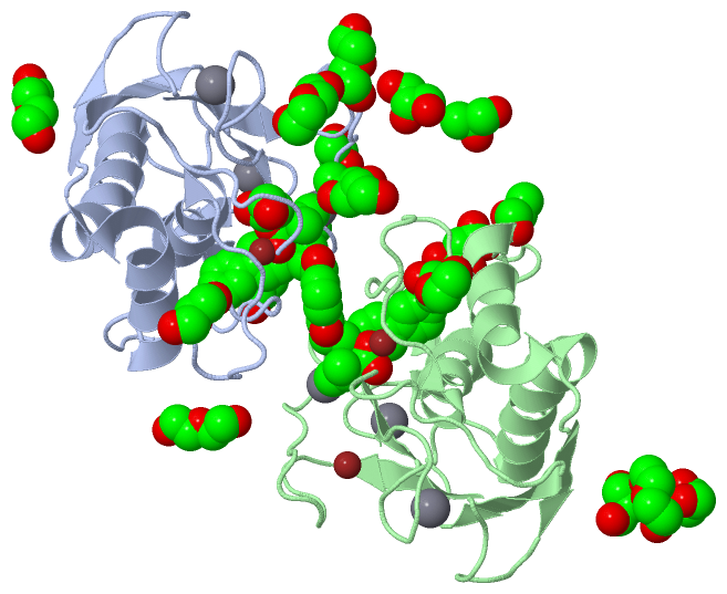
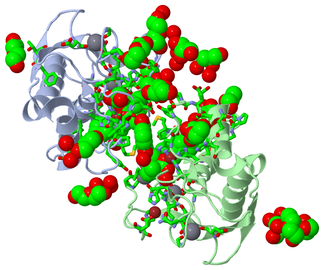
Asymmetric Unit (30, 30)
No.NameEvidenceResiduesDescription
01AC1SOFTWAREHIS A:226 , HIS A:230 , HIS A:236 , 0ZD A:306BINDING SITE FOR RESIDUE ZN A 301
02AC2SOFTWAREHIS A:175 , ASP A:177 , HIS A:190 , HIS A:203BINDING SITE FOR RESIDUE ZN A 302
03AC3SOFTWAREASP A:165 , GLY A:197 , GLN A:199 , ASP A:201 , HOH A:445 , HOH A:446BINDING SITE FOR RESIDUE CA A 303
04AC4SOFTWAREASP A:182 , GLY A:183 , ASP A:185 , LEU A:187 , ASP A:205 , GLU A:208BINDING SITE FOR RESIDUE CA A 304
05AC5SOFTWAREASP A:131 , ASP A:206 , GLU A:208BINDING SITE FOR RESIDUE CA A 305
06AC6SOFTWARELEU A:187 , LEU A:188 , ALA A:189 , LEU A:222 , VAL A:223 , HIS A:226 , GLN A:227 , HIS A:230 , HIS A:236 , LEU A:243 , TYR A:245 , PRO A:246 , MET A:247 , TYR A:248 , ZN A:301 , GOL A:307 , HOH A:417 , HOH A:437 , HOH A:520 , LEU B:187 , LEU B:188 , ALA B:189 , LEU B:222 , HIS B:226 , GLN B:227 , HIS B:230 , HIS B:236 , LEU B:243 , TYR B:245 , PRO B:246 , MET B:247 , TYR B:248 , ZN B:501 , MLT B:506BINDING SITE FOR RESIDUE 0ZD A 306
07AC7SOFTWARETYR A:179 , LEU A:187 , HIS A:190 , 0ZD A:306 , HOH A:440 , PRO B:240 , TYR B:245 , HOH B:615BINDING SITE FOR RESIDUE GOL A 307
08AC8SOFTWAREPHE A:110 , ASN A:262 , PEG A:317 , HOH A:457 , HOH A:479 , HOH A:518BINDING SITE FOR RESIDUE GOL A 308
09AC9SOFTWAREPEG A:314 , HOH A:559 , HOH A:584 , HOH A:587BINDING SITE FOR RESIDUE GOL A 309
10BC1SOFTWAREGLU A:111 , LEU A:114 , GLY A:233 , ASP A:235 , HOH A:577BINDING SITE FOR RESIDUE PGO A 310
11BC2SOFTWAREASP A:207 , HOH A:471 , HOH A:529BINDING SITE FOR RESIDUE PGO A 311
12BC3SOFTWAREGLY A:217 , TYR A:218 , HOH A:566BINDING SITE FOR RESIDUE PGO A 312
13BC4SOFTWAREGLN A:199BINDING SITE FOR RESIDUE PEG A 313
14BC5SOFTWAREGOL A:309 , HOH A:557 , HOH A:559BINDING SITE FOR RESIDUE PEG A 314
15BC6SOFTWAREPHE A:156 , THR A:157 , PGO B:510BINDING SITE FOR RESIDUE PEG A 315
16BC7SOFTWAREHOH A:444 , HOH A:511 , HOH A:537BINDING SITE FOR RESIDUE PEG A 316
17BC8SOFTWARELYS A:184 , GOL A:308 , HOH A:518 , HOH A:594BINDING SITE FOR RESIDUE PEG A 317
18BC9SOFTWARE0ZD A:306 , HIS B:226 , HIS B:230 , HIS B:236BINDING SITE FOR RESIDUE ZN B 501
19CC1SOFTWAREHIS B:175 , ASP B:177 , HIS B:190 , HIS B:203BINDING SITE FOR RESIDUE ZN B 502
20CC2SOFTWAREASP B:165 , GLY B:197 , GLN B:199 , ASP B:201 , HOH B:611 , HOH B:612BINDING SITE FOR RESIDUE CA B 503
21CC3SOFTWAREASP B:182 , GLY B:183 , ASP B:185 , LEU B:187 , ASP B:205 , GLU B:208BINDING SITE FOR RESIDUE CA B 504
22CC4SOFTWAREASP B:131 , ASP B:206 , GLU B:208 , HOH B:681BINDING SITE FOR RESIDUE CA B 505
23CC5SOFTWARE0ZD A:306 , GLU B:241 , THR B:251 , PRO B:254 , PRO B:255 , HIS B:257 , PGO B:509 , HOH B:617BINDING SITE FOR RESIDUE MLT B 506
24CC6SOFTWAREPEG B:512 , HOH B:701BINDING SITE FOR RESIDUE GOL B 507
25CC7SOFTWAREHOH B:701 , HOH B:742 , HOH B:743 , HOH B:746BINDING SITE FOR RESIDUE GOL B 508
26CC8SOFTWARETHR B:251 , GLY B:253 , PRO B:254 , MLT B:506BINDING SITE FOR RESIDUE PGO B 509
27CC9SOFTWAREPEG A:315 , HOH B:714 , HOH B:739BINDING SITE FOR RESIDUE PGO B 510
28DC1SOFTWARESER B:238 , HOH B:729BINDING SITE FOR RESIDUE PGO B 511
29DC2SOFTWAREGOL B:507 , HOH B:743BINDING SITE FOR RESIDUE PEG B 512
30DC3SOFTWAREASP B:259 , ASN B:262 , GLY B:263 , HIS B:266 , HOH B:735BINDING SITE FOR RESIDUE PEG B 513

(-) SS Bonds  (0, 0)

(no "SS Bond" information available for 4HMA)

(-) Cis Peptide Bonds  (1, 1)

Asymmetric/Biological Unit
No.Residues
1Gly A:112 -Asp A:113

 Sequence-Structure Mapping

(-) SAPs(SNPs)/Variants  (0, 0)

(no "SAP(SNP)/Variant" information available for 4HMA)

(-) PROSITE Motifs  (0, 0)

(no "PROSITE Motif" information available for 4HMA)

(-) Exons   (0, 0)

(no "Exon" information available for 4HMA)

(-) Sequences/Alignments

Asymmetric/Biological Unit
   Reformat: Number of residues per line =  ('0' or empty: single-line sequence representation)
  Number of residues per labelling interval =   
  UniProt sequence: complete  aligned part    
   Show mapping: SCOP domains CATH domains Pfam domains Secondary structure (by author)
SAPs(SNPs) PROSITE motifs Exons
(details for a mapped element are shown in a popup box when the mouse pointer rests over it)
Chain A from PDB  Type:PROTEIN  Length:160
                                                                                                                                                                                                
               SCOP domains d4hmaa_ A: Gelatinase B (MMP-9)                                                                                                                                  SCOP domains
               CATH domains ---------------------------------------------------------------------------------------------------------------------------------------------------------------- CATH domains
               Pfam domains ---------------------------------------------------------------------------------------------------------------------------------------------------------------- Pfam domains
         Sec.struct. author ..........eeeeee.......hhhhhhhhhhhhhhhhhhhh..eeee.......eeeeee.................eee..........eeee....ee......eehhhhhhhhhhhhh........................hhhhhhhhhhhhh Sec.struct. author
                 SAPs(SNPs) ---------------------------------------------------------------------------------------------------------------------------------------------------------------- SAPs(SNPs)
                    PROSITE ---------------------------------------------------------------------------------------------------------------------------------------------------------------- PROSITE
                 Transcript ---------------------------------------------------------------------------------------------------------------------------------------------------------------- Transcript
                 4hma A 110 FEGDLKWHHHNITYWIQNYSEDLPRAVIDDAFARAFALWSAVTPLTFTRVYSRDADIVIQFGVAEHGDGYPFDGKDGLLAHAFPPGPGIQGDAHFDDDELWSLGKGQGYSLFLVAAHQFGHALGLDHSSVPEALMYPMYRFTEGPPLHKDDVNGIRHLYG 269
                                   119       129       139       149       159       169       179       189       199       209       219       229       239       249       259       269

Chain B from PDB  Type:PROTEIN  Length:160
                                                                                                                                                                                                
               SCOP domains d4hmab_ B: Gelatinase B (MMP-9)                                                                                                                                  SCOP domains
               CATH domains ---------------------------------------------------------------------------------------------------------------------------------------------------------------- CATH domains
               Pfam domains ---------------------------------------------------------------------------------------------------------------------------------------------------------------- Pfam domains
         Sec.struct. author ..........eeeeee.......hhhhhhhhhhhhhhhhhhh...eeee.......eeeeee.................eee..........eeee....ee......eehhhhhhhhhhhhhh.......................hhhhhhhhhhhhh Sec.struct. author
                 SAPs(SNPs) ---------------------------------------------------------------------------------------------------------------------------------------------------------------- SAPs(SNPs)
                    PROSITE ---------------------------------------------------------------------------------------------------------------------------------------------------------------- PROSITE
                 Transcript ---------------------------------------------------------------------------------------------------------------------------------------------------------------- Transcript
                 4hma B 110 FEGDLKWHHHNITYWIQNYSEDLPRAVIDDAFARAFALWSAVTPLTFTRVYSRDADIVIQFGVAEHGDGYPFDGKDGLLAHAFPPGPGIQGDAHFDDDELWSLGKGQGYSLFLVAAHQFGHALGLDHSSVPEALMYPMYRFTEGPPLHKDDVNGIRHLYG 269
                                   119       129       139       149       159       169       179       189       199       209       219       229       239       249       259       269

   Legend:   → Mismatch (orange background)
  - → Gap (green background, '-', border residues have a numbering label)
    → Modified Residue (blue background, lower-case, 'x' indicates undefined single-letter code, labelled with number + name)
  x → Chemical Group (purple background, 'x', labelled with number + name, e.g. ACE or NH2)
  extra numbering lines below/above indicate numbering irregularities and modified residue names etc., number ends below/above '|'

 Classification and Annotation

(-) SCOP Domains  (1, 2)

Asymmetric/Biological Unit

(-) CATH Domains  (0, 0)

(no "CATH Domain" information available for 4HMA)

(-) Pfam Domains  (0, 0)

(no "Pfam Domain" information available for 4HMA)

(-) Gene Ontology  (38, 38)

Asymmetric/Biological Unit(hide GO term definitions)

 Visualization

(-) Interactive Views

Asymmetric/Biological Unit
  Complete Structure
    Jena3D(integrated viewing of ligand, site, SAP, PROSITE, SCOP information)
    WebMol | AstexViewer[tm]@PDBe
(Java Applets, require no local installation except for Java; loading may be slow)
    STRAP
(Java WebStart application, automatic local installation, requires Java; full application with system access!)
    RasMol
(require local installation)
    Molscript (VRML)
(requires installation of a VRML viewer; select preferred view via VRML and generate a mono or stereo PDF format file)
 
  Ligands, Modified Residues, Ions
    0ZD  [ RasMol | Jena3D ]  +environment [ RasMol | Jena3D ]
    CA  [ RasMol | Jena3D ]  +environment [ RasMol | Jena3D ]
    GOL  [ RasMol | Jena3D ]  +environment [ RasMol | Jena3D ]
    MLT  [ RasMol | Jena3D ]  +environment [ RasMol | Jena3D ]
    PEG  [ RasMol | Jena3D ]  +environment [ RasMol | Jena3D ]
    PGO  [ RasMol | Jena3D ]  +environment [ RasMol | Jena3D ]
    ZN  [ RasMol | Jena3D ]  +environment [ RasMol | Jena3D ]
 
  Sites
    AC1  [ RasMol ]  +environment [ RasMol ]
    AC2  [ RasMol ]  +environment [ RasMol ]
    AC3  [ RasMol ]  +environment [ RasMol ]
    AC4  [ RasMol ]  +environment [ RasMol ]
    AC5  [ RasMol ]  +environment [ RasMol ]
    AC6  [ RasMol ]  +environment [ RasMol ]
    AC7  [ RasMol ]  +environment [ RasMol ]
    AC8  [ RasMol ]  +environment [ RasMol ]
    AC9  [ RasMol ]  +environment [ RasMol ]
    BC1  [ RasMol ]  +environment [ RasMol ]
    BC2  [ RasMol ]  +environment [ RasMol ]
    BC3  [ RasMol ]  +environment [ RasMol ]
    BC4  [ RasMol ]  +environment [ RasMol ]
    BC5  [ RasMol ]  +environment [ RasMol ]
    BC6  [ RasMol ]  +environment [ RasMol ]
    BC7  [ RasMol ]  +environment [ RasMol ]
    BC8  [ RasMol ]  +environment [ RasMol ]
    BC9  [ RasMol ]  +environment [ RasMol ]
    CC1  [ RasMol ]  +environment [ RasMol ]
    CC2  [ RasMol ]  +environment [ RasMol ]
    CC3  [ RasMol ]  +environment [ RasMol ]
    CC4  [ RasMol ]  +environment [ RasMol ]
    CC5  [ RasMol ]  +environment [ RasMol ]
    CC6  [ RasMol ]  +environment [ RasMol ]
    CC7  [ RasMol ]  +environment [ RasMol ]
    CC8  [ RasMol ]  +environment [ RasMol ]
    CC9  [ RasMol ]  +environment [ RasMol ]
    DC1  [ RasMol ]  +environment [ RasMol ]
    DC2  [ RasMol ]  +environment [ RasMol ]
    DC3  [ RasMol ]  +environment [ RasMol ]
 
  Cis Peptide Bonds
    Gly A:112 - Asp A:113   [ RasMol ]  
 

(-) Still Images

Jmol
  protein: cartoon or spacefill or dots and stick; nucleic acid: cartoon and stick; ligands: spacefill; active site: stick
Molscript
  protein, nucleic acid: cartoon; ligands: spacefill; active site: ball and stick

 Databases and Analysis Tools

(-) Databases

Access by PDB/NDB ID
  4hma
    Family and Domain InformationProDom | SYSTERS
    General Structural InformationGlycoscienceDB | MMDB | NDB | OCA | PDB | PDBe | PDBj | PDBsum | PDBWiki | PQS | PROTEOPEDIA
    Orientation in MembranesOPM
    Protein SurfaceSURFACE
    Secondary StructureDSSP (structure derived) | HSSP (homology derived)
    Structural GenomicsGeneCensus
    Structural NeighboursCE | VAST
    Structure ClassificationCATH | Dali | SCOP
    Validation and Original DataBMRB Data View | BMRB Restraints Grid | EDS | PROCHECK | RECOORD | WHAT_CHECK
 
Access by UniProt ID/Accession number
  MMP9_HUMAN | P14780
    Comparative Protein Structure ModelsModBase
    Genomic InformationEnsembl
    Protein-protein InteractionDIP
    Sequence, Family and Domain InformationInterPro | Pfam | SMART | UniProtKB/SwissProt
 
Access by Enzyme Classificator   (EC Number)
  3.4.24.35
    General Enzyme InformationBRENDA | EC-PDB | Enzyme | IntEnz
    PathwayKEGG | MetaCyc
 
Access by Disease Identifier   (MIM ID)
  (no 'MIM ID' available)
    Disease InformationOMIM
 
Access by GenAge ID
  (no 'GenAge ID' available)
    Age Related InformationGenAge

(-) Analysis Tools

Access by PDB/NDB ID
    Domain InformationXDom
    Interatomic Contacts of Structural UnitsCSU
    Ligand-protein ContactsLPC
    Protein CavitiescastP
    Sequence and Secondary StructurePDBCartoon
    Structure AlignmentSTRAP(Java WebStart application, automatic local installation, requires Java; full application with system access!)
    Structure and Sequence BrowserSTING
 
Access by UniProt ID/Accession number
  MMP9_HUMAN | P14780
    Protein Disorder PredictionDisEMBL | FoldIndex | GLOBPLOT (for more information see DisProt)

 Related Entries

(-) Entries Sharing at Least One Protein Chain (UniProt ID)

UniProtKB/Swiss-Prot
        MMP9_HUMAN | P147801gkc 1gkd 1itv 1l6j 1lkg 2ovx 2ovz 2ow0 2ow1 2ow2 4h1q 4h2e 4h3x 4h82 4jij 4jqg 4wzv 4xct 5cuh 5i12 5th6 5th9

(-) Related Entries Specified in the PDB File

4h1q CRYSTAL STRUCTURE OF MUTANT MMP-9 CATALYTIC DOMAIN IN COMPLEX WITH A TWIN INHIBITOR.
4h2e CRYSTAL STRUCTURE OF AN MMP TWIN INHIBITOR COMPLEXING TWO MMP-9 CATALYTIC DOMAINS
4h30 CRYSTAL STRUCTURE OF THE CATALYTIC DOMAIN OF MMP-12 IN COMPLEX WITH A TWIN INHIBITOR.
4h3x CRYSTAL STRUCTURE OF AN MMP BROAD SPECTRUM HYDROXAMATE BASED INHIBITOR CC27 IN COMPLEX WITH THE MMP-9 CATALYTIC DOMAIN
4h82 CRYSTAL STRUCTURE OF MUTANT MMP-9 CATALYTIC DOMAIN IN COMPLEX WITH A TWIN INHIBITOR.