Show PDB file:   
         Plain Text   HTML   (compressed file size)
QuickSearch:   
by PDB,NDB,UniProt,PROSITE Code or Search Term(s)  
(-)Asymmetric Unit
(-)Asym. Unit - sites
(-)Biological Unit 1
(-)Biol. Unit 1 - sites
(-)Biological Unit 2
collapse expand < >
Image Asymmetric Unit
Asymmetric Unit  (Jmol Viewer)
Image Asym. Unit - sites
Asym. Unit - sites  (Jmol Viewer)
Image Biological Unit 1
Biological Unit 1  (Jmol Viewer)
Image Biol. Unit 1 - sites
Biol. Unit 1 - sites  (Jmol Viewer)
Image Biological Unit 2
Biological Unit 2  (Jmol Viewer)

(-) Description

Title :  CRYSTAL STRUCTURE MMP-9 COMPLEXES WITH A CONSTRAINED HYDROXAMATE BASED INHIBITOR LT4
 
Authors :  L. Tepshi, L. Vera, E. Nuti, L. Rosalia, A. Rossello, E. A. Stura
Date :  24 Jul 15  (Deposition) - 10 Feb 16  (Release) - 20 Apr 16  (Revision)
Method :  X-RAY DIFFRACTION
Resolution :  1.83
Chains :  Asym. Unit :  A,B
Biol. Unit 1:  A  (1x)
Biol. Unit 2:  B  (1x)
Keywords :  Mmp-9 Hydroxamate-Based Inhibitor Gelatinase, Hydrolase (Keyword Search: [Gene Ontology, PubMed, Web (Google))
 
Reference :  C. Camodeca, E. Nuti, L. Tepshi, S. Boero, T. Tuccinardi, E. A. Stura, A. Poggi, M. R. Zocchi, A. Rossello
Discovery Of A New Selective Inhibitor Of A Disintegrin And Metalloprotease 10 (Adam-10) Able To Reduce The Shedding Of Nkg2D Ligands In Hodgkin'S Lymphoma Cell Models.
Eur. J. Med. Chem. V. 111 193 2016
PubMed-ID: 26871660  |  Reference-DOI: 10.1016/J.EJMECH.2016.01.053

(-) Compounds

Molecule 1 - MATRIX METALLOPROTEINASE-9,MATRIX METALLOPROTEINASE-9
    ChainsA, B
    EC Number3.4.24.35
    EngineeredYES
    Expression SystemESCHERICHIA COLI
    Expression System PlasmidPET-14B
    Expression System StrainBL21 (DE3 STAR)
    Expression System Taxid562
    Expression System Vector TypePLASMID
    FragmentUNP RESIDUES 107-216, 392-444
    GeneMMP9, CLG4B
    Organism CommonHUMAN
    Organism ScientificHOMO SAPIENS
    Organism Taxid9606
    SynonymMMP-9,92 KDA GELATINASE,92 KDA TYPE IV COLLAGENASE, GELATINASE B,GELB,MMP-9,92 KDA GELATINASE,92 KDA TYPE IV COLLAGENASE, GELATINASE B,GELB

 Structural Features

(-) Chains, Units

  12
Asymmetric Unit AB
Biological Unit 1 (1x)A 
Biological Unit 2 (1x) B

Summary Information (see also Sequences/Alignments below)

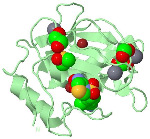
(-) Ligands, Modified Residues, Ions  (6, 19)

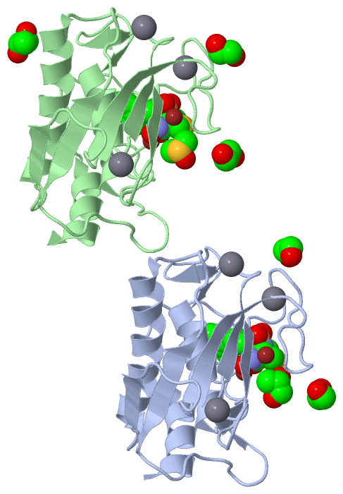
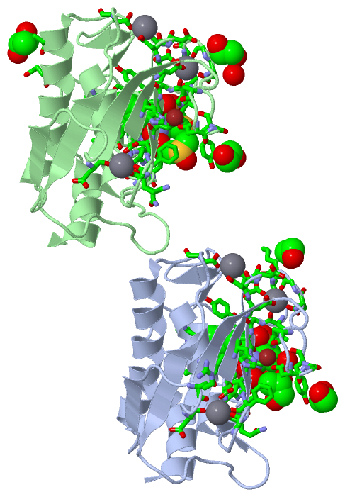
Asymmetric Unit (6, 19)
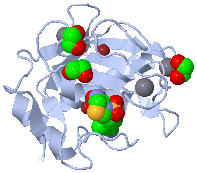
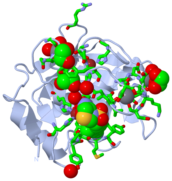
No.NameCountTypeFull Name
1CA6Ligand/IonCALCIUM ION
2DMS1Ligand/IonDIMETHYL SULFOXIDE
3EDO4Ligand/Ion1,2-ETHANEDIOL
4LTQ2Ligand/Ion(4S)-3-{[4-(4-CYANO-2-METHYLPHENYL)PIPERAZIN-1-YL]SULFONYL}-N-HYDROXY-1,3-THIAZOLIDINE-4-CARBOXAMIDE
5PGO2Ligand/IonS-1,2-PROPANEDIOL
6ZN4Ligand/IonZINC ION
Biological Unit 1 (3, 4)
No.NameCountTypeFull Name
1CA-1Ligand/IonCALCIUM ION
2DMS-1Ligand/IonDIMETHYL SULFOXIDE
3EDO2Ligand/Ion1,2-ETHANEDIOL
4LTQ1Ligand/Ion(4S)-3-{[4-(4-CYANO-2-METHYLPHENYL)PIPERAZIN-1-YL]SULFONYL}-N-HYDROXY-1,3-THIAZOLIDINE-4-CARBOXAMIDE
5PGO1Ligand/IonS-1,2-PROPANEDIOL
6ZN-1Ligand/IonZINC ION
Biological Unit 2 (4, 5)
No.NameCountTypeFull Name
1CA-1Ligand/IonCALCIUM ION
2DMS1Ligand/IonDIMETHYL SULFOXIDE
3EDO2Ligand/Ion1,2-ETHANEDIOL
4LTQ1Ligand/Ion(4S)-3-{[4-(4-CYANO-2-METHYLPHENYL)PIPERAZIN-1-YL]SULFONYL}-N-HYDROXY-1,3-THIAZOLIDINE-4-CARBOXAMIDE
5PGO1Ligand/IonS-1,2-PROPANEDIOL
6ZN-1Ligand/IonZINC ION

(-) Sites  (19, 19)

Asymmetric Unit (19, 19)
No.NameEvidenceResiduesDescription
01AC1SOFTWAREHIS A:226 , HIS A:230 , HIS A:236 , LTQ A:306binding site for residue ZN A 301
02AC2SOFTWAREHIS A:175 , ASP A:177 , HIS A:190 , HIS A:203binding site for residue ZN A 302
03AC3SOFTWAREASP A:165 , GLY A:197 , GLN A:199 , ASP A:201 , HOH A:439 , HOH A:441binding site for residue CA A 303
04AC4SOFTWAREASP A:131 , ASP A:206 , GLU A:208 , HOH A:482 , HOH A:512 , HOH A:533binding site for residue CA A 304
05AC5SOFTWAREASP A:182 , GLY A:183 , ASP A:185 , LEU A:187 , ASP A:205 , GLU A:208binding site for residue CA A 305
06AC6SOFTWARELEU A:187 , LEU A:188 , ALA A:189 , LEU A:222 , HIS A:226 , GLU A:227 , HIS A:230 , HIS A:236 , LEU A:243 , TYR A:245 , MET A:247 , TYR A:248 , ZN A:301 , HOH A:419 , TYR B:245 , PRO B:246 , MET B:247binding site for residue LTQ A 306
07AC7SOFTWAREASP A:177 , GLY A:178 , TYR A:179binding site for residue EDO A 307
08AC8SOFTWAREGLY A:183 , LYS A:184 , ASP A:207binding site for residue EDO A 308
09AC9SOFTWAREHIS A:190 , ALA A:191 , PHE A:192 , HOH A:442binding site for residue PGO A 309
10AD1SOFTWAREHIS B:226 , HIS B:230 , HIS B:236 , LTQ B:306binding site for residue ZN B 301
11AD2SOFTWAREHIS B:175 , ASP B:177 , HIS B:190 , HIS B:203binding site for residue ZN B 302
12AD3SOFTWAREASP B:165 , GLY B:197 , GLN B:199 , ASP B:201 , HOH B:429 , HOH B:436binding site for residue CA B 303
13AD4SOFTWAREASP B:131 , ASP B:206 , GLU B:208 , HOH B:528 , HOH B:553binding site for residue CA B 304
14AD5SOFTWAREASP B:182 , GLY B:183 , ASP B:185 , LEU B:187 , ASP B:205 , GLU B:208binding site for residue CA B 305
15AD6SOFTWARETYR A:245 , PRO A:246 , GLY B:186 , LEU B:187 , LEU B:188 , ALA B:189 , LEU B:222 , HIS B:226 , GLU B:227 , HIS B:230 , HIS B:236 , LEU B:243 , TYR B:245 , MET B:247 , TYR B:248 , ARG B:249 , ZN B:301 , HOH B:462binding site for residue LTQ B 306
16AD7SOFTWAREASP B:177 , GLY B:178 , TYR B:179binding site for residue EDO B 307
17AD8SOFTWAREARG A:162 , GLY B:183 , ASP B:207 , HOH B:526binding site for residue EDO B 308
18AD9SOFTWAREHOH A:428 , HIS B:190 , ALA B:191 , PHE B:192binding site for residue DMS B 309
19AE1SOFTWAREALA B:135 , ASP B:139binding site for residue PGO B 310

(-) SS Bonds  (0, 0)

(no "SS Bond" information available for 5CUH)

(-) Cis Peptide Bonds  (0, 0)

(no "Cis Peptide Bond" information available for 5CUH)

 Sequence-Structure Mapping

(-) SAPs(SNPs)/Variants  (0, 0)

(no "SAP(SNP)/Variant" information available for 5CUH)

(-) PROSITE Motifs  (0, 0)

(no "PROSITE Motif" information available for 5CUH)

(-) Exons   (0, 0)

(no "Exon" information available for 5CUH)

(-) Sequences/Alignments

Asymmetric Unit
   Reformat: Number of residues per line =  ('0' or empty: single-line sequence representation)
  Number of residues per labelling interval =   
  UniProt sequence: complete  aligned part    
   Show mapping: SCOP domains CATH domains Pfam domains Secondary structure (by author)
SAPs(SNPs) PROSITE motifs Exons
(details for a mapped element are shown in a popup box when the mouse pointer rests over it)
Chain A from PDB  Type:PROTEIN  Length:164
                                                                                                                                                                                                    
               SCOP domains -------------------------------------------------------------------------------------------------------------------------------------------------------------------- SCOP domains
               CATH domains -------------------------------------------------------------------------------------------------------------------------------------------------------------------- CATH domains
               Pfam domains -------------------------------------------------------------------------------------------------------------------------------------------------------------------- Pfam domains
         Sec.struct. author ..ee..........eeeeee.......hhhhhhhhhhhhhhhhhhh...eeee.......eeeeee.................eee..........eeee....ee......eehhhhhhhhhhhhh.ee.....................hhhhhhhhhhhhh Sec.struct. author
                 SAPs(SNPs) -------------------------------------------------------------------------------------------------------------------------------------------------------------------- SAPs(SNPs)
                    PROSITE -------------------------------------------------------------------------------------------------------------------------------------------------------------------- PROSITE
                 Transcript -------------------------------------------------------------------------------------------------------------------------------------------------------------------- Transcript
                 5cuh A 106 GFQTFEGDLKWHHHNITYWIQNYSEDLPRAVIDDAFARAFALWSAVTPLTFTRVYSRDADIVIQFGVAEHGDGYPFDGKDGLLAHAFPPGPGIQGDAHFDDDELWSLGKGVGYSLFLVAAHEFGHALGLDHSSVPEALMYPMYRFTEGPPLHKDDVNGIRHLYG 269
                                   115       125       135       145       155       165       175       185       195       205       215       225       235       245       255       265    

Chain B from PDB  Type:PROTEIN  Length:164
                                                                                                                                                                                                    
               SCOP domains -------------------------------------------------------------------------------------------------------------------------------------------------------------------- SCOP domains
               CATH domains -------------------------------------------------------------------------------------------------------------------------------------------------------------------- CATH domains
               Pfam domains -------------------------------------------------------------------------------------------------------------------------------------------------------------------- Pfam domains
         Sec.struct. author ..............eeeeee.......hhhhhhhhhhhhhhhhhh....eeee.......eeeeee.................eee..........eeee....ee......eehhhhhhhhhhhhh........................hhhhhhhhhhhhh Sec.struct. author
                 SAPs(SNPs) -------------------------------------------------------------------------------------------------------------------------------------------------------------------- SAPs(SNPs)
                    PROSITE -------------------------------------------------------------------------------------------------------------------------------------------------------------------- PROSITE
                 Transcript -------------------------------------------------------------------------------------------------------------------------------------------------------------------- Transcript
                 5cuh B 106 GFQTFEGDLKWHHHNITYWIQNYSEDLPRAVIDDAFARAFALWSAVTPLTFTRVYSRDADIVIQFGVAEHGDGYPFDGKDGLLAHAFPPGPGIQGDAHFDDDELWSLGKGVGYSLFLVAAHEFGHALGLDHSSVPEALMYPMYRFTEGPPLHKDDVNGIRHLYG 269
                                   115       125       135       145       155       165       175       185       195       205       215       225       235       245       255       265    

   Legend:   → Mismatch (orange background)
  - → Gap (green background, '-', border residues have a numbering label)
    → Modified Residue (blue background, lower-case, 'x' indicates undefined single-letter code, labelled with number + name)
  x → Chemical Group (purple background, 'x', labelled with number + name, e.g. ACE or NH2)
  extra numbering lines below/above indicate numbering irregularities and modified residue names etc., number ends below/above '|'

 Classification and Annotation

(-) SCOP Domains  (0, 0)

(no "SCOP Domain" information available for 5CUH)

(-) CATH Domains  (0, 0)

(no "CATH Domain" information available for 5CUH)

(-) Pfam Domains  (0, 0)

(no "Pfam Domain" information available for 5CUH)

(-) Gene Ontology  (38, 38)

Asymmetric Unit(hide GO term definitions)

 Visualization

(-) Interactive Views

Asymmetric Unit
  Complete Structure
    Jena3D(integrated viewing of ligand, site, SAP, PROSITE, SCOP information)
    WebMol | AstexViewer[tm]@PDBe
(Java Applets, require no local installation except for Java; loading may be slow)
    STRAP
(Java WebStart application, automatic local installation, requires Java; full application with system access!)
    RasMol
(require local installation)
    Molscript (VRML)
(requires installation of a VRML viewer; select preferred view via VRML and generate a mono or stereo PDF format file)
 
  Ligands, Modified Residues, Ions
    CA  [ RasMol | Jena3D ]  +environment [ RasMol | Jena3D ]
    DMS  [ RasMol | Jena3D ]  +environment [ RasMol | Jena3D ]
    EDO  [ RasMol | Jena3D ]  +environment [ RasMol | Jena3D ]
    LTQ  [ RasMol | Jena3D ]  +environment [ RasMol | Jena3D ]
    PGO  [ RasMol | Jena3D ]  +environment [ RasMol | Jena3D ]
    ZN  [ RasMol | Jena3D ]  +environment [ RasMol | Jena3D ]
 
  Sites
    AC1  [ RasMol ]  +environment [ RasMol ]
    AC2  [ RasMol ]  +environment [ RasMol ]
    AC3  [ RasMol ]  +environment [ RasMol ]
    AC4  [ RasMol ]  +environment [ RasMol ]
    AC5  [ RasMol ]  +environment [ RasMol ]
    AC6  [ RasMol ]  +environment [ RasMol ]
    AC7  [ RasMol ]  +environment [ RasMol ]
    AC8  [ RasMol ]  +environment [ RasMol ]
    AC9  [ RasMol ]  +environment [ RasMol ]
    AD1  [ RasMol ]  +environment [ RasMol ]
    AD2  [ RasMol ]  +environment [ RasMol ]
    AD3  [ RasMol ]  +environment [ RasMol ]
    AD4  [ RasMol ]  +environment [ RasMol ]
    AD5  [ RasMol ]  +environment [ RasMol ]
    AD6  [ RasMol ]  +environment [ RasMol ]
    AD7  [ RasMol ]  +environment [ RasMol ]
    AD8  [ RasMol ]  +environment [ RasMol ]
    AD9  [ RasMol ]  +environment [ RasMol ]
    AE1  [ RasMol ]  +environment [ RasMol ]
 
  Cis Peptide Bonds
(no "Cis Peptide Bonds" information available for 5cuh)
 
Biological Units
  Complete Structure
    Biological Unit 1  [ Jena3D ]
    Biological Unit 2  [ Jena3D ]

(-) Still Images

Jmol
  protein: cartoon or spacefill or dots and stick; nucleic acid: cartoon and stick; ligands: spacefill; active site: stick
Molscript
  protein, nucleic acid: cartoon; ligands: spacefill; active site: ball and stick

 Databases and Analysis Tools

(-) Databases

Access by PDB/NDB ID
  5cuh
    Family and Domain InformationProDom | SYSTERS
    General Structural InformationGlycoscienceDB | MMDB | NDB | OCA | PDB | PDBe | PDBj | PDBsum | PDBWiki | PQS | PROTEOPEDIA
    Orientation in MembranesOPM
    Protein SurfaceSURFACE
    Secondary StructureDSSP (structure derived) | HSSP (homology derived)
    Structural GenomicsGeneCensus
    Structural NeighboursCE | VAST
    Structure ClassificationCATH | Dali | SCOP
    Validation and Original DataBMRB Data View | BMRB Restraints Grid | EDS | PROCHECK | RECOORD | WHAT_CHECK
 
Access by UniProt ID/Accession number
  MMP9_HUMAN | P14780
    Comparative Protein Structure ModelsModBase
    Genomic InformationEnsembl
    Protein-protein InteractionDIP
    Sequence, Family and Domain InformationInterPro | Pfam | SMART | UniProtKB/SwissProt
 
Access by Enzyme Classificator   (EC Number)
  3.4.24.35
    General Enzyme InformationBRENDA | EC-PDB | Enzyme | IntEnz
    PathwayKEGG | MetaCyc
 
Access by Disease Identifier   (MIM ID)
  (no 'MIM ID' available)
    Disease InformationOMIM
 
Access by GenAge ID
  (no 'GenAge ID' available)
    Age Related InformationGenAge

(-) Analysis Tools

Access by PDB/NDB ID
    Domain InformationXDom
    Interatomic Contacts of Structural UnitsCSU
    Ligand-protein ContactsLPC
    Protein CavitiescastP
    Sequence and Secondary StructurePDBCartoon
    Structure AlignmentSTRAP(Java WebStart application, automatic local installation, requires Java; full application with system access!)
    Structure and Sequence BrowserSTING
 
Access by UniProt ID/Accession number
  MMP9_HUMAN | P14780
    Protein Disorder PredictionDisEMBL | FoldIndex | GLOBPLOT (for more information see DisProt)

 Related Entries

(-) Entries Sharing at Least One Protein Chain (UniProt ID)

UniProtKB/Swiss-Prot
        MMP9_HUMAN | P147801gkc 1gkd 1itv 1l6j 1lkg 2ovx 2ovz 2ow0 2ow1 2ow2 4h1q 4h2e 4h3x 4h82 4hma 4jij 4jqg 4wzv 4xct 5i12 5th6 5th9

(-) Related Entries Specified in the PDB File

(no "Related Entries Specified in the PDB File" available for 5CUH)