Show PDB file:   
         Plain Text   HTML   (compressed file size)
QuickSearch:   
by PDB,NDB,UniProt,PROSITE Code or Search Term(s)  
(-)Asymmetric Unit
(-)Asym. Unit - sites
(-)Biological Unit 1
(-)Biol. Unit 1 - sites
(-)Biological Unit 2
collapse expand < >
Image Asymmetric Unit
Asymmetric Unit  (Jmol Viewer)
Image Asym. Unit - sites
Asym. Unit - sites  (Jmol Viewer)
Image Biological Unit 1
Biological Unit 1  (Jmol Viewer)
Image Biol. Unit 1 - sites
Biol. Unit 1 - sites  (Jmol Viewer)
Image Biological Unit 2
Biological Unit 2  (Jmol Viewer)

(-) Description

Title :  CRYSTAL STRUCTURE OF AN MMP BROAD SPECTRUM HYDROXAMATE BASED INHIBITOR CC27 IN COMPLEX WITH THE MMP-9 CATALYTIC DOMAIN
 
Authors :  E. A. Stura, L. Vera, E. Cassar-Lajeunesse, E. Nuti, V. Dive, A. Rossell
Date :  14 Sep 12  (Deposition) - 24 Apr 13  (Release) - 31 May 17  (Revision)
Method :  X-RAY DIFFRACTION
Resolution :  1.76
Chains :  Asym. Unit :  A,B
Biol. Unit 1:  A  (1x)
Biol. Unit 2:  B  (1x)
Keywords :  Hydrolase/Hydroxamate Inhibitor, Zincin-Like, Gelatinase, Collagenase (Catalytic Domain), Hydrolase-Hydrolase Inhibitor Complex (Keyword Search: [Gene Ontology, PubMed, Web (Google))
 
Reference :  C. Antoni, L. Vera, L. Devel, M. P. Catalani, B. Czarny, E. Cassar-Lajeunesse, E. Nuti, A. Rossello, V. Dive, E. A. Stura
Crystallization Of Bi-Functional Ligand Protein Complexes.
J. Struct. Biol. V. 182 246 2013
PubMed-ID: 23567804  |  Reference-DOI: 10.1016/J.JSB.2013.03.015
(for further references see the PDB file header)

(-) Compounds

Molecule 1 - MATRIX METALLOPROTEINASE-9
    ChainsA, B
    EC Number3.4.24.35, 3.4.24.35
    EngineeredYES
    Expression SystemESCHERICHIA COLI
    Expression System PlasmidPET-14B
    Expression System StrainBL21 (DE3 STAR)
    Expression System Taxid469008
    Expression System Vector TypePT7 PROMOTER
    FragmentHUMAN WILD-TYPE MMP-9 CATALYTIC DOMAIN UNP RESIDUES 107- 215/391-443
    GeneMMP9, CLG4B
    MutationYES
    Organism CommonHUMAN
    Organism ScientificHOMO SAPIENS
    Organism Taxid9606
    SynonymMMP-9,92 KDA GELATINASE,92 KDA TYPE IV COLLAGENASE, GELATINASE B,GELB

 Structural Features

(-) Chains, Units

  12
Asymmetric Unit AB
Biological Unit 1 (1x)A 
Biological Unit 2 (1x) B

Summary Information (see also Sequences/Alignments below)

(-) Ligands, Modified Residues, Ions  (6, 22)

Asymmetric Unit (6, 22)
No.NameCountTypeFull Name
110B2Ligand/IonN-2-(BIPHENYL-4-YLSULFONYL)-N-2-(ISOPROPYLOXY)-ACETOHYDROXAMIC ACID
2CA6Ligand/IonCALCIUM ION
3GOL7Ligand/IonGLYCEROL
4PEG2Ligand/IonDI(HYDROXYETHYL)ETHER
5PGO1Ligand/IonS-1,2-PROPANEDIOL
6ZN4Ligand/IonZINC ION
Biological Unit 1 (3, 8)
No.NameCountTypeFull Name
110B1Ligand/IonN-2-(BIPHENYL-4-YLSULFONYL)-N-2-(ISOPROPYLOXY)-ACETOHYDROXAMIC ACID
2CA-1Ligand/IonCALCIUM ION
3GOL6Ligand/IonGLYCEROL
4PEG-1Ligand/IonDI(HYDROXYETHYL)ETHER
5PGO1Ligand/IonS-1,2-PROPANEDIOL
6ZN-1Ligand/IonZINC ION
Biological Unit 2 (3, 4)
No.NameCountTypeFull Name
110B1Ligand/IonN-2-(BIPHENYL-4-YLSULFONYL)-N-2-(ISOPROPYLOXY)-ACETOHYDROXAMIC ACID
2CA-1Ligand/IonCALCIUM ION
3GOL1Ligand/IonGLYCEROL
4PEG2Ligand/IonDI(HYDROXYETHYL)ETHER
5PGO-1Ligand/IonS-1,2-PROPANEDIOL
6ZN-1Ligand/IonZINC ION

(-) Sites  (22, 22)

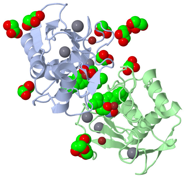
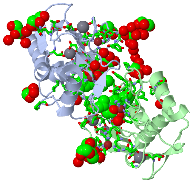
Asymmetric Unit (22, 22)
No.NameEvidenceResiduesDescription
01AC1SOFTWAREHIS A:226 , HIS A:230 , HIS A:236 , 10B A:306BINDING SITE FOR RESIDUE ZN A 301
02AC2SOFTWAREHIS A:175 , ASP A:177 , HIS A:190 , HIS A:203BINDING SITE FOR RESIDUE ZN A 302
03AC3SOFTWAREASP A:182 , GLY A:183 , ASP A:185 , LEU A:187 , ASP A:205 , GLU A:208BINDING SITE FOR RESIDUE CA A 303
04AC4SOFTWAREASP A:131 , ASP A:206 , GLU A:208 , HOH A:452 , HOH A:453BINDING SITE FOR RESIDUE CA A 304
05AC5SOFTWAREASP A:165 , GLY A:197 , GLN A:199 , ASP A:201 , HOH A:406 , HOH A:411BINDING SITE FOR RESIDUE CA A 305
06AC6SOFTWAREGLY A:186 , LEU A:187 , LEU A:188 , ALA A:189 , LEU A:222 , HIS A:226 , GLU A:227 , HIS A:230 , HIS A:236 , LEU A:243 , TYR A:245 , MET A:247 , ZN A:301 , HOH A:415 , HOH A:492 , TYR B:245 , PRO B:246BINDING SITE FOR RESIDUE 10B A 306
07AC7SOFTWAREPRO A:254 , HOH A:421 , PRO B:254 , PRO B:255 , HOH B:448 , HOH B:451BINDING SITE FOR RESIDUE GOL A 307
08AC8SOFTWAREASP A:177 , GLY A:178 , GOL A:311 , HOH A:476 , HOH A:515 , HOH A:553 , HOH A:559 , HOH A:560 , HOH A:590BINDING SITE FOR RESIDUE GOL A 308
09AC9SOFTWARETYR A:179 , LEU A:187 , HIS A:190 , HOH A:594 , HOH A:595 , PRO B:240 , TYR B:245BINDING SITE FOR RESIDUE GOL A 309
10BC1SOFTWAREALA A:173 , GLU A:174BINDING SITE FOR RESIDUE PGO A 310
11BC2SOFTWAREGOL A:308 , HOH A:480 , HOH A:515 , HOH A:529 , HOH A:553BINDING SITE FOR RESIDUE GOL A 311
12BC3SOFTWAREASN A:120 , THR A:122 , THR A:157 , HOH A:536BINDING SITE FOR RESIDUE GOL A 312
13BC4SOFTWAREHOH A:434 , HOH A:483BINDING SITE FOR RESIDUE GOL A 313
14BC5SOFTWAREHIS B:226 , HIS B:230 , HIS B:236 , 10B B:306BINDING SITE FOR RESIDUE ZN B 301
15BC6SOFTWAREHIS B:175 , ASP B:177 , HIS B:190 , HIS B:203BINDING SITE FOR RESIDUE ZN B 302
16BC7SOFTWAREASP B:182 , GLY B:183 , ASP B:185 , LEU B:187 , ASP B:205 , GLU B:208BINDING SITE FOR RESIDUE CA B 303
17BC8SOFTWAREASP B:131 , ASP B:206 , GLU B:208 , HOH B:468 , HOH B:529BINDING SITE FOR RESIDUE CA B 304
18BC9SOFTWAREASP B:165 , GLY B:197 , GLN B:199 , ASP B:201 , HOH B:401 , HOH B:405BINDING SITE FOR RESIDUE CA B 305
19CC1SOFTWARETYR A:245 , PRO A:246 , MET A:247 , GLY B:186 , LEU B:187 , LEU B:188 , ALA B:189 , LEU B:222 , HIS B:226 , GLU B:227 , HIS B:230 , HIS B:236 , LEU B:243 , TYR B:245 , MET B:247 , TYR B:248 , ZN B:301 , HOH B:444BINDING SITE FOR RESIDUE 10B B 306
20CC2SOFTWAREPHE B:250 , THR B:251 , HOH B:423 , HOH B:515 , HOH B:546BINDING SITE FOR RESIDUE GOL B 307
21CC3SOFTWAREPEG B:309 , HOH B:442 , HOH B:443 , HOH B:480BINDING SITE FOR RESIDUE PEG B 308
22CC4SOFTWARETYR B:179 , PEG B:308 , HOH B:442BINDING SITE FOR RESIDUE PEG B 309

(-) SS Bonds  (0, 0)

(no "SS Bond" information available for 4H3X)

(-) Cis Peptide Bonds  (0, 0)

(no "Cis Peptide Bond" information available for 4H3X)

 Sequence-Structure Mapping

(-) SAPs(SNPs)/Variants  (0, 0)

(no "SAP(SNP)/Variant" information available for 4H3X)

(-) PROSITE Motifs  (0, 0)

(no "PROSITE Motif" information available for 4H3X)

(-) Exons   (0, 0)

(no "Exon" information available for 4H3X)

(-) Sequences/Alignments

Asymmetric Unit
   Reformat: Number of residues per line =  ('0' or empty: single-line sequence representation)
  Number of residues per labelling interval =   
  UniProt sequence: complete  aligned part    
   Show mapping: SCOP domains CATH domains Pfam domains Secondary structure (by author)
SAPs(SNPs) PROSITE motifs Exons
(details for a mapped element are shown in a popup box when the mouse pointer rests over it)
Chain A from PDB  Type:PROTEIN  Length:164
                                                                                                                                                                                                    
               SCOP domains d4h3xa_ A: Gelatinase B (MMP-9)                                                                                                                                      SCOP domains
               CATH domains -------------------------------------------------------------------------------------------------------------------------------------------------------------------- CATH domains
               Pfam domains -------------------------------------------------------------------------------------------------------------------------------------------------------------------- Pfam domains
         Sec.struct. author ..ee..........eeeeee.......hhhhhhhhhhhhhhhhhhh...eeee.......eeeeee.................eee..........eeee....ee......eehhhhhhhhhhhhh.ee.....................hhhhhhhhhhhhh Sec.struct. author
                 SAPs(SNPs) -------------------------------------------------------------------------------------------------------------------------------------------------------------------- SAPs(SNPs)
                    PROSITE -------------------------------------------------------------------------------------------------------------------------------------------------------------------- PROSITE
                 Transcript -------------------------------------------------------------------------------------------------------------------------------------------------------------------- Transcript
                 4h3x A 106 GFQTFEGDLKWHHHNITYWIQNYSEDLPRAVIDDAFARAFALWSAVTPLTFTRVYSRDADIVIQFGVAEHGDGYPFDGKDGLLAHAFPPGPGIQGDAHFDDDELWSLGKGVGYSLFLVAAHEFGHALGLDHSSVPEALMYPMYRFTEGPPLHKDDVNGIRHLYG 269
                                   115       125       135       145       155       165       175       185       195       205       215       225       235       245       255       265    

Chain B from PDB  Type:PROTEIN  Length:164
                                                                                                                                                                                                    
               SCOP domains d4h3xb_ B: Gelatinase B (MMP-9)                                                                                                                                      SCOP domains
               CATH domains -------------------------------------------------------------------------------------------------------------------------------------------------------------------- CATH domains
               Pfam domains -------------------------------------------------------------------------------------------------------------------------------------------------------------------- Pfam domains
         Sec.struct. author ..............eeeeee.......hhhhhhhhhhhhhhhhhh....eeee.......eeeeee.................eee..........eeee....ee......eehhhhhhhhhhhhhh.......................hhhhhhhhhhhhh Sec.struct. author
                 SAPs(SNPs) -------------------------------------------------------------------------------------------------------------------------------------------------------------------- SAPs(SNPs)
                    PROSITE -------------------------------------------------------------------------------------------------------------------------------------------------------------------- PROSITE
                 Transcript -------------------------------------------------------------------------------------------------------------------------------------------------------------------- Transcript
                 4h3x B 106 GFQTFEGDLKWHHHNITYWIQNYSEDLPRAVIDDAFARAFALWSAVTPLTFTRVYSRDADIVIQFGVAEHGDGYPFDGKDGLLAHAFPPGPGIQGDAHFDDDELWSLGKGVGYSLFLVAAHEFGHALGLDHSSVPEALMYPMYRFTEGPPLHKDDVNGIRHLYG 269
                                   115       125       135       145       155       165       175       185       195       205       215       225       235       245       255       265    

   Legend:   → Mismatch (orange background)
  - → Gap (green background, '-', border residues have a numbering label)
    → Modified Residue (blue background, lower-case, 'x' indicates undefined single-letter code, labelled with number + name)
  x → Chemical Group (purple background, 'x', labelled with number + name, e.g. ACE or NH2)
  extra numbering lines below/above indicate numbering irregularities and modified residue names etc., number ends below/above '|'

 Classification and Annotation

(-) SCOP Domains  (1, 2)

Asymmetric Unit

(-) CATH Domains  (0, 0)

(no "CATH Domain" information available for 4H3X)

(-) Pfam Domains  (0, 0)

(no "Pfam Domain" information available for 4H3X)

(-) Gene Ontology  (38, 38)

Asymmetric Unit(hide GO term definitions)

 Visualization

(-) Interactive Views

Asymmetric Unit
  Complete Structure
    Jena3D(integrated viewing of ligand, site, SAP, PROSITE, SCOP information)
    WebMol | AstexViewer[tm]@PDBe
(Java Applets, require no local installation except for Java; loading may be slow)
    STRAP
(Java WebStart application, automatic local installation, requires Java; full application with system access!)
    RasMol
(require local installation)
    Molscript (VRML)
(requires installation of a VRML viewer; select preferred view via VRML and generate a mono or stereo PDF format file)
 
  Ligands, Modified Residues, Ions
    10B  [ RasMol | Jena3D ]  +environment [ RasMol | Jena3D ]
    CA  [ RasMol | Jena3D ]  +environment [ RasMol | Jena3D ]
    GOL  [ RasMol | Jena3D ]  +environment [ RasMol | Jena3D ]
    PEG  [ RasMol | Jena3D ]  +environment [ RasMol | Jena3D ]
    PGO  [ RasMol | Jena3D ]  +environment [ RasMol | Jena3D ]
    ZN  [ RasMol | Jena3D ]  +environment [ RasMol | Jena3D ]
 
  Sites
    AC1  [ RasMol ]  +environment [ RasMol ]
    AC2  [ RasMol ]  +environment [ RasMol ]
    AC3  [ RasMol ]  +environment [ RasMol ]
    AC4  [ RasMol ]  +environment [ RasMol ]
    AC5  [ RasMol ]  +environment [ RasMol ]
    AC6  [ RasMol ]  +environment [ RasMol ]
    AC7  [ RasMol ]  +environment [ RasMol ]
    AC8  [ RasMol ]  +environment [ RasMol ]
    AC9  [ RasMol ]  +environment [ RasMol ]
    BC1  [ RasMol ]  +environment [ RasMol ]
    BC2  [ RasMol ]  +environment [ RasMol ]
    BC3  [ RasMol ]  +environment [ RasMol ]
    BC4  [ RasMol ]  +environment [ RasMol ]
    BC5  [ RasMol ]  +environment [ RasMol ]
    BC6  [ RasMol ]  +environment [ RasMol ]
    BC7  [ RasMol ]  +environment [ RasMol ]
    BC8  [ RasMol ]  +environment [ RasMol ]
    BC9  [ RasMol ]  +environment [ RasMol ]
    CC1  [ RasMol ]  +environment [ RasMol ]
    CC2  [ RasMol ]  +environment [ RasMol ]
    CC3  [ RasMol ]  +environment [ RasMol ]
    CC4  [ RasMol ]  +environment [ RasMol ]
 
  Cis Peptide Bonds
(no "Cis Peptide Bonds" information available for 4h3x)
 
Biological Units
  Complete Structure
    Biological Unit 1  [ Jena3D ]
    Biological Unit 2  [ Jena3D ]

(-) Still Images

Jmol
  protein: cartoon or spacefill or dots and stick; nucleic acid: cartoon and stick; ligands: spacefill; active site: stick
Molscript
  protein, nucleic acid: cartoon; ligands: spacefill; active site: ball and stick

 Databases and Analysis Tools

(-) Databases

Access by PDB/NDB ID
  4h3x
    Family and Domain InformationProDom | SYSTERS
    General Structural InformationGlycoscienceDB | MMDB | NDB | OCA | PDB | PDBe | PDBj | PDBsum | PDBWiki | PQS | PROTEOPEDIA
    Orientation in MembranesOPM
    Protein SurfaceSURFACE
    Secondary StructureDSSP (structure derived) | HSSP (homology derived)
    Structural GenomicsGeneCensus
    Structural NeighboursCE | VAST
    Structure ClassificationCATH | Dali | SCOP
    Validation and Original DataBMRB Data View | BMRB Restraints Grid | EDS | PROCHECK | RECOORD | WHAT_CHECK
 
Access by UniProt ID/Accession number
  MMP9_HUMAN | P14780
    Comparative Protein Structure ModelsModBase
    Genomic InformationEnsembl
    Protein-protein InteractionDIP
    Sequence, Family and Domain InformationInterPro | Pfam | SMART | UniProtKB/SwissProt
 
Access by Enzyme Classificator   (EC Number)
  3.4.24.35
    General Enzyme InformationBRENDA | EC-PDB | Enzyme | IntEnz
    PathwayKEGG | MetaCyc
 
Access by Disease Identifier   (MIM ID)
  (no 'MIM ID' available)
    Disease InformationOMIM
 
Access by GenAge ID
  (no 'GenAge ID' available)
    Age Related InformationGenAge

(-) Analysis Tools

Access by PDB/NDB ID
    Domain InformationXDom
    Interatomic Contacts of Structural UnitsCSU
    Ligand-protein ContactsLPC
    Protein CavitiescastP
    Sequence and Secondary StructurePDBCartoon
    Structure AlignmentSTRAP(Java WebStart application, automatic local installation, requires Java; full application with system access!)
    Structure and Sequence BrowserSTING
 
Access by UniProt ID/Accession number
  MMP9_HUMAN | P14780
    Protein Disorder PredictionDisEMBL | FoldIndex | GLOBPLOT (for more information see DisProt)

 Related Entries

(-) Entries Sharing at Least One Protein Chain (UniProt ID)

UniProtKB/Swiss-Prot
        MMP9_HUMAN | P147801gkc 1gkd 1itv 1l6j 1lkg 2ovx 2ovz 2ow0 2ow1 2ow2 4h1q 4h2e 4h82 4hma 4jij 4jqg 4wzv 4xct 5cuh 5i12 5th6 5th9

(-) Related Entries Specified in the PDB File

2ow1 MMP-9 ACTIVE SITE MUTANT WITH TRIFLUOROMETHYL HYDROXAMATE INHIBITOR
4h1q CRYSTAL STRUCTURE OF MUTANT MMP-9 CATALYTIC DOMAIN IN COMPLEX WITH A TWIN INHIBITOR.
4h2e CRYSTAL STRUCTURE OF AN MMP TWIN INHIBITOR COMPLEXING TWO MMP-9 CATALYTIC DOMAINS