Show PDB file:   
         Plain Text   HTML   (compressed file size)
QuickSearch:   
by PDB,NDB,UniProt,PROSITE Code or Search Term(s)  
(-)Asymmetric Unit
(-)Asym. Unit - sites
(-)Biological Unit 1
(-)Biol. Unit 1 - sites
(-)Biological Unit 2
(-)Biological Unit 3
(-)Biological Unit 4
collapse expand < >
Image Asymmetric Unit
Asymmetric Unit  (Jmol Viewer)
Image Asym. Unit - sites
Asym. Unit - sites  (Jmol Viewer)
Image Biological Unit 1
Biological Unit 1  (Jmol Viewer)
Image Biol. Unit 1 - sites
Biol. Unit 1 - sites  (Jmol Viewer)
Image Biological Unit 2
Biological Unit 2  (Jmol Viewer)
Image Biological Unit 3
Biological Unit 3  (Jmol Viewer)
Image Biological Unit 4
Biological Unit 4  (Jmol Viewer)

(-) Description

Title :  CRYSTAL STRUCTURE OF THE ZAIRE EBOLAVIRUS VP35 INTERFERON INHIBITORY DOMAIN R312A/K319A/R322A MUTANT
 
Authors :  J. B. Binning, T. Wang, D. W. Leung, W. Xu, D. Borek, G. K. Amarasinghe
Date :  21 Dec 12  (Deposition) - 09 Oct 13  (Release) - 11 Dec 13  (Revision)
Method :  X-RAY DIFFRACTION
Resolution :  1.90
Chains :  Asym. Unit :  A,B,C,D
Biol. Unit 1:  A  (1x)
Biol. Unit 2:  B  (1x)
Biol. Unit 3:  C  (1x)
Biol. Unit 4:  D  (1x)
Keywords :  Ifn Inhibition, Polymerase Cofactor, Rna Binding Protein, Interferon Antagonism, Virus, Viral Protein (Keyword Search: [Gene Ontology, PubMed, Web (Google))
 
Reference :  J. M. Binning, T. Wang, P. Luthra, R. S. Shabman, D. M. Borek, G. Liu, W. Xu, D. W. Leung, C. F. Basler, G. K. Amarasinghe
Development Of Rna Aptamers Targeting Ebola Virus Vp35.
Biochemistry V. 52 8406 2013
PubMed-ID: 24067086  |  Reference-DOI: 10.1021/BI400704D

(-) Compounds

Molecule 1 - POLYMERASE COFACTOR VP35
    ChainsA, B, C, D
    EngineeredYES
    Expression SystemESCHERICHIA COLI
    Expression System PlasmidPET15B
    Expression System StrainBL21(DE3)
    Expression System Taxid469008
    Expression System Vector TypePLASMID
    FragmentINTERFERON INHIBITORY DOMAIN (UNP RESIDUES 218-340)
    GeneVP35
    MutationYES
    Organism CommonZEBOV
    Organism ScientificZAIRE EBOLAVIRUS
    Organism Taxid128952
    StrainMAYINGA-76

 Structural Features

(-) Chains, Units

  1234
Asymmetric Unit ABCD
Biological Unit 1 (1x)A   
Biological Unit 2 (1x) B  
Biological Unit 3 (1x)  C 
Biological Unit 4 (1x)   D

Summary Information (see also Sequences/Alignments below)

(-) Ligands, Modified Residues, Ions  (3, 9)

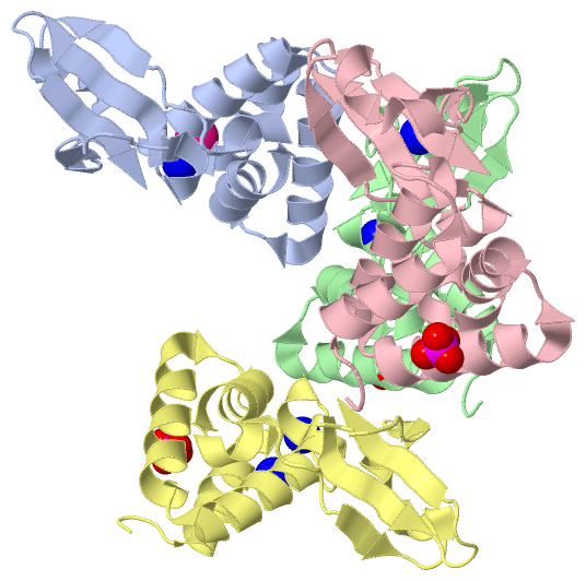
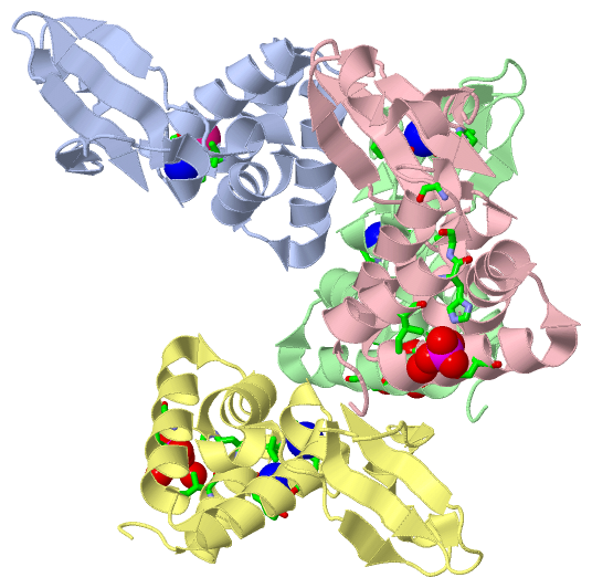
Asymmetric Unit (3, 9)
No.NameCountTypeFull Name
1K1Ligand/IonPOTASSIUM ION
2NA5Ligand/IonSODIUM ION
3PO43Ligand/IonPHOSPHATE ION
Biological Unit 1 (0, 0)
No.NameCountTypeFull Name
1K-1Ligand/IonPOTASSIUM ION
2NA-1Ligand/IonSODIUM ION
3PO4-1Ligand/IonPHOSPHATE ION
Biological Unit 2 (1, 1)
No.NameCountTypeFull Name
1K-1Ligand/IonPOTASSIUM ION
2NA-1Ligand/IonSODIUM ION
3PO41Ligand/IonPHOSPHATE ION
Biological Unit 3 (1, 1)
No.NameCountTypeFull Name
1K-1Ligand/IonPOTASSIUM ION
2NA-1Ligand/IonSODIUM ION
3PO41Ligand/IonPHOSPHATE ION
Biological Unit 4 (1, 1)
No.NameCountTypeFull Name
1K-1Ligand/IonPOTASSIUM ION
2NA-1Ligand/IonSODIUM ION
3PO41Ligand/IonPHOSPHATE ION

(-) Sites  (9, 9)

Asymmetric Unit (9, 9)
No.NameEvidenceResiduesDescription
1AC1SOFTWAREPRO A:233 , THR A:237 , HOH A:509BINDING SITE FOR RESIDUE NA A 401
2AC2SOFTWARELEU A:232 , PRO A:233 , GLY A:234 , HOH A:503 , HOH A:583 , LEU C:232 , GLY C:234BINDING SITE FOR RESIDUE K A 402
3AC3SOFTWAREPRO B:233 , THR B:237 , HOH B:507BINDING SITE FOR RESIDUE NA B 401
4AC4SOFTWAREPRO B:315 , PRO B:316 , HOH B:547 , PRO C:315 , PRO C:316 , HOH C:537BINDING SITE FOR RESIDUE NA B 402
5AC5SOFTWAREILE B:227 , HIS B:231 , GLN B:264 , HOH B:565 , HOH B:580BINDING SITE FOR RESIDUE PO4 B 403
6AC6SOFTWAREILE C:227 , HIS C:231 , GLN C:264 , HOH C:583BINDING SITE FOR RESIDUE PO4 C 401
7AC7SOFTWAREGLY B:234 , TYR D:229 , LEU D:232 , GLY D:234 , HIS D:240 , HOH D:586 , HOH D:590BINDING SITE FOR RESIDUE NA D 401
8AC8SOFTWAREPRO D:233 , THR D:237 , HOH D:507BINDING SITE FOR RESIDUE NA D 402
9AC9SOFTWAREILE D:227 , HIS D:231 , GLN D:264 , HOH D:534 , HOH D:545BINDING SITE FOR RESIDUE PO4 D 403

(-) SS Bonds  (0, 0)

(no "SS Bond" information available for 4IJE)

(-) Cis Peptide Bonds  (0, 0)

(no "Cis Peptide Bond" information available for 4IJE)

 Sequence-Structure Mapping

(-) SAPs(SNPs)/Variants  (0, 0)

(no "SAP(SNP)/Variant" information available for 4IJE)

(-) PROSITE Motifs  (0, 0)

(no "PROSITE Motif" information available for 4IJE)

(-) Exons   (0, 0)

(no "Exon" information available for 4IJE)

(-) Sequences/Alignments

Asymmetric Unit
   Reformat: Number of residues per line =  ('0' or empty: single-line sequence representation)
  Number of residues per labelling interval =   
  UniProt sequence: complete  aligned part    
   Show mapping: SCOP domains CATH domains Pfam domains Secondary structure (by author)
SAPs(SNPs) PROSITE motifs Exons
(details for a mapped element are shown in a popup box when the mouse pointer rests over it)
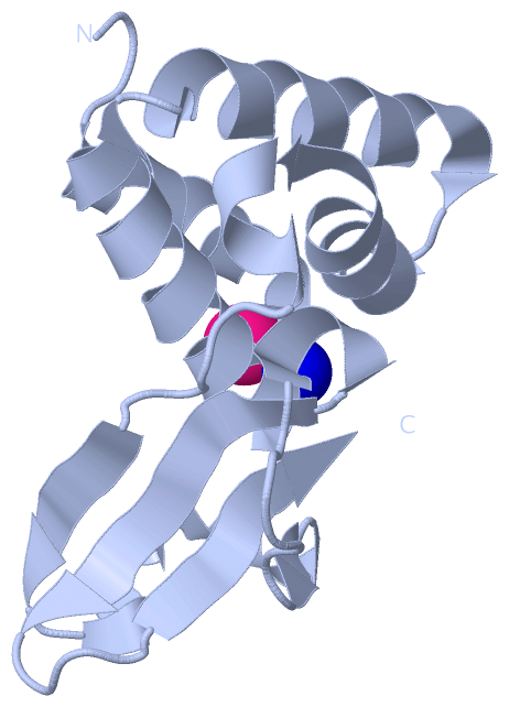
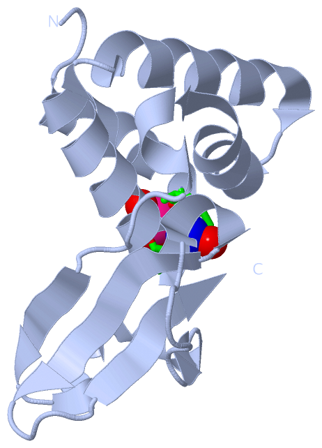
Chain A from PDB  Type:PROTEIN  Length:124
                                                                                                                                                            
               SCOP domains ---------------------------------------------------------------------------------------------------------------------------- SCOP domains
               CATH domains ---------------------------------------------------------------------------------------------------------------------------- CATH domains
               Pfam domains ---------------------------------------------------------------------------------------------------------------------------- Pfam domains
         Sec.struct. author ...hhhhhhhhhhhhh....hhhhhhhhhhhhhhhhh.hhhhhhhhhhhhhhhh.hhhhhhhhhhhhhhhhh.....eeee.hhhhhhhhhhh.ee......hhhh.eeeeeee....eeeee. Sec.struct. author
                 SAPs(SNPs) ---------------------------------------------------------------------------------------------------------------------------- SAPs(SNPs)
                    PROSITE ---------------------------------------------------------------------------------------------------------------------------- PROSITE
                 Transcript ---------------------------------------------------------------------------------------------------------------------------- Transcript
                 4ije A 217 PDISAKDLRNIMYDHLPGFGTAFHQLVQVICKLGKDSNSLDIIHAEFQASLAEGDSPQCALIQITKRVPIFQDAAPPVIHIRSRGDIPRACQKSLAPVPPSPAIDAGWVCVFQLQDGKTLGLKI 340
                                   226       236       246       256       266       276       286       296       306       316       326       336    

Chain B from PDB  Type:PROTEIN  Length:124
                                                                                                                                                            
               SCOP domains ---------------------------------------------------------------------------------------------------------------------------- SCOP domains
               CATH domains ---------------------------------------------------------------------------------------------------------------------------- CATH domains
               Pfam domains ---------------------------------------------------------------------------------------------------------------------------- Pfam domains
         Sec.struct. author ...hhhhhhhhhhh......hhhhhhhhhhhhhhhhh.hhhhhhhhhhhhhhhh.hhhhhhhhhhhhhhhhh.....eee..hhhhhhhhhhh.ee......hhhh.eeeeee.....eeeee. Sec.struct. author
                 SAPs(SNPs) ---------------------------------------------------------------------------------------------------------------------------- SAPs(SNPs)
                    PROSITE ---------------------------------------------------------------------------------------------------------------------------- PROSITE
                 Transcript ---------------------------------------------------------------------------------------------------------------------------- Transcript
                 4ije B 217 PDISAKDLRNIMYDHLPGFGTAFHQLVQVICKLGKDSNSLDIIHAEFQASLAEGDSPQCALIQITKRVPIFQDAAPPVIHIRSRGDIPRACQKSLAPVPPSPAIDAGWVCVFQLQDGKTLGLKI 340
                                   226       236       246       256       266       276       286       296       306       316       326       336    

Chain C from PDB  Type:PROTEIN  Length:124
                                                                                                                                                            
               SCOP domains ---------------------------------------------------------------------------------------------------------------------------- SCOP domains
               CATH domains ---------------------------------------------------------------------------------------------------------------------------- CATH domains
               Pfam domains ---------------------------------------------------------------------------------------------------------------------------- Pfam domains
         Sec.struct. author ...hhhhhhhhhhhhh....hhhhhhhhhhhhhhhhh.hhhhhhhhhhhhhhhh.hhhhhhhhhhhhhhhhh.....eeee.hhhhhhhhhh..ee......hhhh.eeeeeee....eeeee. Sec.struct. author
                 SAPs(SNPs) ---------------------------------------------------------------------------------------------------------------------------- SAPs(SNPs)
                    PROSITE ---------------------------------------------------------------------------------------------------------------------------- PROSITE
                 Transcript ---------------------------------------------------------------------------------------------------------------------------- Transcript
                 4ije C 217 PDISAKDLRNIMYDHLPGFGTAFHQLVQVICKLGKDSNSLDIIHAEFQASLAEGDSPQCALIQITKRVPIFQDAAPPVIHIRSRGDIPRACQKSLAPVPPSPAIDAGWVCVFQLQDGKTLGLKI 340
                                   226       236       246       256       266       276       286       296       306       316       326       336    

Chain D from PDB  Type:PROTEIN  Length:124
                                                                                                                                                            
               SCOP domains ---------------------------------------------------------------------------------------------------------------------------- SCOP domains
               CATH domains ---------------------------------------------------------------------------------------------------------------------------- CATH domains
               Pfam domains ---------------------------------------------------------------------------------------------------------------------------- Pfam domains
         Sec.struct. author ...hhhhhhhhhhhhh....hhhhhhhhhhhhhhhhh.hhhhhhhhhhhhhhhh.hhhhhhhhhhhhhhhhh.....eee..hhhhhhhhhhh.ee......hhhh.eeeeee.....eeeee. Sec.struct. author
                 SAPs(SNPs) ---------------------------------------------------------------------------------------------------------------------------- SAPs(SNPs)
                    PROSITE ---------------------------------------------------------------------------------------------------------------------------- PROSITE
                 Transcript ---------------------------------------------------------------------------------------------------------------------------- Transcript
                 4ije D 217 PDISAKDLRNIMYDHLPGFGTAFHQLVQVICKLGKDSNSLDIIHAEFQASLAEGDSPQCALIQITKRVPIFQDAAPPVIHIRSRGDIPRACQKSLAPVPPSPAIDAGWVCVFQLQDGKTLGLKI 340
                                   226       236       246       256       266       276       286       296       306       316       326       336    

   Legend:   → Mismatch (orange background)
  - → Gap (green background, '-', border residues have a numbering label)
    → Modified Residue (blue background, lower-case, 'x' indicates undefined single-letter code, labelled with number + name)
  x → Chemical Group (purple background, 'x', labelled with number + name, e.g. ACE or NH2)
  extra numbering lines below/above indicate numbering irregularities and modified residue names etc., number ends below/above '|'

 Classification and Annotation

(-) SCOP Domains  (0, 0)

(no "SCOP Domain" information available for 4IJE)

(-) CATH Domains  (0, 0)

(no "CATH Domain" information available for 4IJE)

(-) Pfam Domains  (0, 0)

(no "Pfam Domain" information available for 4IJE)

(-) Gene Ontology  (21, 21)

Asymmetric Unit(hide GO term definitions)

 Visualization

(-) Interactive Views

Asymmetric Unit
  Complete Structure
    Jena3D(integrated viewing of ligand, site, SAP, PROSITE, SCOP information)
    WebMol | AstexViewer[tm]@PDBe
(Java Applets, require no local installation except for Java; loading may be slow)
    STRAP
(Java WebStart application, automatic local installation, requires Java; full application with system access!)
    RasMol
(require local installation)
    Molscript (VRML)
(requires installation of a VRML viewer; select preferred view via VRML and generate a mono or stereo PDF format file)
 
  Ligands, Modified Residues, Ions
    K  [ RasMol | Jena3D ]  +environment [ RasMol | Jena3D ]
    NA  [ RasMol | Jena3D ]  +environment [ RasMol | Jena3D ]
    PO4  [ RasMol | Jena3D ]  +environment [ RasMol | Jena3D ]
 
  Sites
    AC1  [ RasMol ]  +environment [ RasMol ]
    AC2  [ RasMol ]  +environment [ RasMol ]
    AC3  [ RasMol ]  +environment [ RasMol ]
    AC4  [ RasMol ]  +environment [ RasMol ]
    AC5  [ RasMol ]  +environment [ RasMol ]
    AC6  [ RasMol ]  +environment [ RasMol ]
    AC7  [ RasMol ]  +environment [ RasMol ]
    AC8  [ RasMol ]  +environment [ RasMol ]
    AC9  [ RasMol ]  +environment [ RasMol ]
 
  Cis Peptide Bonds
(no "Cis Peptide Bonds" information available for 4ije)
 
Biological Units
  Complete Structure
    Biological Unit 1  [ Jena3D ]
    Biological Unit 2  [ Jena3D ]
    Biological Unit 3  [ Jena3D ]
    Biological Unit 4  [ Jena3D ]

(-) Still Images

Jmol
  protein: cartoon or spacefill or dots and stick; nucleic acid: cartoon and stick; ligands: spacefill; active site: stick
Molscript
  protein, nucleic acid: cartoon; ligands: spacefill; active site: ball and stick

 Databases and Analysis Tools

(-) Databases

Access by PDB/NDB ID
  4ije
    Family and Domain InformationProDom | SYSTERS
    General Structural InformationGlycoscienceDB | MMDB | NDB | OCA | PDB | PDBe | PDBj | PDBsum | PDBWiki | PQS | PROTEOPEDIA
    Orientation in MembranesOPM
    Protein SurfaceSURFACE
    Secondary StructureDSSP (structure derived) | HSSP (homology derived)
    Structural GenomicsGeneCensus
    Structural NeighboursCE | VAST
    Structure ClassificationCATH | Dali | SCOP
    Validation and Original DataBMRB Data View | BMRB Restraints Grid | EDS | PROCHECK | RECOORD | WHAT_CHECK
 
Access by UniProt ID/Accession number
  VP35_EBOZM | Q05127
    Comparative Protein Structure ModelsModBase
    Genomic InformationEnsembl
    Protein-protein InteractionDIP
    Sequence, Family and Domain InformationInterPro | Pfam | SMART | UniProtKB/SwissProt
 
Access by Enzyme Classificator   (EC Number)
  (no 'Enzyme Classificator' available)
    General Enzyme InformationBRENDA | EC-PDB | Enzyme | IntEnz
    PathwayKEGG | MetaCyc
 
Access by Disease Identifier   (MIM ID)
  (no 'MIM ID' available)
    Disease InformationOMIM
 
Access by GenAge ID
  (no 'GenAge ID' available)
    Age Related InformationGenAge

(-) Analysis Tools

Access by PDB/NDB ID
    Domain InformationXDom
    Interatomic Contacts of Structural UnitsCSU
    Ligand-protein ContactsLPC
    Protein CavitiescastP
    Sequence and Secondary StructurePDBCartoon
    Structure AlignmentSTRAP(Java WebStart application, automatic local installation, requires Java; full application with system access!)
    Structure and Sequence BrowserSTING
 
Access by UniProt ID/Accession number
  VP35_EBOZM | Q05127
    Protein Disorder PredictionDisEMBL | FoldIndex | GLOBPLOT (for more information see DisProt)

 Related Entries

(-) Entries Sharing at Least One Protein Chain (UniProt ID)

UniProtKB/Swiss-Prot
        VP35_EBOZM | Q051273fke 3l25 3l26 3l27 3l28 3l29 4ibb 4ibc 4ibd 4ibe 4ibf 4ibg 4ibi 4ibj 4ibk 4ijf 4ypi 4zta 4ztg 4zti

(-) Related Entries Specified in the PDB File

3fke ZAIRE EBOV VP35 IID WILDTYPE
3l25
3l26
3l27
3l28
3l29
4ijf