Show PDB file:   
         Plain Text   HTML   (compressed file size)
QuickSearch:   
by PDB,NDB,UniProt,PROSITE Code or Search Term(s)  
(-)Asymmetric Unit
(-)Asym. Unit - sites
(-)Biological Unit 1
(-)Biol. Unit 1 - sites
(-)Biological Unit 2
collapse expand < >
Image Asymmetric Unit
Asymmetric Unit  (Jmol Viewer)
Image Asym. Unit - sites
Asym. Unit - sites  (Jmol Viewer)
Image Biological Unit 1
Biological Unit 1  (Jmol Viewer)
Image Biol. Unit 1 - sites
Biol. Unit 1 - sites  (Jmol Viewer)
Image Biological Unit 2
Biological Unit 2  (Jmol Viewer)

(-) Description

Title :  EBOLA VIRUS VP35 BOUND TO SMALL MOLECULE
 
Authors :  C. S. Brown, D. W. Leung, W. Xu, D. M. Borek, Z. Otwinowski, P. Ramanan, A. D. S. Peterson, J. M. Binning, G. K. Amarasinghe
Date :  08 Dec 12  (Deposition) - 19 Mar 14  (Release) - 14 May 14  (Revision)
Method :  X-RAY DIFFRACTION
Resolution :  1.47
Chains :  Asym. Unit :  A,B
Biol. Unit 1:  A  (1x)
Biol. Unit 2:  B  (1x)
Keywords :  Interferon Inhibitor Domain, Transcription-Transcription Inhibitor Complex (Keyword Search: [Gene Ontology, PubMed, Web (Google))
 
Reference :  C. S. Brown, M. S. Lee, D. W. Leung, T. Wang, W. Xu, P. Luthra, M. Anantpadma, R. S. Shabman, L. M. Melito, K. S. Macmillan, D. M. Borek Z. Otwinowski, P. Ramanan, A. J. Stubbs, D. S. Peterson, J. M. Binning, M. Tonelli, M. A. Olson, R. A. Davey, J. M. Ready, C. F. Basler, G. K. Amarasinghe
In Silico Derived Small Molecules Bind The Filovirus Vp35 Protein And Inhibit Its Polymerase Cofactor Activity.
J. Mol. Biol. V. 426 2045 2014
PubMed-ID: 24495995  |  Reference-DOI: 10.1016/J.JMB.2014.01.010

(-) Compounds

Molecule 1 - POLYMERASE COFACTOR VP35
    ChainsA, B
    EngineeredYES
    Expression SystemESCHERICHIA COLI
    Expression System PlasmidPET15B
    Expression System StrainBL21(DE3)
    Expression System Taxid469008
    Expression System Vector TypePLASMID
    FragmentUNP RESIDUES 215-340
    GeneVP35
    Organism CommonZEBOV
    Organism ScientificEBOLA VIRUS
    Organism Taxid128952
    StrainMAYINGA-76

 Structural Features

(-) Chains, Units

  12
Asymmetric Unit AB
Biological Unit 1 (1x)A 
Biological Unit 2 (1x) B

Summary Information (see also Sequences/Alignments below)

(-) Ligands, Modified Residues, Ions  (1, 2)

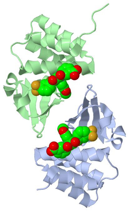
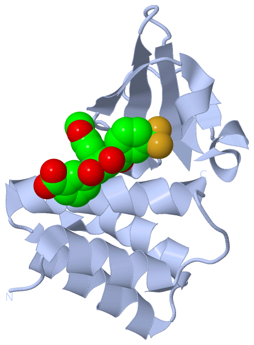
Asymmetric Unit (1, 2)
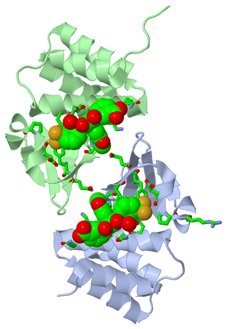
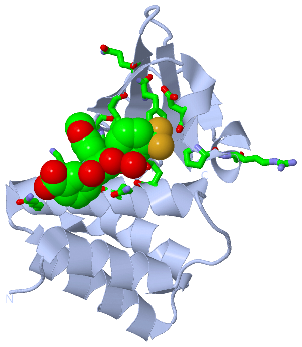
No.NameCountTypeFull Name
11D82Ligand/Ion3-{(2S)-2-(7-CHLORO-1,3-BENZODIOXOL-5-YL)-4-HYDROXY-5-OXO-3-[3-(TRIFLUOROMETHYL)BENZOYL]-2,5-DIHYDRO-1H-PYRROL-1-YL}BENZOIC ACID
Biological Unit 1 (1, 1)
No.NameCountTypeFull Name
11D81Ligand/Ion3-{(2S)-2-(7-CHLORO-1,3-BENZODIOXOL-5-YL)-4-HYDROXY-5-OXO-3-[3-(TRIFLUOROMETHYL)BENZOYL]-2,5-DIHYDRO-1H-PYRROL-1-YL}BENZOIC ACID
Biological Unit 2 (1, 1)
No.NameCountTypeFull Name
11D81Ligand/Ion3-{(2S)-2-(7-CHLORO-1,3-BENZODIOXOL-5-YL)-4-HYDROXY-5-OXO-3-[3-(TRIFLUOROMETHYL)BENZOYL]-2,5-DIHYDRO-1H-PYRROL-1-YL}BENZOIC ACID

(-) Sites  (2, 2)

Asymmetric Unit (2, 2)
No.NameEvidenceResiduesDescription
1AC1SOFTWAREGLN A:241 , GLN A:244 , LYS A:248 , LYS A:251 , PRO A:293 , ILE A:295 , ASP A:302 , PRO A:304 , ARG A:305 , PHE A:328 , HOH A:590 , GLN B:329BINDING SITE FOR RESIDUE 1D8 A 401
2AC2SOFTWAREGLN A:329 , GLN B:241 , GLN B:244 , LYS B:248 , LYS B:251 , PRO B:293 , ASP B:302 , PRO B:304 , PHE B:328 , HOH B:578BINDING SITE FOR RESIDUE 1D8 B 401

(-) SS Bonds  (0, 0)

(no "SS Bond" information available for 4IBI)

(-) Cis Peptide Bonds  (1, 1)

Asymmetric Unit
No.Residues
1Pro B:316 -Ser B:317

 Sequence-Structure Mapping

(-) SAPs(SNPs)/Variants  (0, 0)

(no "SAP(SNP)/Variant" information available for 4IBI)

(-) PROSITE Motifs  (0, 0)

(no "PROSITE Motif" information available for 4IBI)

(-) Exons   (0, 0)

(no "Exon" information available for 4IBI)

(-) Sequences/Alignments

Asymmetric Unit
   Reformat: Number of residues per line =  ('0' or empty: single-line sequence representation)
  Number of residues per labelling interval =   
  UniProt sequence: complete  aligned part    
   Show mapping: SCOP domains CATH domains Pfam domains Secondary structure (by author)
SAPs(SNPs) PROSITE motifs Exons
(details for a mapped element are shown in a popup box when the mouse pointer rests over it)
Chain A from PDB  Type:PROTEIN  Length:123
                                                                                                                                                           
               SCOP domains --------------------------------------------------------------------------------------------------------------------------- SCOP domains
               CATH domains --------------------------------------------------------------------------------------------------------------------------- CATH domains
               Pfam domains --------------------------------------------------------------------------------------------------------------------------- Pfam domains
         Sec.struct. author ..hhhhhhhhhhh......hhhhhhhhhhhhhhhhh.hhhhhhhhhhhhhhh..hhhhhhhhhhhhhhhhh.....eeee.hhhhhhhhhhh.ee......hhhh.eeeeeee....eeeee. Sec.struct. author
                 SAPs(SNPs) --------------------------------------------------------------------------------------------------------------------------- SAPs(SNPs)
                    PROSITE --------------------------------------------------------------------------------------------------------------------------- PROSITE
                 Transcript --------------------------------------------------------------------------------------------------------------------------- Transcript
                 4ibi A 218 DISAKDLRNIMYDHLPGFGTAFHQLVQVICKLGKDSNSLDIIHAEFQASLAEGDSPQCALIQITKRVPIFQDAAPPVIHIRSRGDIPRACQKSLRPVPPSPKIDRGWVCVFQLQDGKTLGLKI 340
                                   227       237       247       257       267       277       287       297       307       317       327       337   

Chain B from PDB  Type:PROTEIN  Length:127
                                                                                                                                                               
               SCOP domains ------------------------------------------------------------------------------------------------------------------------------- SCOP domains
               CATH domains ------------------------------------------------------------------------------------------------------------------------------- CATH domains
               Pfam domains ------------------------------------------------------------------------------------------------------------------------------- Pfam domains
         Sec.struct. author ......hhhhhhhhhhh......hhhhhhhhhhhhhhhhh.hhhhhhhhhhhhhhh..hhhhhhhhhhhhhhhhh.....eeee.hhhhhhhhhhh.ee......hhhh.eeeeeee....eeeee. Sec.struct. author
                 SAPs(SNPs) ------------------------------------------------------------------------------------------------------------------------------- SAPs(SNPs)
                    PROSITE ------------------------------------------------------------------------------------------------------------------------------- PROSITE
                 Transcript ------------------------------------------------------------------------------------------------------------------------------- Transcript
                 4ibi B 214 MGKPDISAKDLRNIMYDHLPGFGTAFHQLVQVICKLGKDSNSLDIIHAEFQASLAEGDSPQCALIQITKRVPIFQDAAPPVIHIRSRGDIPRACQKSLRPVPPSPKIDRGWVCVFQLQDGKTLGLKI 340
                                   223       233       243       253       263       273       283       293       303       313       323       333       

   Legend:   → Mismatch (orange background)
  - → Gap (green background, '-', border residues have a numbering label)
    → Modified Residue (blue background, lower-case, 'x' indicates undefined single-letter code, labelled with number + name)
  x → Chemical Group (purple background, 'x', labelled with number + name, e.g. ACE or NH2)
  extra numbering lines below/above indicate numbering irregularities and modified residue names etc., number ends below/above '|'

 Classification and Annotation

(-) SCOP Domains  (0, 0)

(no "SCOP Domain" information available for 4IBI)

(-) CATH Domains  (0, 0)

(no "CATH Domain" information available for 4IBI)

(-) Pfam Domains  (0, 0)

(no "Pfam Domain" information available for 4IBI)

(-) Gene Ontology  (21, 21)

Asymmetric Unit(hide GO term definitions)

 Visualization

(-) Interactive Views

Asymmetric Unit
  Complete Structure
    Jena3D(integrated viewing of ligand, site, SAP, PROSITE, SCOP information)
    WebMol | AstexViewer[tm]@PDBe
(Java Applets, require no local installation except for Java; loading may be slow)
    STRAP
(Java WebStart application, automatic local installation, requires Java; full application with system access!)
    RasMol
(require local installation)
    Molscript (VRML)
(requires installation of a VRML viewer; select preferred view via VRML and generate a mono or stereo PDF format file)
 
  Ligands, Modified Residues, Ions
    1D8  [ RasMol | Jena3D ]  +environment [ RasMol | Jena3D ]
 
  Sites
    AC1  [ RasMol ]  +environment [ RasMol ]
    AC2  [ RasMol ]  +environment [ RasMol ]
 
  Cis Peptide Bonds
    Pro B:316 - Ser B:317   [ RasMol ]  
 
Biological Units
  Complete Structure
    Biological Unit 1  [ Jena3D ]
    Biological Unit 2  [ Jena3D ]

(-) Still Images

Jmol
  protein: cartoon or spacefill or dots and stick; nucleic acid: cartoon and stick; ligands: spacefill; active site: stick
Molscript
  protein, nucleic acid: cartoon; ligands: spacefill; active site: ball and stick

 Databases and Analysis Tools

(-) Databases

Access by PDB/NDB ID
  4ibi
    Family and Domain InformationProDom | SYSTERS
    General Structural InformationGlycoscienceDB | MMDB | NDB | OCA | PDB | PDBe | PDBj | PDBsum | PDBWiki | PQS | PROTEOPEDIA
    Orientation in MembranesOPM
    Protein SurfaceSURFACE
    Secondary StructureDSSP (structure derived) | HSSP (homology derived)
    Structural GenomicsGeneCensus
    Structural NeighboursCE | VAST
    Structure ClassificationCATH | Dali | SCOP
    Validation and Original DataBMRB Data View | BMRB Restraints Grid | EDS | PROCHECK | RECOORD | WHAT_CHECK
 
Access by UniProt ID/Accession number
  VP35_EBOZM | Q05127
    Comparative Protein Structure ModelsModBase
    Genomic InformationEnsembl
    Protein-protein InteractionDIP
    Sequence, Family and Domain InformationInterPro | Pfam | SMART | UniProtKB/SwissProt
 
Access by Enzyme Classificator   (EC Number)
  (no 'Enzyme Classificator' available)
    General Enzyme InformationBRENDA | EC-PDB | Enzyme | IntEnz
    PathwayKEGG | MetaCyc
 
Access by Disease Identifier   (MIM ID)
  (no 'MIM ID' available)
    Disease InformationOMIM
 
Access by GenAge ID
  (no 'GenAge ID' available)
    Age Related InformationGenAge

(-) Analysis Tools

Access by PDB/NDB ID
    Domain InformationXDom
    Interatomic Contacts of Structural UnitsCSU
    Ligand-protein ContactsLPC
    Protein CavitiescastP
    Sequence and Secondary StructurePDBCartoon
    Structure AlignmentSTRAP(Java WebStart application, automatic local installation, requires Java; full application with system access!)
    Structure and Sequence BrowserSTING
 
Access by UniProt ID/Accession number
  VP35_EBOZM | Q05127
    Protein Disorder PredictionDisEMBL | FoldIndex | GLOBPLOT (for more information see DisProt)

 Related Entries

(-) Entries Sharing at Least One Protein Chain (UniProt ID)

UniProtKB/Swiss-Prot
        VP35_EBOZM | Q051273fke 3l25 3l26 3l27 3l28 3l29 4ibb 4ibc 4ibd 4ibe 4ibf 4ibg 4ibj 4ibk 4ije 4ijf 4ypi 4zta 4ztg 4zti

(-) Related Entries Specified in the PDB File

4ibb4ibc4ibd4ibe4ibf4ibh4ibj4ibk
4ibg