|
|
|
|
 Description
|
|
Compounds
|
||||||||||||||||||||||||||||||||||||||||||||||||||||
Chains, Units
Summary Information (see also Sequences/Alignments below) |
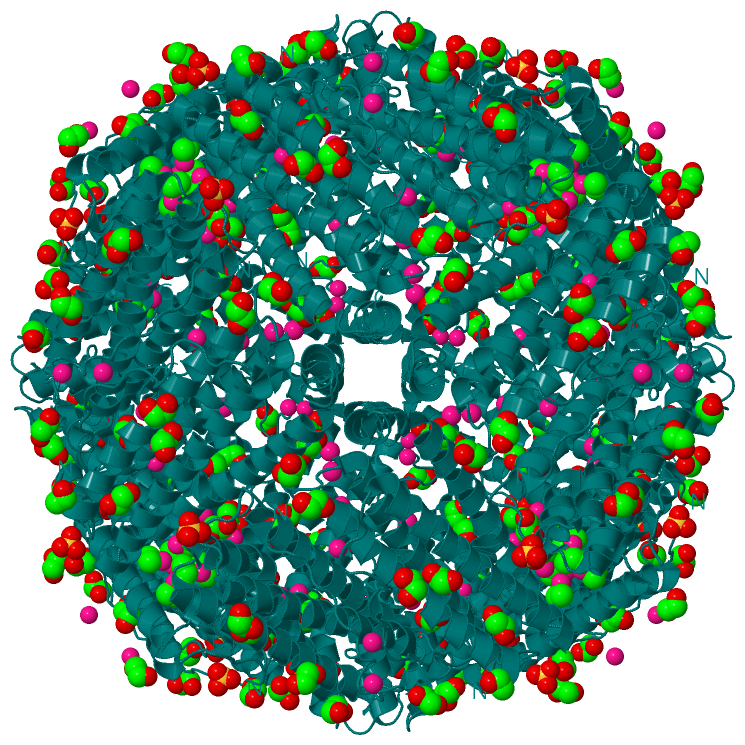
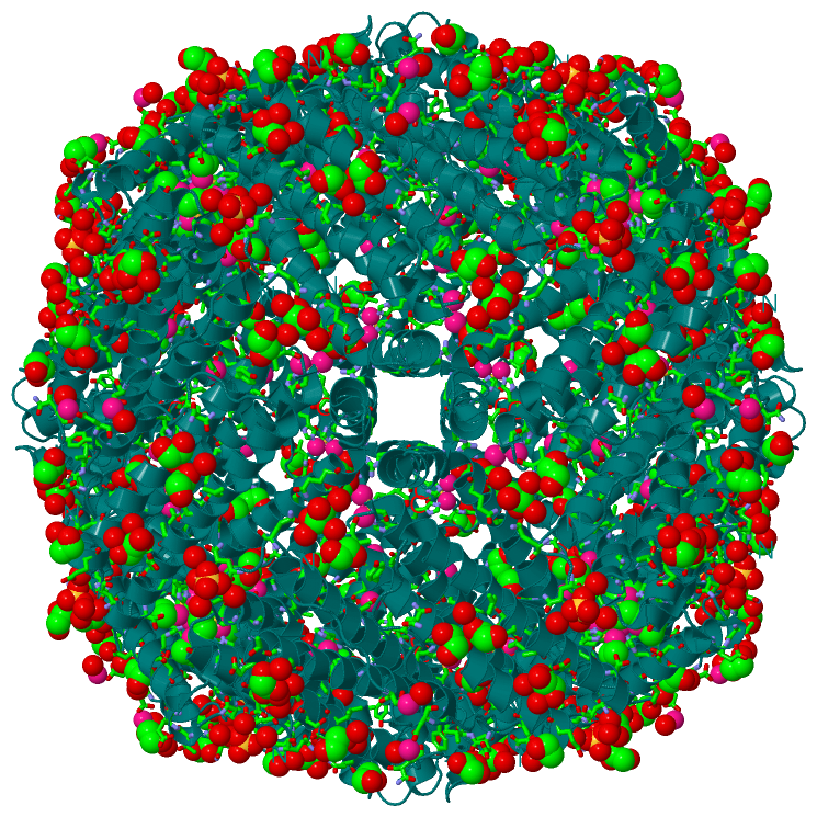
Ligands, Modified Residues, Ions (5, 15)| Asymmetric Unit (5, 15) Biological Unit 1 (3, 264) |
Sites (15, 15)
Asymmetric Unit (15, 15)
|
SS Bonds (0, 0)| (no "SS Bond" information available for 3NP2) |
Cis Peptide Bonds (0, 0)| (no "Cis Peptide Bond" information available for 3NP2) |
SAPs(SNPs)/Variants (0, 0)| (no "SAP(SNP)/Variant" information available for 3NP2) |
PROSITE Motifs (3, 3)
Asymmetric Unit (3, 3)
|
||||||||||||||||||||||||||||||||||||||||||||||||||||||||||||||||||||||||||||||||
Exons (0, 0)| (no "Exon" information available for 3NP2) |
Sequences/Alignments
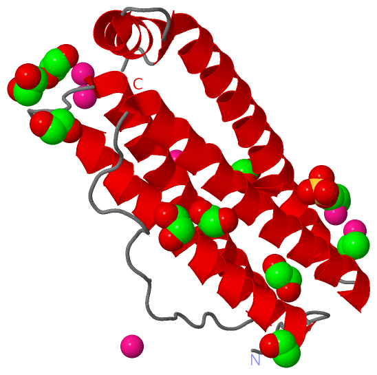
Asymmetric UnitChain X from PDB Type:PROTEIN Length:172 aligned with FRIL_HORSE | P02791 from UniProtKB/Swiss-Prot Length:175 Alignment length:172 12 22 32 42 52 62 72 82 92 102 112 122 132 142 152 162 172 FRIL_HORSE 3 SQIRQNYSTEVEAAVNRLVNLYLRASYTYLSLGFYFDRDDVALEGVCHFFRELAEEKREGAERLLKMQNQRGGRALFQDLQKPSQDEWGTTLDAMKAAIVLEKSLNQALLDLHALGSAQADPHLCDFLESHFLDEEVKLIKKMGDHLTNIQRLVGSQAGLGEYLFERLTLKH 174 SCOP domains d3np2x_ X: automated matches SCOP domains CATH domains ---------------------------------------------------------------------------------------------------------------------------------------------------------------------------- CATH domains Pfam domains ---------------------------------------------------------------------------------------------------------------------------------------------------------------------------- Pfam domains SAPs(SNPs) ---------------------------------------------------------------------------------------------------------------------------------------------------------------------------- SAPs(SNPs) PROSITE (1) ----FERRITIN_LIKE PDB: X:6-155 UniProt: 7-156 ------------------ PROSITE (1) PROSITE (2) -------------------------------------------------------FERRITIN_1 ----------------------------------------------FERRITIN_2 ------------------------------- PROSITE (2) Transcript ---------------------------------------------------------------------------------------------------------------------------------------------------------------------------- Transcript 3np2 X 2 SQIRQNYSTEVEAAVNRLVNLYLRASYTYLSLGFYFDRDDVALCGVAHFFRELAEEKREGAERLLKMQNQRGGRALFQDLQKPSQDEWGTTLDAMKAAIVLEKSLNQALLDLHALGSAQADPHLCDFLESHFLDEEVKLIKKMGDHLTNIQRLVGSQAGLGEYLFERLTLKH 173 11 21 31 41 51 61 71 81 91 101 111 121 131 141 151 161 171
|
||||||||||||||||||||
SCOP Domains (1, 1)
Asymmetric Unit
 |
CATH Domains (0, 0)| (no "CATH Domain" information available for 3NP2) |
Pfam Domains (0, 0)| (no "Pfam Domain" information available for 3NP2) |
Gene Ontology (6, 6)|
Asymmetric Unit(hide GO term definitions) Chain X (FRIL_HORSE | P02791)
|
||||||||||||||||||||||||||||||||||||||||||||||||||||||
Interactive Views
|
||||||||||||||||||||||||||||||||||||||||||||||||||||||||||||||||||||||||||||||||||||||||||||||||||||||||||||||||||||||||||||||||||||||||||||||||||||||||||||||||||||||||||||||||||||||||||||||||||||||||||||||||||||||||||||||||||||||||||||||||||||||||||||||||||||||
Still Images
|
||||||||||||||||
Databases
|
||||||||||||||||||||||||||||||||||||||||||||||||||||||||||||||||||||||||||||||||||||||||||||||||||||||||||||||||||||||||||||||||||||||||||||||||||||||||||||||||
Analysis Tools
|
|||||||||||||||||||||||||||||||||||||||||||||||||||||||||||||
Entries Sharing at Least One Protein Chain (UniProt ID)
Related Entries Specified in the PDB File
|
|