Show PDB file:   
         Plain Text   HTML   (compressed file size)
QuickSearch:   
by PDB,NDB,UniProt,PROSITE Code or Search Term(s)  
(-)Asym./Biol. Unit - manually
(-)Asym./Biol. Unit
(-)Asym./Biol. Unit - sites
collapse expand < >
Image Asym./Biol. Unit - manually
Asym./Biol. Unit - manually  (Jmol Viewer)
Image Asym./Biol. Unit
Asym./Biol. Unit  (Jmol Viewer)
Image Asym./Biol. Unit - sites
Asym./Biol. Unit - sites  (Jmol Viewer)

(-) Description

Title :  CRYSTAL STRUCTURE OF HIS-TAGGED FORM OF PA4794 PROTEIN
 
Authors :  K. A. Majorek, M. Chruszcz, A. Joachimiak, W. Minor, Midwest Center For Structural Genomics (Mcsg)
Date :  06 Nov 09  (Deposition) - 24 Nov 09  (Release) - 24 Nov 09  (Revision)
Method :  X-RAY DIFFRACTION
Resolution :  1.41
Chains :  Asym./Biol. Unit :  A
Keywords :  Acetyltransferase, Gnat Family, Structural Genomics, Psi, Protein Structure Initiative, Midwest Center For Structural Genomics, Mcsg (Keyword Search: [Gene Ontology, PubMed, Web (Google))
 
Reference :  K. A. Majorek, M. Chruszcz, A. Joachimiak, W. Minor
Crystal Structure Of His-Tagged Form Of Pa4794 Protein
To Be Published
PubMed: search
(for further references see the PDB file header)

(-) Compounds

Molecule 1 - PUTATIVE UNCHARACTERIZED PROTEIN
    ChainsA
    EngineeredYES
    Expression SystemESCHERICHIA COLI BL21(DE3)
    Expression System PlasmidP11
    Expression System StrainBL21 (DE3) REAL
    Expression System Taxid469008
    Expression System Vector TypePLASMID
    GenePA4794
    Organism ScientificPSEUDOMONAS AERUGINOSA PAO1
    Organism Taxid208964
    StrainPAO1

 Structural Features

(-) Chains, Units

  1
Asymmetric/Biological Unit A

Summary Information (see also Sequences/Alignments below)

(-) Ligands, Modified Residues, Ions  (2, 19)

Asymmetric/Biological Unit (2, 19)
No.NameCountTypeFull Name
1EDO17Ligand/Ion1,2-ETHANEDIOL
2SO42Ligand/IonSULFATE ION

(-) Sites  (19, 19)

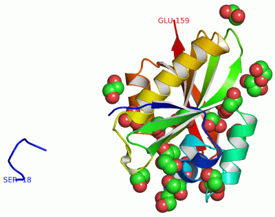
Asymmetric Unit (19, 19)
No.NameEvidenceResiduesDescription
01AC1SOFTWAREHIS A:-16 , HIS A:-17 , SER A:-18 , ARG A:141 , GLN A:153 , HOH A:235 , HOH A:353 , HOH A:370BINDING SITE FOR RESIDUE SO4 A 161
02AC2SOFTWAREGLY A:89 , LEU A:90 , GLY A:91 , VAL A:92 , ALA A:93 , ARG A:94 , LEU A:127 , HOH A:203 , HOH A:212 , HOH A:215 , HOH A:244 , HOH A:291BINDING SITE FOR RESIDUE SO4 A 162
03AC3SOFTWAREGLU A:25 , CYS A:29 , MET A:82 , VAL A:83 , ARG A:88 , HOH A:363 , HOH A:371BINDING SITE FOR RESIDUE EDO A 163
04AC4SOFTWAREGLU A:14 , ALA A:17 , GLY A:18 , PHE A:37 , HIS A:72 , LYS A:109 , ARG A:111 , HOH A:225 , HOH A:226BINDING SITE FOR RESIDUE EDO A 164
05AC5SOFTWARELEU A:126 , THR A:129 , GLN A:130 , GLU A:140 , HOH A:249 , HOH A:331 , HOH A:364BINDING SITE FOR RESIDUE EDO A 165
06AC6SOFTWAREGLN A:69 , TRP A:70 , GLN A:71 , HOH A:308BINDING SITE FOR RESIDUE EDO A 166
07AC7SOFTWARETHR A:15 , GLN A:60 , VAL A:61 , HIS A:142 , GLY A:146 , ARG A:148 , HOH A:216 , HOH A:266BINDING SITE FOR RESIDUE EDO A 167
08AC8SOFTWAREARG A:50 , TRP A:70 , HOH A:204 , HOH A:274BINDING SITE FOR RESIDUE EDO A 168
09AC9SOFTWAREILE A:34 , TRP A:35 , PRO A:144 , HOH A:223 , HOH A:338 , HOH A:383BINDING SITE FOR RESIDUE EDO A 169
10BC1SOFTWARETYR A:30 , ARG A:49 , HOH A:229 , HOH A:352BINDING SITE FOR RESIDUE EDO A 170
11BC2SOFTWAREARG A:23 , ASP A:24 , ASP A:143 , HOH A:231BINDING SITE FOR RESIDUE EDO A 171
12BC3SOFTWAREGLY A:18 , GLN A:21 , LYS A:109 , ARG A:111 , HOH A:288BINDING SITE FOR RESIDUE EDO A 172
13BC4SOFTWAREGLU A:14 , THR A:15 , LYS A:109 , HOH A:315BINDING SITE FOR RESIDUE EDO A 173
14BC5SOFTWAREMET A:81 , ASN A:121 , GLY A:124 , TYR A:128 , HOH A:244 , HOH A:371BINDING SITE FOR RESIDUE EDO A 174
15BC6SOFTWAREHIS A:-16 , HIS A:-17 , CYS A:29 , PRO A:31 , ARG A:49 , TYR A:68 , GLY A:79 , ASN A:80 , HOH A:229 , HOH A:246BINDING SITE FOR RESIDUE EDO A 175
16BC7SOFTWAREGLY A:79 , ASN A:80 , MET A:81 , SER A:116 , TYR A:128 , HOH A:246 , HOH A:264BINDING SITE FOR RESIDUE EDO A 176
17BC8SOFTWAREGLN A:21 , GLU A:25 , ARG A:88 , HOH A:299 , HOH A:382BINDING SITE FOR RESIDUE EDO A 177
18BC9SOFTWAREARG A:50 , GLY A:51 , SER A:52 , HOH A:209 , HOH A:300BINDING SITE FOR RESIDUE EDO A 178
19CC1SOFTWARETHR A:10 , GLY A:11 , LEU A:13 , GLU A:14 , GLY A:89 , ARG A:148 , HOH A:190BINDING SITE FOR RESIDUE EDO A 179

(-) SS Bonds  (0, 0)

(no "SS Bond" information available for 3KKW)

(-) Cis Peptide Bonds  (1, 1)

Asymmetric/Biological Unit
No.Residues
1Trp A:35 -Pro A:36

 Sequence-Structure Mapping

(-) SAPs(SNPs)/Variants  (0, 0)

(no "SAP(SNP)/Variant" information available for 3KKW)

(-) PROSITE Motifs  (0, 0)

(no "PROSITE Motif" information available for 3KKW)

(-) Exons   (0, 0)

(no "Exon" information available for 3KKW)

(-) Sequences/Alignments

Asymmetric/Biological Unit
   Reformat: Number of residues per line =  ('0' or empty: single-line sequence representation)
  Number of residues per labelling interval =   
  UniProt sequence: complete  aligned part    
   Show mapping: SCOP domains CATH domains Pfam domains Secondary structure (by author)
SAPs(SNPs) PROSITE motifs Exons
(details for a mapped element are shown in a popup box when the mouse pointer rests over it)
Chain A from PDB  Type:PROTEIN  Length:166
 aligned with Q9HV14_PSEAE | Q9HV14 from UniProtKB/TrEMBL  Length:160

    Alignment length:166
                                   1                                                                                                                                                              
                                   | 3        13        23        33        43        53        63        73        83        93       103       113       123       133       143       153      
         Q9HV14_PSEAE     - -------MQLSHRPAETGDLETVAGFPQDRDELFYCYPKAIWPFSVAQLAAAIAERRGSTVAVHDGQVLGFANFYQWQHGDFCALGNMMVAPAARGLGVARYLIGVMENLAREQYKARLMKISCFNANAAGLLLYTQLGYQPRAIAERHDPDGRRVALIQMDKPLE 159
               SCOP domains d3kkwa_ A: Putative acetyltransferase PA4794                                                                                                                           SCOP domains
               CATH domains ---------------------------------------------------------------------------------------------------------------------------------------------------------------------- CATH domains
               Pfam domains ---------------------------------------------------------------------------------------------------------------------------------------------------------------------- Pfam domains
         Sec.struct. author ..........eeee.hhhhhhhhhh...hhhhhhhhh.......hhhhhhhhhhhheeeeeeee..eeeeeeeeeeee...eeeeeeeee.hhhh..hhhhhhhhhhhhhhhhhhh..eeeeeee..hhhhhhhhhhh..eeeeeeeee.....eeeeeeeeee.. Sec.struct. author
                 SAPs(SNPs) ---------------------------------------------------------------------------------------------------------------------------------------------------------------------- SAPs(SNPs)
                    PROSITE ---------------------------------------------------------------------------------------------------------------------------------------------------------------------- PROSITE
                 Transcript ---------------------------------------------------------------------------------------------------------------------------------------------------------------------- Transcript
                 3kkw A -18 SHHHHHHMQLSHRPAETGDLETVAGFPQDRDELFYCYPKAIWPFSVAQLAAAIAERRGSTVAVHDGQVLGFANFYQWQHGDFCALGNMMVAPAARGLGVARYLIGVMENLAREQYKARLMKISCFNANAAGLLLYTQLGYQPRAIAERHDPDGRRVALIQMDKPLE 159
                                  || 3        13        23        33        43        53        63        73        83        93       103       113       123       133       143       153      
                                -12|                                                                                                                                                              
                                   1                                                                                                                                                              

   Legend:   → Mismatch (orange background)
  - → Gap (green background, '-', border residues have a numbering label)
    → Modified Residue (blue background, lower-case, 'x' indicates undefined single-letter code, labelled with number + name)
  x → Chemical Group (purple background, 'x', labelled with number + name, e.g. ACE or NH2)
  extra numbering lines below/above indicate numbering irregularities and modified residue names etc., number ends below/above '|'

 Classification and Annotation

(-) SCOP Domains  (1, 1)

Asymmetric/Biological Unit

(-) CATH Domains  (0, 0)

(no "CATH Domain" information available for 3KKW)

(-) Pfam Domains  (0, 0)

(no "Pfam Domain" information available for 3KKW)

(-) Gene Ontology  (4, 4)

Asymmetric/Biological Unit(hide GO term definitions)
Chain A   (Q9HV14_PSEAE | Q9HV14)
molecular function
    GO:0008080    N-acetyltransferase activity    Catalysis of the transfer of an acetyl group to a nitrogen atom on the acceptor molecule.
    GO:0004596    peptide alpha-N-acetyltransferase activity    Catalysis of the reaction: acetyl-CoA + peptide = CoA + N-alpha-acetylpeptide. This reaction is the acetylation of the N-terminal amino acid residue of a peptide or protein.
biological process
    GO:0006474    N-terminal protein amino acid acetylation    The acetylation of the N-terminal amino acid of proteins.
cellular component
    GO:0031248    protein acetyltransferase complex    A complex that catalyzes the transfer of an acetyl group to a protein acceptor molecule.

 Visualization

(-) Interactive Views

Asymmetric/Biological Unit
  Complete Structure
    Jena3D(integrated viewing of ligand, site, SAP, PROSITE, SCOP information)
    WebMol | AstexViewer[tm]@PDBe
(Java Applets, require no local installation except for Java; loading may be slow)
    STRAP
(Java WebStart application, automatic local installation, requires Java; full application with system access!)
    RasMol
(require local installation)
    Molscript (VRML)
(requires installation of a VRML viewer; select preferred view via VRML and generate a mono or stereo PDF format file)
 
  Ligands, Modified Residues, Ions
    EDO  [ RasMol | Jena3D ]  +environment [ RasMol | Jena3D ]
    SO4  [ RasMol | Jena3D ]  +environment [ RasMol | Jena3D ]
 
  Sites
    AC1  [ RasMol ]  +environment [ RasMol ]
    AC2  [ RasMol ]  +environment [ RasMol ]
    AC3  [ RasMol ]  +environment [ RasMol ]
    AC4  [ RasMol ]  +environment [ RasMol ]
    AC5  [ RasMol ]  +environment [ RasMol ]
    AC6  [ RasMol ]  +environment [ RasMol ]
    AC7  [ RasMol ]  +environment [ RasMol ]
    AC8  [ RasMol ]  +environment [ RasMol ]
    AC9  [ RasMol ]  +environment [ RasMol ]
    BC1  [ RasMol ]  +environment [ RasMol ]
    BC2  [ RasMol ]  +environment [ RasMol ]
    BC3  [ RasMol ]  +environment [ RasMol ]
    BC4  [ RasMol ]  +environment [ RasMol ]
    BC5  [ RasMol ]  +environment [ RasMol ]
    BC6  [ RasMol ]  +environment [ RasMol ]
    BC7  [ RasMol ]  +environment [ RasMol ]
    BC8  [ RasMol ]  +environment [ RasMol ]
    BC9  [ RasMol ]  +environment [ RasMol ]
    CC1  [ RasMol ]  +environment [ RasMol ]
 
  Cis Peptide Bonds
    Trp A:35 - Pro A:36   [ RasMol ]  
 

(-) Still Images

Jmol
  protein: cartoon or spacefill or dots and stick; nucleic acid: cartoon and stick; ligands: spacefill; active site: stick
Molscript
  protein, nucleic acid: cartoon; ligands: spacefill; active site: ball and stick

 Databases and Analysis Tools

(-) Databases

Access by PDB/NDB ID
  3kkw
    Family and Domain InformationProDom | SYSTERS
    General Structural InformationGlycoscienceDB | MMDB | NDB | OCA | PDB | PDBe | PDBj | PDBsum | PDBWiki | PQS | PROTEOPEDIA
    Orientation in MembranesOPM
    Protein SurfaceSURFACE
    Secondary StructureDSSP (structure derived) | HSSP (homology derived)
    Structural GenomicsGeneCensus
    Structural NeighboursCE | VAST
    Structure ClassificationCATH | Dali | SCOP
    Validation and Original DataBMRB Data View | BMRB Restraints Grid | EDS | PROCHECK | RECOORD | WHAT_CHECK
 
Access by UniProt ID/Accession number
  Q9HV14_PSEAE | Q9HV14
    Comparative Protein Structure ModelsModBase
    Genomic InformationEnsembl
    Protein-protein InteractionDIP
    Sequence, Family and Domain InformationInterPro | Pfam | SMART | UniProtKB/TrEMBL
 
Access by Enzyme Classificator   (EC Number)
  (no 'Enzyme Classificator' available)
    General Enzyme InformationBRENDA | EC-PDB | Enzyme | IntEnz
    PathwayKEGG | MetaCyc
 
Access by Disease Identifier   (MIM ID)
  (no 'MIM ID' available)
    Disease InformationOMIM
 
Access by GenAge ID
  (no 'GenAge ID' available)
    Age Related InformationGenAge

(-) Analysis Tools

Access by PDB/NDB ID
    Domain InformationXDom
    Interatomic Contacts of Structural UnitsCSU
    Ligand-protein ContactsLPC
    Protein CavitiescastP
    Sequence and Secondary StructurePDBCartoon
    Structure AlignmentSTRAP(Java WebStart application, automatic local installation, requires Java; full application with system access!)
    Structure and Sequence BrowserSTING
 
Access by UniProt ID/Accession number
  Q9HV14_PSEAE | Q9HV14
    Protein Disorder PredictionDisEMBL | FoldIndex | GLOBPLOT (for more information see DisProt)

 Related Entries

(-) Entries Sharing at Least One Protein Chain (UniProt ID)

UniProtKB/TrEMBL
        Q9HV14_PSEAE | Q9HV143pgp 4klv 4klw 4kor 4kos 4kot 4kou 4kov 4kow 4kox 4koy 4kua 4kub 4l89 4l8a 4m3s 4oad 4oae

(-) Related Entries Specified in the PDB File

2i6c RELATED ID: APC5605 RELATED DB: TARGETDB