|
|
|
|
 Description
|
|
Compounds
|
||||||||||||||||||||||||||||||||||||||||||||||||||||
Chains, Units
Summary Information (see also Sequences/Alignments below) |
Ligands, Modified Residues, Ions (0, 0)| (no "Ligand,Modified Residues,Ions" information available for 2Y0T) |
Sites (0, 0)| (no "Site" information available for 2Y0T) |
SS Bonds (0, 0)| (no "SS Bond" information available for 2Y0T) |
Cis Peptide Bonds (0, 0)| (no "Cis Peptide Bond" information available for 2Y0T) |
SAPs(SNPs)/Variants (0, 0)| (no "SAP(SNP)/Variant" information available for 2Y0T) |
PROSITE Motifs (0, 0)| (no "PROSITE Motif" information available for 2Y0T) |
Exons (0, 0)| (no "Exon" information available for 2Y0T) |
Sequences/Alignments
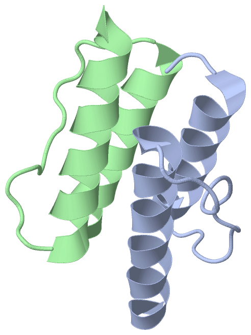
Asymmetric UnitChain A from PDB Type:PROTEIN Length:52 aligned with O28769_ARCFU | O28769 from UniProtKB/TrEMBL Length:338 Alignment length:52 288 298 308 318 328 O28769_ARCFU 279 TITRPIIELSNTADKIAEGNLEAEVPHQNRADEIGILAKSIERLRRSLKVAM 330 SCOP domains d2y0ta_ A: automated matches SCOP domains CATH domains ---------------------------------------------------- CATH domains Pfam domains ---------------------------------------------------- Pfam domains SAPs(SNPs) ---------------------------------------------------- SAPs(SNPs) PROSITE ---------------------------------------------------- PROSITE Transcript ---------------------------------------------------- Transcript 2y0t A 279 TITRPIIELSNTFDKIAEGNLEAEVPHQNRADEIGILAKSIERLRRSLKVAM 330 288 298 308 318 328 Chain B from PDB Type:PROTEIN Length:49 aligned with O28769_ARCFU | O28769 from UniProtKB/TrEMBL Length:338 Alignment length:49 289 299 309 319 O28769_ARCFU 280 ITRPIIELSNTADKIAEGNLEAEVPHQNRADEIGILAKSIERLRRSLKV 328 SCOP domains d2y0tb_ B: automated matches SCOP domains CATH domains ------------------------------------------------- CATH domains Pfam domains (1) HAMP-2y0tB01 B:280-328 Pfam domains (1) Pfam domains (2) HAMP-2y0tB02 B:280-328 Pfam domains (2) SAPs(SNPs) ------------------------------------------------- SAPs(SNPs) PROSITE ------------------------------------------------- PROSITE Transcript ------------------------------------------------- Transcript 2y0t B 280 ITRPIIELSNTFDKIAEGNLEAEVPHQNRADEIGILAKSIERLRRSLKV 328 289 299 309 319
|
||||||||||||||||||||
SCOP Domains (1, 2)
Asymmetric Unit
 |
CATH Domains (0, 0)| (no "CATH Domain" information available for 2Y0T) |
Pfam Domains (1, 2)
Asymmetric Unit
|
Gene Ontology (5, 5)|
Asymmetric Unit(hide GO term definitions) Chain A,B (O28769_ARCFU | O28769)
|
||||||||||||||||||||||||||||||||||||||||||||||||
Interactive Views
|
||||||||||||||||||||||||||||||||||||||||||||||||||||||||||||||||||||||||||||||||||||||||||||||||||||||||||||||||||||||||||||||||||||||
Still Images
|
||||||||||||||||
Databases
|
||||||||||||||||||||||||||||||||||||||||||||||||||||||||||||||||||||||||||||||||||||||||||||||||||||||||||||||||||||||||||||||||||||||||||||||||||||||||||||||||
Analysis Tools
|
|||||||||||||||||||||||||||||||||||||||||||||||||||||||||||||
Entries Sharing at Least One Protein Chain (UniProt ID)
Related Entries Specified in the PDB File
|
|