|
|
|
|
 Description
|
|
Compounds
|
||||||||||||||||||||||||||||||||||||||||
Chains, Units
Summary Information (see also Sequences/Alignments below) |
Ligands, Modified Residues, Ions (0, 0)| (no "Ligand,Modified Residues,Ions" information available for 2L7H) |
Sites (0, 0)| (no "Site" information available for 2L7H) |
SS Bonds (0, 0)| (no "SS Bond" information available for 2L7H) |
Cis Peptide Bonds (0, 0)| (no "Cis Peptide Bond" information available for 2L7H) |
SAPs(SNPs)/Variants (0, 0)| (no "SAP(SNP)/Variant" information available for 2L7H) |
PROSITE Motifs (0, 0)| (no "PROSITE Motif" information available for 2L7H) |
Exons (0, 0)| (no "Exon" information available for 2L7H) |
Sequences/Alignments
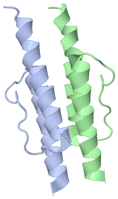
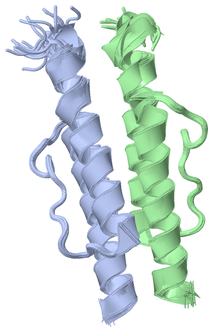
NMR StructureChain A from PDB Type:PROTEIN Length:58 aligned with O28769_ARCFU | O28769 from UniProtKB/TrEMBL Length:338 Alignment length:92 249 259 269 279 289 299 309 319 329 O28769_ARCFU 240 ASEISKSVEGAIQQVYYALGIAAAIAIVFVIVLAVFTTSTITRPIIELSNTADKIAEGNLEAEVPHQNRADEIGILAKSIERLRRSLKVAME 331 SCOP domains d2l7h a_ A: Hypothetical protein AF1503 SCOP domains CATH domains -------------------------------------------------------------------------------------------- CATH domains Pfam domains -------------------------------------------------------------------------------------------- Pfam domains SAPs(SNPs) -------------------------------------------------------------------------------------------- SAPs(SNPs) PROSITE -------------------------------------------------------------------------------------------- PROSITE Transcript -------------------------------------------------------------------------------------------- Transcript 2l7h A 274 GSHMS----------------------------------TITRPIIELSNTADKIAEGNLEAEVPHQNRADEIGILAKSIERLRRSLKVAME 331 | - - - 279 289 299 309 319 329 278 279 Chain B from PDB Type:PROTEIN Length:58 aligned with O28769_ARCFU | O28769 from UniProtKB/TrEMBL Length:338 Alignment length:92 249 259 269 279 289 299 309 319 329 O28769_ARCFU 240 ASEISKSVEGAIQQVYYALGIAAAIAIVFVIVLAVFTTSTITRPIIELSNTADKIAEGNLEAEVPHQNRADEIGILAKSIERLRRSLKVAME 331 SCOP domains d2l7h b_ B: Hypothetical protein AF1503 SCOP domains CATH domains -------------------------------------------------------------------------------------------- CATH domains Pfam domains (1) ---------------------------------------HAMP-2l7hB01 B:279-328 --- Pfam domains (1) Pfam domains (2) ---------------------------------------HAMP-2l7hB02 B:279-328 --- Pfam domains (2) SAPs(SNPs) -------------------------------------------------------------------------------------------- SAPs(SNPs) PROSITE -------------------------------------------------------------------------------------------- PROSITE Transcript -------------------------------------------------------------------------------------------- Transcript 2l7h B 274 GSHMS----------------------------------TITRPIIELSNTADKIAEGNLEAEVPHQNRADEIGILAKSIERLRRSLKVAME 331 | - - - 279 289 299 309 319 329 278 279
|
||||||||||||||||||||
SCOP Domains (1, 2)
NMR Structure
 |
CATH Domains (0, 0)| (no "CATH Domain" information available for 2L7H) |
Pfam Domains (1, 2)
NMR Structure
|
Gene Ontology (5, 5)|
NMR Structure(hide GO term definitions) Chain A,B (O28769_ARCFU | O28769)
|
||||||||||||||||||||||||||||||||||||||||||||||||
Interactive Views
|
||||||||||||||||||||||||||||||||||||||||||||||||||||||||||||||||||||||||||||||||||||||||||||||||||||||||||||||||||||
Still Images
|
||||||||||||||||
Databases
|
||||||||||||||||||||||||||||||||||||||||||||||||||||||||||||||||||||||||||||||||||||||||||||||||||||||||||||||||||||||||||||||||||||||||||||||||||||||||||||||||
Analysis Tools
|
|||||||||||||||||||||||||||||||||||||||||||||||||||||||||||||
Entries Sharing at Least One Protein Chain (UniProt ID)
Related Entries Specified in the PDB File
|
|