Show PDB file:   
         Plain Text   HTML   (compressed file size)
QuickSearch:   
by PDB,NDB,UniProt,PROSITE Code or Search Term(s)  
(-)Asymmetric Unit
(-)Biological Unit 1
(-)Biological Unit 2
collapse expand < >
Image Asymmetric Unit
Asymmetric Unit  (Jmol Viewer)
Image Biological Unit 1
Biological Unit 1  (Jmol Viewer)
Image Biological Unit 2
Biological Unit 2  (Jmol Viewer)

(-) Description

Title :  STRUCTURAL AND BIOCHEMICAL CHARACTERIZATION OF FIBRINOGEN BINDING TO CLFA FROM STAPHYLOCCCUS AUREUS
 
Authors :  V. K. Ganesh, J. J. Rivera, E. Smeds, M. G. Bowden, E. R. Wann, S. Gurusidappa, J. R. Fitzgerald, M. Hook
Date :  25 Mar 08  (Deposition) - 19 May 09  (Release) - 19 May 09  (Revision)
Method :  X-RAY DIFFRACTION
Resolution :  1.95
Chains :  Asym. Unit :  A,B,C,D
Biol. Unit 1:  A,C  (1x)
Biol. Unit 2:  B,D  (1x)
Keywords :  Platelet Aggregation, Peptidoglycan-Anchor, Cell Adhesion, Staphylococcus Aureus, Fibrinogen Gamma-Chain, Secreted, Cell Wall, Virulence, Clumping Factor (Keyword Search: [Gene Ontology, PubMed, Web (Google))
 
Reference :  V. K. Ganesh, J. J. Rivera, E. Smeds, Y. P. Ko, M. G. Bowden, E. R. Wann, S. Gurusiddappa, J. R. Fitzgerald, M. Hook
A Structural Model Of The Staphylococcus Aureus Clfa-Fibrinogen Interaction Opens New Avenues For The Design Of Anti-Staphylococcal Therapeutics.
Plos Pathog. V. 4 226 2008
PubMed-ID: 19043557  |  Reference-DOI: 10.1371/JOURNAL.PPAT.1000226

(-) Compounds

Molecule 1 - CLUMPING FACTOR A
    ChainsA, B
    EngineeredYES
    Expression SystemESCHERICHIA COLI
    Expression System Taxid562
    FragmentN2N3, RESIDUES 229-545
    MutationYES
    Organism ScientificSTAPHYLOCOCCUS AUREUS
    Organism Taxid1280
    Other DetailsENGINEERED DISULFIDE BOND BETWEEN 327 AND 541
    SynonymFIBRINOGEN-BINDING PROTEIN A, FIBRINOGEN RECEPTOR A
 
Molecule 2 - FIBRINOGEN GAMMA-CHAIN
    ChainsC, D
    FragmentC-TERMINAL GAMMA-CHAIN PEPTIDE ANALOG, RESIDUES 425-437
    MutationYES
    Organism CommonHUMAN
    Organism ScientificHOMO SAPIENS
    Organism Taxid9606
    SyntheticYES

 Structural Features

(-) Chains, Units

  1234
Asymmetric Unit ABCD
Biological Unit 1 (1x)A C 
Biological Unit 2 (1x) B D

Summary Information (see also Sequences/Alignments below)

(-) Ligands, Modified Residues, Ions  (0, 0)

(no "Ligand,Modified Residues,Ions" information available for 2VR3)

(-) Sites  (0, 0)

(no "Site" information available for 2VR3)

(-) SS Bonds  (2, 2)

Asymmetric Unit
No.Residues
1A:327 -A:541
2B:327 -B:541

(-) Cis Peptide Bonds  (0, 0)

(no "Cis Peptide Bond" information available for 2VR3)

 Sequence-Structure Mapping

(-) SAPs(SNPs)/Variants  (0, 0)

(no "SAP(SNP)/Variant" information available for 2VR3)

(-) PROSITE Motifs  (0, 0)

(no "PROSITE Motif" information available for 2VR3)

(-) Exons   (1, 2)

Asymmetric Unit (1, 2)
 ENSEMBLUniProtKBPDB
No.Transcript IDExonExon IDGenome LocationLengthIDLocationLengthCountLocationLength
1.1fENST000003360981fENSE00001820766chr4:155533800-155533684117FIBG_HUMAN1-26260--
1.2aENST000003360982aENSE00001781463chr4:155533587-15553354345FIBG_HUMAN27-41150--
1.2dENST000003360982dENSE00001624767chr4:155533353-155533170184FIBG_HUMAN42-103620--
1.2gENST000003360982gENSE00001652916chr4:155533050-15553295794FIBG_HUMAN103-134320--
1.3aENST000003360983aENSE00001127239chr4:155531349-155531219131FIBG_HUMAN134-178450--
1.3fENST000003360983fENSE00002146570chr4:155530915-155530782134FIBG_HUMAN178-222450--
1.4bENST000003360984bENSE00001127220chr4:155529802-155529618185FIBG_HUMAN223-284620--
1.5ENST000003360985ENSE00001127212chr4:155528134-155527857278FIBG_HUMAN284-377940--
1.6cENST000003360986cENSE00001344514chr4:155526218-155525728491FIBG_HUMAN377-453772C:400-411 (gaps)
D:399-411 (gaps)
16
17

(-) Sequences/Alignments

Asymmetric Unit
   Reformat: Number of residues per line =  ('0' or empty: single-line sequence representation)
  Number of residues per labelling interval =   
  UniProt sequence: complete  aligned part    
   Show mapping: SCOP domains CATH domains Pfam domains Secondary structure (by author)
SAPs(SNPs) PROSITE motifs Exons
(details for a mapped element are shown in a popup box when the mouse pointer rests over it)
Chain A from PDB  Type:PROTEIN  Length:308
 aligned with CLFA_STAA8 | Q2G015 from UniProtKB/Swiss-Prot  Length:927

    Alignment length:316
                                   239       249       259       269       279       289       299       309       319       329       339       349       359       369       379       389       399       409       419       429       439       449       459       469       479       489       499       509       519       529       539      
           CLFA_STAA8   230 TDITNQLTNVTVGIDSGTTVYPHQAGYVKLNYGFSVPNSAVKGDTFKITVPKELNLNGVTSTAKVPPIMAGDQVLANGVIDSDGNVIYTFTDYVNTKDDVKATLTMPAYIDPENVKKTGNVTLATGIGSTTANKTVLVDYEKYGKFYNLSIKGTIDQIDKTNNTYRQTIYVNPSGDNVIAPVLTGNLKPNTDSNALIDQQNTSIKVYKVDNAADLSESYFVNPENFEDVTNSVNITFPNPNQYKVEFNTPDDQITTPYIVVVNGHIDPNSKGDLALRSTLYGYNSNIIWRSMSWDNEVAFNNGSGSGDGIDKPVVP 545
               SCOP domains ---------------------------------------------------------------------------------------------------------------------------------------------------------------------------------------------------------------------------------------------------------------------------------------------------------------------------- SCOP domains
               CATH domains ---------------------------------------------------------------------------------------------------------------------------------------------------------------------------------------------------------------------------------------------------------------------------------------------------------------------------- CATH domains
               Pfam domains ---------------------------------------------------------------------------------------------------------------------------------------------------------------------------------------------------------------------------------------------------------------------------------------------------------------------------- Pfam domains
         Sec.struct. author ..hhhhheeeeeeee...ee.......eeeeeeeee........eeeee....ee............ee.---..eeeee.....eeeee.hhhhheeeeeeeeeeeeee........eeeeeeeee..eeeeeeeee.....eee..eeeeeeeeeee....eeeeeeee......eeeeeeeeee.......ee......eeeeee..hhhhh........---..hhhhheeeeee..eeeee.--...ee...eeeeeeeee.........eeeeeeee.....eeeeeeeeeeeee..eeeeeeee..... Sec.struct. author
                 SAPs(SNPs) ---------------------------------------------------------------------------------------------------------------------------------------------------------------------------------------------------------------------------------------------------------------------------------------------------------------------------- SAPs(SNPs)
                    PROSITE ---------------------------------------------------------------------------------------------------------------------------------------------------------------------------------------------------------------------------------------------------------------------------------------------------------------------------- PROSITE
                 Transcript ---------------------------------------------------------------------------------------------------------------------------------------------------------------------------------------------------------------------------------------------------------------------------------------------------------------------------- Transcript
                 2vr3 A 230 TDITNQLTNVTVGIDSGTTVYPHQAGYVKLNYGFSVPNSAVKGDTFKITVPKELNLNGVTSTAKVPPIMA---VLANGVIDSDGNVIYTFTDYVNTKCDVKATLTMPAYIDPENVKKTGNVTLATGIGSTTANKTVLVDYEKYGKFYNLSIKGTIDQIDKTNNTYRQTIYVNPSGDNVIAPVLTGNLKPNTDSNALIDQQNTSIKVYKVDNAADLSESYFVNP---EDVTNSVNITFPNPNQYKVEF--PDDQITTPYIVVVNGHIDPNSKGDLALRSTLYGYNSNIIWRSMSWDNEVAFNNGSGSGDGIDCPVVP 545
                                   239       249       259       269       279       289       299   |   309       319       329       339       349       359       369       379       389       399       409       419       429       439       449  |   |459       469      |479       489       499       509       519       529       539      
                                                                                               299 303                                                                                                                                                  452 456                 476  |                                                                  
                                                                                                                                                                                                                                                                                   479                                                                  

Chain B from PDB  Type:PROTEIN  Length:311
 aligned with CLFA_STAA8 | Q2G015 from UniProtKB/Swiss-Prot  Length:927

    Alignment length:314
                                   239       249       259       269       279       289       299       309       319       329       339       349       359       369       379       389       399       409       419       429       439       449       459       469       479       489       499       509       519       529       539    
           CLFA_STAA8   230 TDITNQLTNVTVGIDSGTTVYPHQAGYVKLNYGFSVPNSAVKGDTFKITVPKELNLNGVTSTAKVPPIMAGDQVLANGVIDSDGNVIYTFTDYVNTKDDVKATLTMPAYIDPENVKKTGNVTLATGIGSTTANKTVLVDYEKYGKFYNLSIKGTIDQIDKTNNTYRQTIYVNPSGDNVIAPVLTGNLKPNTDSNALIDQQNTSIKVYKVDNAADLSESYFVNPENFEDVTNSVNITFPNPNQYKVEFNTPDDQITTPYIVVVNGHIDPNSKGDLALRSTLYGYNSNIIWRSMSWDNEVAFNNGSGSGDGIDKPV 543
               SCOP domains -------------------------------------------------------------------------------------------------------------------------------------------------------------------------------------------------------------------------------------------------------------------------------------------------------------------------- SCOP domains
               CATH domains -------------------------------------------------------------------------------------------------------------------------------------------------------------------------------------------------------------------------------------------------------------------------------------------------------------------------- CATH domains
           Pfam domains (1) --------------------------------------------------------------------------------------------------------------------------------------------SdrG_C_C-2vr3B01 B:370-531                                                                                                                                        ------------ Pfam domains (1)
           Pfam domains (2) --------------------------------------------------------------------------------------------------------------------------------------------SdrG_C_C-2vr3B02 B:370-531                                                                                                                                        ------------ Pfam domains (2)
         Sec.struct. author ..hhhhheeeeeeee...eehhh....eeeeeeeee........eeeee....ee............eee..eeeeeeee.....eeeee.hhhh.eeeeeeeeeeeeee........eeeeeeeee..eeeeeeeee.....eee..eeeeeeeeeee....eeeeeeee......eeeeeeeeee.......ee......eeeeee.-hhhhh......hhhhhee.hhhheeeeee..eeeee.--...ee...eeeeeeeee........eeeeeeeee.....eeeeeeeeeeeee..eeeeeeee... Sec.struct. author
                 SAPs(SNPs) -------------------------------------------------------------------------------------------------------------------------------------------------------------------------------------------------------------------------------------------------------------------------------------------------------------------------- SAPs(SNPs)
                    PROSITE -------------------------------------------------------------------------------------------------------------------------------------------------------------------------------------------------------------------------------------------------------------------------------------------------------------------------- PROSITE
                 Transcript -------------------------------------------------------------------------------------------------------------------------------------------------------------------------------------------------------------------------------------------------------------------------------------------------------------------------- Transcript
                 2vr3 B 230 TDITNQLTNVTVGIDSGTTVYPHQAGYVKLNYGFSVPNSAVKGDTFKITVPKELNLNGVTSTAKVPPIMAGDQVLANGVIDSDGNVIYTFTDYVNTKCDVKATLTMPAYIDPENVKKTGNVTLATGIGSTTANKTVLVDYEKYGKFYNLSIKGTIDQIDKTNNTYRQTIYVNPSGDNVIAPVLTGNLKPNTDSNALIDQQNTSIKVYKV-NAADLSESYFVNPENFEDVTNSVNITFPNPNQYKVEF--PDDQITTPYIVVVNGHIDPNSKGDLALRSTLYGYNSNIIWRSMSWDNEVAFNNGSGSGDGIDCPV 543
                                   239       249       259       269       279       289       299       309       319       329       339       349       359       369       379       389       399       409       419       429        |-|      449       459       469      |479       489       499       509       519       529       539    
                                                                                                                                                                                                                                          438 |                                 476  |                                                                
                                                                                                                                                                                                                                            440                                    479                                                                

Chain C from PDB  Type:PROTEIN  Length:12
 aligned with FIBG_HUMAN | P02679 from UniProtKB/Swiss-Prot  Length:453

    Alignment length:16
                                   435      
           FIBG_HUMAN   426 HHLGGAKQVRPEHPAE 441
               SCOP domains ---------------- SCOP domains
               CATH domains ---------------- CATH domains
               Pfam domains ---------------- Pfam domains
         Sec.struct. author .eeeeeeeee----e. Sec.struct. author
                 SAPs(SNPs) ---------------- SAPs(SNPs)
                    PROSITE ---------------- PROSITE
               Transcript 1 Exon 1.6c        Transcript 1
                 2vr3 C 400 HHLGGAKQAG----AV 411
                                   409    | 
                                   409  410 

Chain D from PDB  Type:PROTEIN  Length:13
 aligned with FIBG_HUMAN | P02679 from UniProtKB/Swiss-Prot  Length:453

    Alignment length:17
                                   434       
           FIBG_HUMAN   425 QHHLGGAKQVRPEHPAE 441
               SCOP domains ----------------- SCOP domains
               CATH domains ----------------- CATH domains
               Pfam domains ----------------- Pfam domains
         Sec.struct. author .eeeeeeeeee----e. Sec.struct. author
                 SAPs(SNPs) ----------------- SAPs(SNPs)
                    PROSITE ----------------- PROSITE
               Transcript 1 Exon 1.6c         Transcript 1
                 2vr3 D 399 QHHLGGAKQAG----AV 411
                                   408|    | 
                                    409  410 

   Legend:   → Mismatch (orange background)
  - → Gap (green background, '-', border residues have a numbering label)
    → Modified Residue (blue background, lower-case, 'x' indicates undefined single-letter code, labelled with number + name)
  x → Chemical Group (purple background, 'x', labelled with number + name, e.g. ACE or NH2)
  extra numbering lines below/above indicate numbering irregularities and modified residue names etc., number ends below/above '|'

 Classification and Annotation

(-) SCOP Domains  (0, 0)

(no "SCOP Domain" information available for 2VR3)

(-) CATH Domains  (0, 0)

(no "CATH Domain" information available for 2VR3)

(-) Pfam Domains  (1, 2)

Asymmetric Unit

(-) Gene Ontology  (46, 47)

Asymmetric Unit(hide GO term definitions)
Chain A,B   (CLFA_STAA8 | Q2G015)
biological process
    GO:0007155    cell adhesion    The attachment of a cell, either to another cell or to an underlying substrate such as the extracellular matrix, via cell adhesion molecules.
    GO:0009405    pathogenesis    The set of specific processes that generate the ability of an organism to induce an abnormal, generally detrimental state in another organism.
cellular component
    GO:0005618    cell wall    The rigid or semi-rigid envelope lying outside the cell membrane of plant, fungal, most prokaryotic cells and some protozoan parasites, maintaining their shape and protecting them from osmotic lysis. In plants it is made of cellulose and, often, lignin; in fungi it is composed largely of polysaccharides; in bacteria it is composed of peptidoglycan; in protozoan parasites such as Giardia species, it's made of carbohydrates and proteins.
    GO:0005576    extracellular region    The space external to the outermost structure of a cell. For cells without external protective or external encapsulating structures this refers to space outside of the plasma membrane. This term covers the host cell environment outside an intracellular parasite.
    GO:0016020    membrane    A lipid bilayer along with all the proteins and protein complexes embedded in it an attached to it.

Chain C,D   (FIBG_HUMAN | P02679)
molecular function
    GO:0050839    cell adhesion molecule binding    Interacting selectively and non-covalently with a cell adhesion molecule.
    GO:0046872    metal ion binding    Interacting selectively and non-covalently with any metal ion.
    GO:0005515    protein binding    Interacting selectively and non-covalently with any protein or protein complex (a complex of two or more proteins that may include other nonprotein molecules).
    GO:0030674    protein binding, bridging    The binding activity of a molecule that brings together two or more protein molecules, or a protein and another macromolecule or complex, through a selective, non-covalent, often stoichiometric interaction, permitting those molecules to function in a coordinated way.
    GO:0005102    receptor binding    Interacting selectively and non-covalently with one or more specific sites on a receptor molecule, a macromolecule that undergoes combination with a hormone, neurotransmitter, drug or intracellular messenger to initiate a change in cell function.
    GO:0005198    structural molecule activity    The action of a molecule that contributes to the structural integrity of a complex or its assembly within or outside a cell.
biological process
    GO:0007596    blood coagulation    The sequential process in which the multiple coagulation factors of the blood interact, ultimately resulting in the formation of an insoluble fibrin clot; it may be divided into three stages: stage 1, the formation of intrinsic and extrinsic prothrombin converting principle; stage 2, the formation of thrombin; stage 3, the formation of stable fibrin polymers.
    GO:0072378    blood coagulation, fibrin clot formation    A protein activation cascade that contributes to blood coagulation and consists of the cascade of enzymatic reactions initiated by physical damage to the wall of a blood vessel, leading to the formation of a formation of a fibrin clot at the site of the injury. The process also includes numerous positive and negative regulatory events.
    GO:0007160    cell-matrix adhesion    The binding of a cell to the extracellular matrix via adhesion molecules.
    GO:0043623    cellular protein complex assembly    The aggregation, arrangement and bonding together of a set of components to form a protein complex, occurring at the level of an individual cell.
    GO:0030198    extracellular matrix organization    A process that is carried out at the cellular level which results in the assembly, arrangement of constituent parts, or disassembly of an extracellular matrix.
    GO:0042730    fibrinolysis    A process that solubilizes fibrin in the bloodstream of a multicellular organism, chiefly by the proteolytic action of plasmin.
    GO:0007599    hemostasis    The stopping of bleeding (loss of body fluid) or the arrest of the circulation to an organ or part.
    GO:2000352    negative regulation of endothelial cell apoptotic process    Any process that stops, prevents or reduces the frequency, rate or extent of endothelial cell apoptotic process.
    GO:1902042    negative regulation of extrinsic apoptotic signaling pathway via death domain receptors    Any process that stops, prevents or reduces the frequency, rate or extent of extrinsic apoptotic signaling pathway via death domain receptors.
    GO:0031639    plasminogen activation    The process in which inactive plasminogen is processed to active plasmin. This process includes cleavage at an internal Arg-Val site to form an N-terminal A-chain and C-terminal B-chain held together by a disulfide bond, and can include further proteolytic cleavage events to remove the preactivation peptide.
    GO:0030168    platelet activation    A series of progressive, overlapping events triggered by exposure of the platelets to subendothelial tissue. These events include shape change, adhesiveness, aggregation, and release reactions. When carried through to completion, these events lead to the formation of a stable hemostatic plug.
    GO:0070527    platelet aggregation    The adhesion of one platelet to one or more other platelets via adhesion molecules.
    GO:0002576    platelet degranulation    The regulated exocytosis of secretory granules containing preformed mediators such as histamine and serotonin by a platelet.
    GO:0070374    positive regulation of ERK1 and ERK2 cascade    Any process that activates or increases the frequency, rate or extent of signal transduction mediated by the ERK1 and ERK2 cascade.
    GO:0045921    positive regulation of exocytosis    Any process that activates or increases the frequency, rate or extent of exocytosis.
    GO:0034116    positive regulation of heterotypic cell-cell adhesion    Any process that activates or increases the frequency, rate, or extent of heterotypic cell-cell adhesion.
    GO:0090277    positive regulation of peptide hormone secretion    Any process that increases the rate, frequency, or extent of the regulated release of a peptide hormone from secretory granules.
    GO:0050714    positive regulation of protein secretion    Any process that activates or increases the frequency, rate or extent of the controlled release of a protein from a cell.
    GO:1900026    positive regulation of substrate adhesion-dependent cell spreading    Any process that activates or increases the frequency, rate or extent of substrate adhesion-dependent cell spreading.
    GO:0045907    positive regulation of vasoconstriction    Any process that activates or increases the frequency, rate or extent of vasoconstriction.
    GO:0051258    protein polymerization    The process of creating protein polymers, compounds composed of a large number of component monomers; polymeric proteins may be made up of different or identical monomers. Polymerization occurs by the addition of extra monomers to an existing poly- or oligomeric protein.
    GO:0009306    protein secretion    The controlled release of proteins from a cell.
    GO:0051592    response to calcium ion    Any process that results in a change in state or activity of a cell or an organism (in terms of movement, secretion, enzyme production, gene expression, etc.) as a result of a calcium ion stimulus.
    GO:0007165    signal transduction    The cellular process in which a signal is conveyed to trigger a change in the activity or state of a cell. Signal transduction begins with reception of a signal (e.g. a ligand binding to a receptor or receptor activation by a stimulus such as light), or for signal transduction in the absence of ligand, signal-withdrawal or the activity of a constitutively active receptor. Signal transduction ends with regulation of a downstream cellular process, e.g. regulation of transcription or regulation of a metabolic process. Signal transduction covers signaling from receptors located on the surface of the cell and signaling via molecules located within the cell. For signaling between cells, signal transduction is restricted to events at and within the receiving cell.
cellular component
    GO:0072562    blood microparticle    A phospholipid microvesicle that is derived from any of several cell types, such as platelets, blood cells, endothelial cells, or others, and contains membrane receptors as well as other proteins characteristic of the parental cell. Microparticles are heterogeneous in size, and are characterized as microvesicles free of nucleic acids.
    GO:0005938    cell cortex    The region of a cell that lies just beneath the plasma membrane and often, but not always, contains a network of actin filaments and associated proteins.
    GO:0009986    cell surface    The external part of the cell wall and/or plasma membrane.
    GO:0005737    cytoplasm    All of the contents of a cell excluding the plasma membrane and nucleus, but including other subcellular structures.
    GO:0009897    external side of plasma membrane    The leaflet of the plasma membrane that faces away from the cytoplasm and any proteins embedded or anchored in it or attached to its surface.
    GO:0070062    extracellular exosome    A vesicle that is released into the extracellular region by fusion of the limiting endosomal membrane of a multivesicular body with the plasma membrane. Extracellular exosomes, also simply called exosomes, have a diameter of about 40-100 nm.
    GO:0005576    extracellular region    The space external to the outermost structure of a cell. For cells without external protective or external encapsulating structures this refers to space outside of the plasma membrane. This term covers the host cell environment outside an intracellular parasite.
    GO:0005615    extracellular space    That part of a multicellular organism outside the cells proper, usually taken to be outside the plasma membranes, and occupied by fluid.
    GO:0005577    fibrinogen complex    A highly soluble, elongated protein complex found in blood plasma and involved in clot formation. It is converted into fibrin monomer by the action of thrombin. In the mouse, fibrinogen is a hexamer, 46 nm long and 9 nm maximal diameter, containing two sets of nonidentical chains (alpha, beta, and gamma) linked together by disulfide bonds.
    GO:0005886    plasma membrane    The membrane surrounding a cell that separates the cell from its external environment. It consists of a phospholipid bilayer and associated proteins.
    GO:0031091    platelet alpha granule    A secretory organelle found in blood platelets, which is unique in that it exhibits further compartmentalization and acquires its protein content via two distinct mechanisms: (1) biosynthesis predominantly at the megakaryocyte (MK) level (with some vestigial platelet synthesis) (e.g. platelet factor 4) and (2) endocytosis and pinocytosis at both the MK and circulating platelet levels (e.g. fibrinogen (Fg) and IgG).
    GO:0031093    platelet alpha granule lumen    The volume enclosed by the membrane of the platelet alpha granule.
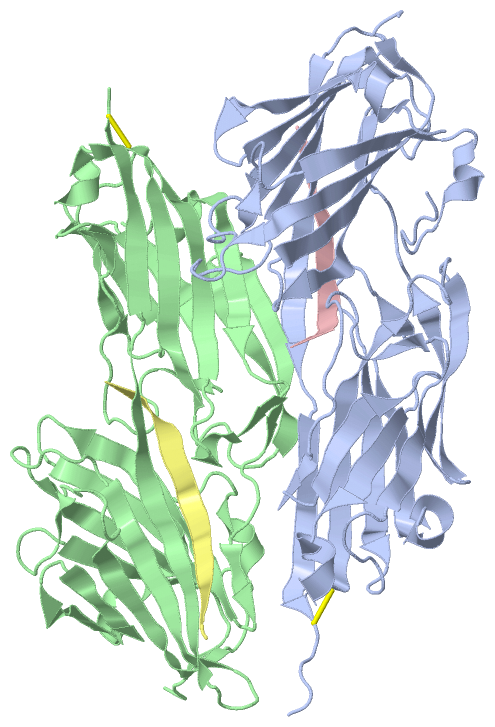
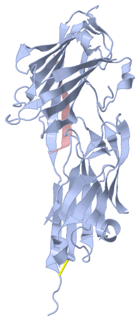
 Visualization

(-) Interactive Views

Asymmetric Unit
  Complete Structure
    Jena3D(integrated viewing of ligand, site, SAP, PROSITE, SCOP information)
    WebMol | AstexViewer[tm]@PDBe
(Java Applets, require no local installation except for Java; loading may be slow)
    STRAP
(Java WebStart application, automatic local installation, requires Java; full application with system access!)
    RasMol
(require local installation)
    Molscript (VRML)
(requires installation of a VRML viewer; select preferred view via VRML and generate a mono or stereo PDF format file)
 
  Ligands, Modified Residues, Ions
(no "Ligands, Modified Residues, Ions" information available for 2vr3)
 
  Sites
(no "Sites" information available for 2vr3)
 
  Cis Peptide Bonds
(no "Cis Peptide Bonds" information available for 2vr3)
 
Biological Units
  Complete Structure
    Biological Unit 1  [ Jena3D ]
    Biological Unit 2  [ Jena3D ]

(-) Still Images

Jmol
  protein: cartoon or spacefill or dots and stick; nucleic acid: cartoon and stick; ligands: spacefill; active site: stick
Molscript
  protein, nucleic acid: cartoon; ligands: spacefill; active site: ball and stick

 Databases and Analysis Tools

(-) Databases

Access by PDB/NDB ID
  2vr3
    Family and Domain InformationProDom | SYSTERS
    General Structural InformationGlycoscienceDB | MMDB | NDB | OCA | PDB | PDBe | PDBj | PDBsum | PDBWiki | PQS | PROTEOPEDIA
    Orientation in MembranesOPM
    Protein SurfaceSURFACE
    Secondary StructureDSSP (structure derived) | HSSP (homology derived)
    Structural GenomicsGeneCensus
    Structural NeighboursCE | VAST
    Structure ClassificationCATH | Dali | SCOP
    Validation and Original DataBMRB Data View | BMRB Restraints Grid | EDS | PROCHECK | RECOORD | WHAT_CHECK
 
Access by UniProt ID/Accession number
  CLFA_STAA8 | Q2G015
    Comparative Protein Structure ModelsModBase
    Genomic InformationEnsembl
    Protein-protein InteractionDIP
    Sequence, Family and Domain InformationInterPro | Pfam | SMART | UniProtKB/SwissProt
  FIBG_HUMAN | P02679
    Comparative Protein Structure ModelsModBase
    Genomic InformationEnsembl
    Protein-protein InteractionDIP
    Sequence, Family and Domain InformationInterPro | Pfam | SMART | UniProtKB/SwissProt
 
Access by Enzyme Classificator   (EC Number)
  (no 'Enzyme Classificator' available)
    General Enzyme InformationBRENDA | EC-PDB | Enzyme | IntEnz
    PathwayKEGG | MetaCyc
 
Access by Disease Identifier   (MIM ID)
  (no 'MIM ID' available)
    Disease InformationOMIM
 
Access by GenAge ID
  (no 'GenAge ID' available)
    Age Related InformationGenAge

(-) Analysis Tools

Access by PDB/NDB ID
    Domain InformationXDom
    Interatomic Contacts of Structural UnitsCSU
    Ligand-protein ContactsLPC
    Protein CavitiescastP
    Sequence and Secondary StructurePDBCartoon
    Structure AlignmentSTRAP(Java WebStart application, automatic local installation, requires Java; full application with system access!)
    Structure and Sequence BrowserSTING
 
Access by UniProt ID/Accession number
  CLFA_STAA8 | Q2G015
    Protein Disorder PredictionDisEMBL | FoldIndex | GLOBPLOT (for more information see DisProt)
  FIBG_HUMAN | P02679
    Protein Disorder PredictionDisEMBL | FoldIndex | GLOBPLOT (for more information see DisProt)

 Related Entries

(-) Entries Sharing at Least One Protein Chain (UniProt ID)

UniProtKB/Swiss-Prot
        FIBG_HUMAN | P026791dug 1fib 1fic 1fid 1fza 1fzb 1fzc 1fze 1fzf 1fzg 1lt9 1ltj 1n86 1n8e 1re3 1re4 1rf0 1rf1 2a45 2ffd 2fib 2h43 2hlo 2hod 2hpc 2hwl 2oyh 2oyi 2q9i 2vdo 2vdp 2vdq 2vdr 2xnx 2xny 2y7l 2z4e 3bvh 3e1i 3fib 3ghg 3h32 3hus 4b60

(-) Related Entries Specified in the PDB File

1dug STRUCTURE OF THE FIBRINOGEN G CHAIN INTEGRIN BINDING ANDFACTOR XIIIA CROSSLINKING SITES OBTAINED THROUGH CARRIERPROTEIN DRIVEN CRYSTALLIZATION
1fib RECOMBINANT HUMAN GAMMA-FIBRINOGEN CARBOXYL TERMINAL FRAGMENT (RESIDUES 143-411) BOUND TO CALCIUM AT PH 6.0
1fic STRUCTURE OF HUMAN GAMMA FIBRINOGEN 30 KD CARBOXYL TERMINAL FRAGMENT
1fid STRUCTURE OF HUMAN GAMMA FIBRINOGEN 30 KD CARBOXYL TERMINAL FRAGMENT
1fza CRYSTAL STRUCTURE OF FIBRINOGEN FRAGMENT D
1fzb CRYSTAL STRUCTURE OF CROSSLINKED FRAGMENT D
1fzc CRYSTAL STRUCTURE OF FRAGMENT DOUBLE-D FROM HUMAN FIBRIN WITH TWO DIFFERENT BOUND LIGANDS
1fze CRYSTAL STRUCTURE OF FRAGMENT DOUBLE-D FROM HUMAN FIBRIN
1fzf CRYSTAL STRUCTURE OF FRAGMENT DOUBLE-D FROM HUMAN FIBRIN WITH THE PEPTIDE LIGAND GLY- HIS-ARG-PRO-AMIDE
1fzg CRYSTAL STRUCTURE OF FRAGMENT D FROM HUMAN FIBRINOGEN WITH THE PEPTIDE LIGAND GLY-HIS- ARG-PRO-AMIDE
1lt9 CRYSTAL STRUCTURE OF RECOMBINANT HUMAN FIBRINOGEN FRAGMENT D
1ltj CRYSTAL STRUCTURE OF RECOMBINANT HUMAN FIBRINOGEN FRAGMENTD WITH THE PEPTIDE LIGANDS GLY-PRO-ARG-PRO-AMIDE AND GLY-HIS-ARG -PRO-AMIDE
1n86 CRYSTAL STRUCTURE OF HUMAN D-DIMER FROM CROSS-LINKED FIBRINCOMPLEXED WITH GPR AND GHRPLDK PEPTIDE LIGANDS.
1n8e FRAGMENT DOUBLE-D FROM HUMAN FIBRIN
1re3 CRYSTAL STRUCTURE OF FRAGMENT D OF BBETAD398A FIBRINOGENWITH THE PEPTIDE LIGAND GLY-HIS-ARG-PRO-AMIDE
1re4 CRYSTAL STRUCTURE OF FRAGMENT D OF BBETAD398A FIBRINOGEN
1rf0 CRYSTAL STRUCTURE OF FRAGMENT D OF GAMMAE132A FIBRINOGEN
1rf1 CRYSTAL STRUCTURE OF FRAGMENT D OF GAMMAE132A FIBRINOGENWITH THE PEPTIDE LIGAND GLY-HIS-ARG-PRO-AMIDE
2ffd FIBRINOGEN FRAGMENT D WITH "A" KNOB PEPTIDE MIMIC GPRVVE
2fib RECOMBINANT HUMAN GAMMA-FIBRINOGEN CARBOXYL TERMINAL FRAGMENT (RESIDUES 143-411) COMPLEXED TO THE PEPTIDE GLY-PRO-ARG-PRO AT PH 6.0
3fib RECOMBINANT HUMAN GAMMA-FIBRINOGEN CARBOXYL TERMINAL FRAGMENT (RESIDUES 143-411) BOUND TO CALCIUM AT PH 6.0: A FURTHER REFINEMENT OF PDB ENTRY 1FIB, AND DIFFERS FROM 1FIB BY THE MODELLING OF A CIS PEPTIDE BOND BETWEEN RESIDUES K338 AND C339