Show PDB file:   
         Plain Text   HTML   (compressed file size)
QuickSearch:   
by PDB,NDB,UniProt,PROSITE Code or Search Term(s)  
(-)Asymmetric Unit
(-)Asym. Unit - sites
(-)Biological Unit 1
(-)Biol. Unit 1 - sites
collapse expand < >
Image Asymmetric Unit
Asymmetric Unit  (Jmol Viewer)
Image Asym. Unit - sites
Asym. Unit - sites  (Jmol Viewer)
Image Biological Unit 1
Biological Unit 1  (Jmol Viewer)
Image Biol. Unit 1 - sites
Biol. Unit 1 - sites  (Jmol Viewer)

(-) Description

Title :  BACTERIORHODOPSIN-M PHOTOINTERMEDIATE STATE OF THE E204Q MUTANT AT 1.8 ANGSTROM RESOLUTION
 
Authors :  H. Luecke, B. Schobert, J. P. Cartailler, H. T. Richter, A. Rosengarth, R. Needleman, J. K. Lanyi
Date :  10 Jun 00  (Deposition) - 09 Aug 00  (Release) - 13 Jul 11  (Revision)
Method :  X-RAY DIFFRACTION
Resolution :  1.80
Chains :  Asym. Unit :  A
Biol. Unit 1:  A  (3x)
Keywords :  Ion Pump, Membrane Protein, Retinal Protein, Lipids, Photoreceptor, Haloarchaea, 7-Transmembrane, Ion Transport, Merohedral Twinning, E204Q Mutant M State, Photocycle Intermediate, Proton Transport (Keyword Search: [Gene Ontology, PubMed, Web (Google))
 
Reference :  H. Luecke, B. Schobert, J. P. Cartailler, H. T. Richter, A. Rosengarth R. Needleman, J. K. Lanyi
Coupling Photoisomerization Of Retinal To Directional Transport In Bacteriorhodopsin.
J. Mol. Biol. V. 300 1237 2000
PubMed-ID: 10903866  |  Reference-DOI: 10.1006/JMBI.2000.3884

(-) Compounds

Molecule 1 - BACTERIORHODOPSIN
    ChainsA
    EngineeredYES
    Expression SystemHALOBACTERIUM SALINARUM
    Expression System Taxid2242
    MutationYES
    Organism ScientificHALOBACTERIUM SALINARUM
    Organism Taxid2242
    SynonymBR

 Structural Features

(-) Chains, Units

  1
Asymmetric Unit A
Biological Unit 1 (3x)A

Summary Information (see also Sequences/Alignments below)

(-) Ligands, Modified Residues, Ions  (3, 15)

Asymmetric Unit (3, 15)
No.NameCountTypeFull Name
1LI113Ligand/Ion1-[2,6,10.14-TETRAMETHYL-HEXADECAN-16-YL]-2-[2,10,14-TRIMETHYLHEXADECAN-16-YL]GLYCEROL
2RET1Ligand/IonRETINAL
3SQU1Ligand/Ion2,10,23-TRIMETHYL-TETRACOSANE
Biological Unit 1 (3, 45)
No.NameCountTypeFull Name
1LI139Ligand/Ion1-[2,6,10.14-TETRAMETHYL-HEXADECAN-16-YL]-2-[2,10,14-TRIMETHYLHEXADECAN-16-YL]GLYCEROL
2RET3Ligand/IonRETINAL
3SQU3Ligand/Ion2,10,23-TRIMETHYL-TETRACOSANE

(-) Sites  (15, 15)

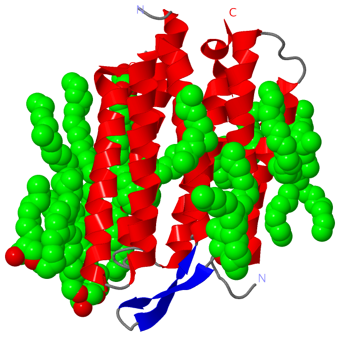
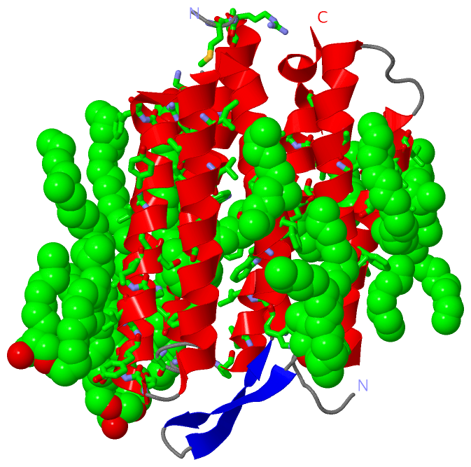
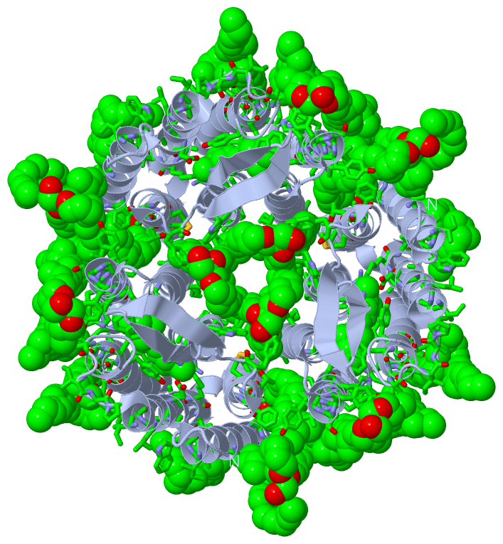
Asymmetric Unit (15, 15)
No.NameEvidenceResiduesDescription
01AC1SOFTWARETYR A:133 , VAL A:136 , ILE A:140 , LI1 A:606 , LI1 A:608 , LI1 A:612 , SQU A:701BINDING SITE FOR RESIDUE LI1 A 601
02AC2SOFTWARETRP A:12 , TYR A:131 , SER A:132 , PHE A:135 , ALA A:139 , MET A:163 , ARG A:164 , PRO A:165 , ALA A:196 , ILE A:203 , LEU A:206 , LEU A:207 , LI1 A:603BINDING SITE FOR RESIDUE LI1 A 602
03AC3SOFTWAREVAL A:187 , LEU A:190 , ILE A:198 , VAL A:199 , PRO A:200 , LI1 A:602 , LI1 A:604 , LI1 A:609 , LI1 A:610BINDING SITE FOR RESIDUE LI1 A 603
04AC4SOFTWAREILE A:198 , LI1 A:603 , LI1 A:610BINDING SITE FOR RESIDUE LI1 A 604
05AC5SOFTWAREILE A:11 , ALA A:18 , LI1 A:606 , LI1 A:611BINDING SITE FOR RESIDUE LI1 A 605
06AC6SOFTWAREALA A:18 , LI1 A:601 , LI1 A:605BINDING SITE FOR RESIDUE LI1 A 606
07AC7SOFTWAREILE A:52 , THR A:55 , MET A:56 , TYR A:64 , TRP A:80 , PHE A:88BINDING SITE FOR RESIDUE LI1 A 607
08AC8SOFTWAREILE A:222 , LI1 A:601 , LI1 A:611 , SQU A:701BINDING SITE FOR RESIDUE LI1 A 608
09AC9SOFTWARETHR A:142 , LEU A:146 , PHE A:154 , VAL A:173 , ASN A:176 , LI1 A:603 , LI1 A:610BINDING SITE FOR RESIDUE LI1 A 609
10BC1SOFTWAREPHE A:153 , LYS A:172 , ARG A:175 , ASN A:176 , VAL A:179 , VAL A:180 , VAL A:187 , LI1 A:603 , LI1 A:604 , LI1 A:609BINDING SITE FOR RESIDUE LI1 A 610
11BC2SOFTWARELEU A:25 , VAL A:29 , LI1 A:605 , LI1 A:608 , SQU A:701BINDING SITE FOR RESIDUE LI1 A 611
12BC3SOFTWARETHR A:24 , LEU A:28 , ALA A:44 , THR A:47 , PHE A:54 , ALA A:110 , TYR A:147 , HOH A:512 , LI1 A:601BINDING SITE FOR RESIDUE LI1 A 612
13BC4SOFTWAREPRO A:91 , LEU A:95 , ILE A:108 , VAL A:112BINDING SITE FOR RESIDUE LI1 A 613
14BC5SOFTWARELEU A:19 , TYR A:26 , SER A:214 , VAL A:217 , LEU A:221 , ARG A:225 , LI1 A:601 , LI1 A:608 , LI1 A:611BINDING SITE FOR RESIDUE SQU A 701
15BC6SOFTWARETRP A:86 , THR A:90 , MET A:118 , TRP A:138 , SER A:141 , THR A:142 , TRP A:182 , TYR A:185 , PRO A:186 , TRP A:189 , ASP A:212 , ALA A:215 , LYS A:216BINDING SITE FOR RESIDUE RET A 301

(-) SS Bonds  (0, 0)

(no "SS Bond" information available for 1F4Z)

(-) Cis Peptide Bonds  (0, 0)

(no "Cis Peptide Bond" information available for 1F4Z)

 Sequence-Structure Mapping

(-) SAPs(SNPs)/Variants  (0, 0)

(no "SAP(SNP)/Variant" information available for 1F4Z)

(-) PROSITE Motifs  (2, 2)

Asymmetric Unit (2, 2)
 PROSITEUniProtKBPDB
No.IDACDescriptionIDLocationCountLocation
1BACTERIAL_OPSIN_1PS00950 Bacterial rhodopsins signature 1.BACR_HALSA95-107  1A:82-94
2BACTERIAL_OPSIN_RETPS00327 Bacterial rhodopsins retinal binding site.BACR_HALSA221-232  1A:208-219
Biological Unit 1 (2, 6)
 PROSITEUniProtKBPDB
No.IDACDescriptionIDLocationCountLocation
1BACTERIAL_OPSIN_1PS00950 Bacterial rhodopsins signature 1.BACR_HALSA95-107  3A:82-94
2BACTERIAL_OPSIN_RETPS00327 Bacterial rhodopsins retinal binding site.BACR_HALSA221-232  3A:208-219

(-) Exons   (0, 0)

(no "Exon" information available for 1F4Z)

(-) Sequences/Alignments

Asymmetric Unit
   Reformat: Number of residues per line =  ('0' or empty: single-line sequence representation)
  Number of residues per labelling interval =   
  UniProt sequence: complete  aligned part    
   Show mapping: SCOP domains CATH domains Pfam domains Secondary structure (by author)
SAPs(SNPs) PROSITE motifs Exons
(details for a mapped element are shown in a popup box when the mouse pointer rests over it)
Chain A from PDB  Type:PROTEIN  Length:222
 aligned with BACR_HALSA | P02945 from UniProtKB/Swiss-Prot  Length:262

    Alignment length:227
                                    27        37        47        57        67        77        87        97       107       117       127       137       147       157       167       177       187       197       207       217       227       237       
           BACR_HALSA    18 TGRPEWIWLALGTALMGLGTLYFLVKGMGVSDPDAKKFYAITTLVPAIAFTMYLSMLLGYGLTMVPFGGEQNPIYWARYADWLFTTPLLLLDLALLVDADQGTILALVGADGIMIGTGLVGALTKVYSYRFVWWAISTAAMLYILYVLFFGFTSKAESMRPEVASTFKVLRNVTVVLWSAYPVVWLIGSEGAGIVPLNIETLLFMVLDVSAKVGFGLILLRSRAIFG 244
               SCOP domains d1f4za_ A: Bacteriorhodopsin                                                                                                                                                                                                        SCOP domains
               CATH domains 1f4zA00 A:5-231 Rhopdopsin 7-helix transmembrane proteins                                                                                                                                                                           CATH domains
               Pfam domains ----------------------------------------------------------------------------------------------------------------------------------------------------------------------------------------------------------------------------------- Pfam domains
         Sec.struct. author ....hhhhhhhhhhhhhhhhhhhhhhh....hhhhhhhhhhhhhhhhhhhhhhhhhhh....eeeee..eeeee.hhhhhhhhhhhhhhhhhhhhhhh.hhhhhhhhhhhhhhhhhhhhhhhhh.hhhhhhhhhhhhhhhhhhhhhhhhhh.-----..hhhhhhhhhhhhhhhhhhhhhhhhhhhhh.......hhhhhhhhhhhhhhhhhhhhhhhhhhhhhhh. Sec.struct. author
                 SAPs(SNPs) ----------------------------------------------------------------------------------------------------------------------------------------------------------------------------------------------------------------------------------- SAPs(SNPs)
                    PROSITE -----------------------------------------------------------------------------BACTERIAL_OPS-----------------------------------------------------------------------------------------------------------------BACTERIAL_OP------------ PROSITE
                 Transcript ----------------------------------------------------------------------------------------------------------------------------------------------------------------------------------------------------------------------------------- Transcript
                 1f4z A   5 TGRPEWIWLALGTALMGLGTLYFLVKGMGVSDPDAKKFYAITTLVPAIAFTMYLSMLLGYGLTMVPFGGEQNPIYWARYADWLFTTPLLLLDLALLVDADQGTILALVGADGIMIGTGLVGALTKVYSYRFVWWAISTAAMLYILYVLFFGF-----SMRPEVASTFKVLRNVTVVLWSAYPVVWLIGSEGAGIVPLNIQTLLFMVLDVSAKVGFGLILLRSRAIFG 231
                                    14        24        34        44        54        64        74        84        94       104       114       124       134       144       154 |     164       174       184       194       204       214       224       
                                                                                                                                                                                 156   162                                                                     

   Legend:   → Mismatch (orange background)
  - → Gap (green background, '-', border residues have a numbering label)
    → Modified Residue (blue background, lower-case, 'x' indicates undefined single-letter code, labelled with number + name)
  x → Chemical Group (purple background, 'x', labelled with number + name, e.g. ACE or NH2)
  extra numbering lines below/above indicate numbering irregularities and modified residue names etc., number ends below/above '|'

 Classification and Annotation

(-) SCOP Domains  (1, 1)

Asymmetric Unit

(-) CATH Domains  (1, 1)

Asymmetric Unit

(-) Pfam Domains  (0, 0)

(no "Pfam Domain" information available for 1F4Z)

(-) Gene Ontology  (12, 12)

Asymmetric Unit(hide GO term definitions)
Chain A   (BACR_HALSA | P02945)
molecular function
    GO:0005216    ion channel activity    Enables the facilitated diffusion of an ion (by an energy-independent process) by passage through a transmembrane aqueous pore or channel without evidence for a carrier-mediated mechanism. May be either selective (it enables passage of a specific ion only) or non-selective (it enables passage of two or more ions of same charge but different size).
    GO:0009881    photoreceptor activity    The function of absorbing and responding to incidental electromagnetic radiation, particularly visible light. The response may involve a change in conformation.
biological process
    GO:0034220    ion transmembrane transport    A process in which an ion is transported from one side of a membrane to the other by means of some agent such as a transporter or pore.
    GO:0006811    ion transport    The directed movement of charged atoms or small charged molecules into, out of or within a cell, or between cells, by means of some agent such as a transporter or pore.
    GO:0007602    phototransduction    The sequence of reactions within a cell required to convert absorbed photons into a molecular signal.
    GO:0018298    protein-chromophore linkage    The covalent or noncovalent attachment of a chromophore to a protein.
    GO:0015992    proton transport    The directed movement of protons (hydrogen ions) into, out of or within a cell, or between cells, by means of some agent such as a transporter or pore.
    GO:0050896    response to stimulus    Any process that results in a change in state or activity of a cell or an organism (in terms of movement, secretion, enzyme production, gene expression, etc.) as a result of a stimulus. The process begins with detection of the stimulus and ends with a change in state or activity or the cell or organism.
    GO:0006810    transport    The directed movement of substances (such as macromolecules, small molecules, ions) or cellular components (such as complexes and organelles) into, out of or within a cell, or between cells, or within a multicellular organism by means of some agent such as a transporter, pore or motor protein.
cellular component
    GO:0016021    integral component of membrane    The component of a membrane consisting of the gene products and protein complexes having at least some part of their peptide sequence embedded in the hydrophobic region of the membrane.
    GO:0016020    membrane    A lipid bilayer along with all the proteins and protein complexes embedded in it an attached to it.
    GO:0005886    plasma membrane    The membrane surrounding a cell that separates the cell from its external environment. It consists of a phospholipid bilayer and associated proteins.

 Visualization

(-) Interactive Views

Asymmetric Unit
  Complete Structure
    Jena3D(integrated viewing of ligand, site, SAP, PROSITE, SCOP information)
    WebMol | AstexViewer[tm]@PDBe
(Java Applets, require no local installation except for Java; loading may be slow)
    STRAP
(Java WebStart application, automatic local installation, requires Java; full application with system access!)
    RasMol
(require local installation)
    Molscript (VRML)
(requires installation of a VRML viewer; select preferred view via VRML and generate a mono or stereo PDF format file)
 
  Ligands, Modified Residues, Ions
    LI1  [ RasMol | Jena3D ]  +environment [ RasMol | Jena3D ]
    RET  [ RasMol | Jena3D ]  +environment [ RasMol | Jena3D ]
    SQU  [ RasMol | Jena3D ]  +environment [ RasMol | Jena3D ]
 
  Sites
    AC1  [ RasMol ]  +environment [ RasMol ]
    AC2  [ RasMol ]  +environment [ RasMol ]
    AC3  [ RasMol ]  +environment [ RasMol ]
    AC4  [ RasMol ]  +environment [ RasMol ]
    AC5  [ RasMol ]  +environment [ RasMol ]
    AC6  [ RasMol ]  +environment [ RasMol ]
    AC7  [ RasMol ]  +environment [ RasMol ]
    AC8  [ RasMol ]  +environment [ RasMol ]
    AC9  [ RasMol ]  +environment [ RasMol ]
    BC1  [ RasMol ]  +environment [ RasMol ]
    BC2  [ RasMol ]  +environment [ RasMol ]
    BC3  [ RasMol ]  +environment [ RasMol ]
    BC4  [ RasMol ]  +environment [ RasMol ]
    BC5  [ RasMol ]  +environment [ RasMol ]
    BC6  [ RasMol ]  +environment [ RasMol ]
 
  Cis Peptide Bonds
(no "Cis Peptide Bonds" information available for 1f4z)
 
Biological Unit
  Complete Structure
    Biological Unit 1  [ Jena3D ]

(-) Still Images

Jmol
  protein: cartoon or spacefill or dots and stick; nucleic acid: cartoon and stick; ligands: spacefill; active site: stick
Molscript
  protein, nucleic acid: cartoon; ligands: spacefill; active site: ball and stick

 Databases and Analysis Tools

(-) Databases

Access by PDB/NDB ID
  1f4z
    Family and Domain InformationProDom | SYSTERS
    General Structural InformationGlycoscienceDB | MMDB | NDB | OCA | PDB | PDBe | PDBj | PDBsum | PDBWiki | PQS | PROTEOPEDIA
    Orientation in MembranesOPM
    Protein SurfaceSURFACE
    Secondary StructureDSSP (structure derived) | HSSP (homology derived)
    Structural GenomicsGeneCensus
    Structural NeighboursCE | VAST
    Structure ClassificationCATH | Dali | SCOP
    Validation and Original DataBMRB Data View | BMRB Restraints Grid | EDS | PROCHECK | RECOORD | WHAT_CHECK
 
Access by UniProt ID/Accession number
  BACR_HALSA | P02945
    Comparative Protein Structure ModelsModBase
    Genomic InformationEnsembl
    Protein-protein InteractionDIP
    Sequence, Family and Domain InformationInterPro | Pfam | SMART | UniProtKB/SwissProt
 
Access by Enzyme Classificator   (EC Number)
  (no 'Enzyme Classificator' available)
    General Enzyme InformationBRENDA | EC-PDB | Enzyme | IntEnz
    PathwayKEGG | MetaCyc
 
Access by Disease Identifier   (MIM ID)
  (no 'MIM ID' available)
    Disease InformationOMIM
 
Access by GenAge ID
  (no 'GenAge ID' available)
    Age Related InformationGenAge

(-) Analysis Tools

Access by PDB/NDB ID
    Domain InformationXDom
    Interatomic Contacts of Structural UnitsCSU
    Ligand-protein ContactsLPC
    Protein CavitiescastP
    Sequence and Secondary StructurePDBCartoon
    Structure AlignmentSTRAP(Java WebStart application, automatic local installation, requires Java; full application with system access!)
    Structure and Sequence BrowserSTING
 
Access by UniProt ID/Accession number
  BACR_HALSA | P02945
    Protein Disorder PredictionDisEMBL | FoldIndex | GLOBPLOT (for more information see DisProt)

 Related Entries

(-) Entries Sharing at Least One Protein Chain (UniProt ID)

UniProtKB/Swiss-Prot
        BACR_HALSA | P029451ap9 1at9 1bac 1bad 1bct 1bha 1bhb 1bm1 1brd 1brr 1brx 1c3w 1c8r 1c8s 1cwq 1dze 1e0p 1f50 1fbb 1fbk 1iw6 1iw9 1ixf 1jv6 1jv7 1kg8 1kg9 1kgb 1kme 1l0m 1m0k 1m0l 1m0m 1mgy 1o0a 1p8h 1p8i 1p8u 1pxr 1pxs 1py6 1q5i 1q5j 1qhj 1qko 1qkp 1qm8 1r2n 1r84 1s51 1s52 1s53 1s54 1s8j 1s8l 1tn0 1tn5 1ucq 1vjm 1x0i 1x0k 1x0s 1xji 2at9 2brd 2i1x 2i20 2i21 2ntu 2ntw 2wjk 2wjl 2zfe 2zzl 3coc 3cod 3han 3hao 3hap 3haq 3har 3has 3mbv 3ns0 3nsb 3t45 3utv 3utw 3utx 3uty 3vhz 3vi0 4fpd 4hwl 4hyx 4md1 4md2 4ov0 4x31 4x32 4xxj 5a44 5a45 5b34 5b35 5b6v 5b6w 5b6x 5b6y 5b6z 5br2 5br5 5h2h 5h2i 5h2j 5h2k 5h2l 5h2m 5h2n 5h2o 5h2p 5j7a 5vn7 5vn9

(-) Related Entries Specified in the PDB File

1c3w 1C3W.PDB CONTAINS THE WILD-TYPE BACTERIORHODOPSIN AT 1.55 A RESOLUTION
1c8r BACTERIORHODOPSIN ("BR" STATE INTERMEDIATE) - D96N MUTANT, 1.80 A RESOLUTION
1c8s BACTERIORHODOPSIN ("M" STATE INTERMEDIATE) - D96N MUTANT, 2.00 A RESOLUTION
1f50 BACTERIORHODOPSIN ("BR" STATE) - E204Q MUTANT, 1.70 A RESOLUTION