|
|
|
|
 Description
|
|
Compounds
|
||||||||||||||||||||||||||||||||||||||||||||
Chains, Units
Summary Information (see also Sequences/Alignments below) |
Ligands, Modified Residues, Ions (1, 2)
Asymmetric/Biological Unit (1, 2)
|
Sites (2, 2)
Asymmetric Unit (2, 2)
|
SS Bonds (0, 0)| (no "SS Bond" information available for 1D0B) |
Cis Peptide Bonds (0, 0)| (no "Cis Peptide Bond" information available for 1D0B) |
SAPs(SNPs)/Variants (0, 0)| (no "SAP(SNP)/Variant" information available for 1D0B) |
PROSITE Motifs (1, 6)
Asymmetric/Biological Unit (1, 6)
|
||||||||||||||||||||||||
Exons (0, 0)| (no "Exon" information available for 1D0B) |
Sequences/Alignments
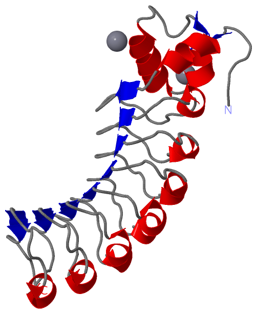
Asymmetric/Biological UnitChain A from PDB Type:PROTEIN Length:207 aligned with INLB_LISMO | P25147 from UniProtKB/Swiss-Prot Length:630 Alignment length:207 45 55 65 75 85 95 105 115 125 135 145 155 165 175 185 195 205 215 225 235 INLB_LISMO 36 ETITVPTPIKQIFSDDAFAETIKDNLKKKSVTDAVTQNELNSIDQIIANNSDIKSVQGIQYLPNVTKLFLNGNKLTDIKPLANLKNLGWLFLDENKVKDLSSLKDLKKLKSLSLEHNGISDINGLVHLPQLESLYLGNNKITDITVLSRLTKLDTLSLEDNQISDIVPLAGLTKLQNLYLSKNHISDLRALAGLKNLDVLELFSQEC 242 SCOP domains d1d0ba_ A: Internalin B SCOP domains CATH domains 1d0bA00 A:36-242 Ribonuclease Inhibitor CATH domains Pfam domains --------------------------------------------------------------------------------------------------------------------------------------------------------------------------------------------------------------- Pfam domains SAPs(SNPs) --------------------------------------------------------------------------------------------------------------------------------------------------------------------------------------------------------------- SAPs(SNPs) PROSITE ---------------------------------------------------------------LRR PDB: A:99-120 LRR PDB: A:121-142 LRR PDB: A:143-164 LRR PDB: A:165-186 LRR PDB: A:187-208 LRR PDB: A:209-230 ------------ PROSITE Transcript --------------------------------------------------------------------------------------------------------------------------------------------------------------------------------------------------------------- Transcript 1d0b A 36 ETITVSTPIKQIFPDDAFAETIKDNLKKKSVTDAVTQNELNSIDQIIANNSDIKSVQGIQYLPNVTKLFLNGNKLTDIKPLTNLKNLGWLFLDENKIKDLSSLKDLKKLKSLSLEHNGISDINGLVHLPQLESLYLGNNKITDITVLSRLTKLDTLSLEDNQISDIVPLAGLTKLQNLYLSKNHISDLRALAGLKNLDVLELFSQEC 242 45 55 65 75 85 95 105 115 125 135 145 155 165 175 185 195 205 215 225 235
|
||||||||||||||||||||
SCOP Domains (1, 1)
Asymmetric/Biological Unit
 |
CATH Domains (1, 1)
Asymmetric/Biological Unit
|
Pfam Domains (0, 0)| (no "Pfam Domain" information available for 1D0B) |
Gene Ontology (1, 1)|
Asymmetric/Biological Unit(hide GO term definitions) Chain A (INLB_LISMO | P25147)
|
||||||||||||
Interactive Views
|
|||||||||||||||||||||||||||||||||||||||||||||||||||||||||||||||||||||||||||||||||||||||||||||||||||||||||||||||||||||||||||||
Still Images
|
||||||||||||||||
Databases
|
||||||||||||||||||||||||||||||||||||||||||||||||||||||||||||||||||||||||||||||||||||||||||||||||||||||||||||||||||||||||||||||||||||||||||||||||||||||||||||||||
Analysis Tools
|
|||||||||||||||||||||||||||||||||||||||||||||||||||||||||||||
Entries Sharing at Least One Protein Chain (UniProt ID)
Related Entries Specified in the PDB File
|
|