|
|
|
|
 Description
|
|
Compounds
|
||||||||||||||||||||||||||||||||||||||||||||||||||||||||||||||||
Chains, Units
Summary Information (see also Sequences/Alignments below) |
Ligands, Modified Residues, Ions (2, 2)| Asymmetric Unit (2, 2) Biological Unit 1 (0, 0) |
Sites (2, 2)
Asymmetric Unit (2, 2)
|
SS Bonds (0, 0)| (no "SS Bond" information available for 1AA0) |
Cis Peptide Bonds (0, 0)| (no "Cis Peptide Bond" information available for 1AA0) |
SAPs(SNPs)/Variants (0, 0)| (no "SAP(SNP)/Variant" information available for 1AA0) |
PROSITE Motifs (0, 0)| (no "PROSITE Motif" information available for 1AA0) |
Exons (0, 0)| (no "Exon" information available for 1AA0) |
Sequences/Alignments
Asymmetric UnitChain A from PDB Type:PROTEIN Length:113 aligned with WAC_BPT4 | P10104 from UniProtKB/Swiss-Prot Length:487 Alignment length:113 381 391 401 411 421 431 441 451 461 471 481 WAC_BPT4 372 VSGLNNAVQNLQVEIGNNSAGIKGQVVALNTLVNGTNPNGSTVEERGLTNSIKANETNIASVTQEVNTAKGNISSLQGDVQALQEAGYIPEAPRDGQAYVRKDGEWVFLSTFL 484 SCOP domains d1aa0a_ A: Fibritin SCOP domains CATH domains 1aa0A00 A:371-483 6-Phosphogluconate Dehydrogenase, domain 3 CATH domains Pfam domains ----------------------------------------------------------------------------------------------------------------- Pfam domains SAPs(SNPs) ----------------------------------------------------------------------------------------------------------------- SAPs(SNPs) PROSITE ----------------------------------------------------------------------------------------------------------------- PROSITE Transcript ----------------------------------------------------------------------------------------------------------------- Transcript 1aa0 A 371 VSGLNNAVQNLQVEIGNNSAGIKGQVVALNTLVNGTNPNGSTVEERGLTNSIKANETNIASVTQEVNTAKGNISSLQGDVQALQEAGYIPEAPRDGQAYVRKDGEWVLLSTFL 483 380 390 400 410 420 430 440 450 460 470 480
|
||||||||||||||||||||
SCOP Domains (1, 1)
Asymmetric Unit
 |
CATH Domains (1, 1)
Asymmetric Unit
|
Pfam Domains (0, 0)| (no "Pfam Domain" information available for 1AA0) |
Gene Ontology (1, 1)|
Asymmetric Unit(hide GO term definitions) Chain A (WAC_BPT4 | P10104)
|
||||||||||||
Interactive Views
|
||||||||||||||||||||||||||||||||||||||||||||||||||||||||||||||||||||||||||||||||||||||||||||||||||||||||||||||||||||||||||||||||||||||||||||||||||||||
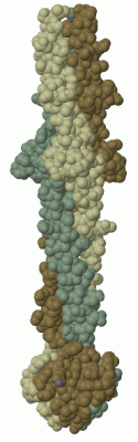
Still Images
|
||||||||||||||||
Databases
|
||||||||||||||||||||||||||||||||||||||||||||||||||||||||||||||||||||||||||||||||||||||||||||||||||||||||||||||||||||||||||||||||||||||||||||||||||||||||||||||||
Analysis Tools
|
|||||||||||||||||||||||||||||||||||||||||||||||||||||||||||||
Entries Sharing at Least One Protein Chain (UniProt ID)
Related Entries Specified in the PDB File
|
|