Show PDB file:   
         Plain Text   HTML   (compressed file size)
QuickSearch:   
by PDB,NDB,UniProt,PROSITE Code or Search Term(s)  
(-)Biol.Unit 1 - manually
(-)Asymmetric Unit
(-)Asym. Unit - sites
(-)Biological Unit 1
(-)Biol. Unit 1 - sites
(-)Biological Unit 2
collapse expand < >
Image Biol.Unit 1 - manually
Biol.Unit 1 - manually  (Jmol Viewer)
Image Asymmetric Unit
Asymmetric Unit  (Jmol Viewer)
Image Asym. Unit - sites
Asym. Unit - sites  (Jmol Viewer)
Image Biological Unit 1
Biological Unit 1  (Jmol Viewer)
Image Biol. Unit 1 - sites
Biol. Unit 1 - sites  (Jmol Viewer)
Image Biological Unit 2
Biological Unit 2  (Jmol Viewer)

(-) Description

Title :  ANTENNAPEDIA HOMEODOMAIN-DNA COMPLEX
 
Authors :  E. Fraenkel, C. O. Pabo
Date :  02 Jul 98  (Deposition) - 14 Oct 98  (Release) - 24 Feb 09  (Revision)
Method :  X-RAY DIFFRACTION
Resolution :  2.40
Chains :  Asym. Unit :  A,B,C,D,E,F
Biol. Unit 1:  A,C,D  (1x)
Biol. Unit 2:  B,E,F  (1x)
Keywords :  Homeodomain, Dna-Binding Protein, Complex (Homeodomain/Dna), Transcription/Dna Complex (Keyword Search: [Gene Ontology, PubMed, Web (Google))
 
Reference :  E. Fraenkel, C. O. Pabo
Comparison Of X-Ray And Nmr Structures For The Antennapedia Homeodomain-Dna Complex.
Nat. Struct. Biol. V. 5 692 1998
PubMed-ID: 9699632
(for further references see the PDB file header)

(-) Compounds

Molecule 1 - DNA (5'- D(*AP*GP*AP*AP*AP*GP*CP*CP*AP*TP*TP*AP*GP*AP*G)-3')
    ChainsC, E
    EngineeredYES
 
Molecule 2 - DNA (5'- D(*TP*CP*TP*CP*TP*AP*AP*TP*GP*GP*CP*TP*TP*TP*C)-3')
    ChainsD, F
    EngineeredYES
 
Molecule 3 - ANTENNAPEDIA HOMEODOMAIN
    ChainsA, B
    EngineeredYES
    Expression SystemESCHERICHIA COLI
    Expression System Taxid562
    FragmentHOMEODOMAIN
    MutationYES
    Organism CommonFRUIT FLY
    Organism ScientificDROSOPHILA MELANOGASTER
    Organism Taxid7227
    SynonymHD

 Structural Features

(-) Chains, Units

  123456
Asymmetric Unit ABCDEF
Biological Unit 1 (1x)A CD  
Biological Unit 2 (1x) B  EF

Summary Information (see also Sequences/Alignments below)

(-) Ligands, Modified Residues, Ions  (1, 1)

Asymmetric Unit (1, 1)
No.NameCountTypeFull Name
1NI1Ligand/IonNICKEL (II) ION
Biological Unit 1 (0, 0)
No.NameCountTypeFull Name
1NI-1Ligand/IonNICKEL (II) ION
Biological Unit 2 (0, 0)
No.NameCountTypeFull Name
1NI-1Ligand/IonNICKEL (II) ION

(-) Sites  (1, 1)

Asymmetric Unit (1, 1)
No.NameEvidenceResiduesDescription
1AC1SOFTWAREHIS A:21 , HIS B:21 , ASN B:60 , HOH B:721 , HOH B:722BINDING SITE FOR RESIDUE NI B 601

(-) SS Bonds  (0, 0)

(no "SS Bond" information available for 9ANT)

(-) Cis Peptide Bonds  (0, 0)

(no "Cis Peptide Bond" information available for 9ANT)

 Sequence-Structure Mapping

(-) SAPs(SNPs)/Variants  (0, 0)

(no "SAP(SNP)/Variant" information available for 9ANT)

(-) PROSITE Motifs  (0, 0)

(no "PROSITE Motif" information available for 9ANT)

(-) Exons   (0, 0)

(no "Exon" information available for 9ANT)

(-) Sequences/Alignments

Asymmetric Unit
   Reformat: Number of residues per line =  ('0' or empty: single-line sequence representation)
  Number of residues per labelling interval =   
  UniProt sequence: complete  aligned part    
   Show mapping: SCOP domains CATH domains Pfam domains Secondary structure (by author)
SAPs(SNPs) PROSITE motifs Exons
(details for a mapped element are shown in a popup box when the mouse pointer rests over it)
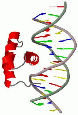
Chain A from PDB  Type:PROTEIN  Length:56
                                                                                        
               SCOP domains d9anta_ A: Antennapedia Homeodomain                      SCOP domains
               CATH domains 9antA00 A:5-60 Homeodomain-like                          CATH domains
               Pfam domains -------------------------------------------------------- Pfam domains
         Sec.struct. author .....hhhhhhhhhhhhh.....hhhhhhhhhhh...hhhhhhhhhhhhhhhhh.. Sec.struct. author
                 SAPs(SNPs) -------------------------------------------------------- SAPs(SNPs)
                    PROSITE -------------------------------------------------------- PROSITE
                 Transcript -------------------------------------------------------- Transcript
                 9ant A   5 RQTYTRYQTLELEKEFHFNRYLTRRRRIEIAHALSLTERQIKIWFQNRRMKWKKEN  60
                                    14        24        34        44        54      

Chain B from PDB  Type:PROTEIN  Length:56
                                                                                        
               SCOP domains d9antb_ B: Antennapedia Homeodomain                      SCOP domains
               CATH domains 9antB00 B:5-60 Homeodomain-like                          CATH domains
               Pfam domains -------------------------------------------------------- Pfam domains
         Sec.struct. author .....hhhhhhhhhhhhh.....hhhhhhhhhhh...hhhhhhhhhhhhhhhhh.. Sec.struct. author
                 SAPs(SNPs) -------------------------------------------------------- SAPs(SNPs)
                    PROSITE -------------------------------------------------------- PROSITE
                 Transcript -------------------------------------------------------- Transcript
                 9ant B   5 RQTYTRYQTLELEKEFHFNRYLTRRRRIEIAHALSLTERQIKIWFQNRRMKWKKEN  60
                                    14        24        34        44        54      

Chain C from PDB  Type:DNA  Length:15
                                               
                 9ant C 100 AGAAAGCCATTAGAG 114
                                   109     

Chain D from PDB  Type:DNA  Length:15
                                               
                 9ant D 214 TCTCTAATGGCTTTC 228
                                   223     

Chain E from PDB  Type:DNA  Length:15
                                               
                 9ant E 400 AGAAAGCCATTAGAG 414
                                   409     

Chain F from PDB  Type:DNA  Length:15
                                               
                 9ant F 514 TCTCTAATGGCTTTC 528
                                   523     

   Legend:   → Mismatch (orange background)
  - → Gap (green background, '-', border residues have a numbering label)
    → Modified Residue (blue background, lower-case, 'x' indicates undefined single-letter code, labelled with number + name)
  x → Chemical Group (purple background, 'x', labelled with number + name, e.g. ACE or NH2)
  extra numbering lines below/above indicate numbering irregularities and modified residue names etc., number ends below/above '|'

 Classification and Annotation

(-) SCOP Domains  (1, 2)

Asymmetric Unit

(-) CATH Domains  (1, 2)

Asymmetric Unit

(-) Pfam Domains  (0, 0)

(no "Pfam Domain" information available for 9ANT)

(-) Gene Ontology  (20, 20)

Asymmetric Unit(hide GO term definitions)

 Visualization

(-) Interactive Views

Asymmetric Unit
  Complete Structure
    Jena3D(integrated viewing of ligand, site, SAP, PROSITE, SCOP information)
    WebMol | AstexViewer[tm]@PDBe
(Java Applets, require no local installation except for Java; loading may be slow)
    STRAP
(Java WebStart application, automatic local installation, requires Java; full application with system access!)
    RasMol
(require local installation)
    Molscript (VRML)
(requires installation of a VRML viewer; select preferred view via VRML and generate a mono or stereo PDF format file)
 
  Ligands, Modified Residues, Ions
    NI  [ RasMol | Jena3D ]  +environment [ RasMol | Jena3D ]
 
  Sites
    AC1  [ RasMol ]  +environment [ RasMol ]
 
  Cis Peptide Bonds
(no "Cis Peptide Bonds" information available for 9ant)
 
Biological Units
  Complete Structure
    Biological Unit 1  [ Jena3D ]
    Biological Unit 2  [ Jena3D ]

(-) Still Images

Jmol
  protein: cartoon or spacefill or dots and stick; nucleic acid: cartoon and stick; ligands: spacefill; active site: stick
Molscript
  protein, nucleic acid: cartoon; ligands: spacefill; active site: ball and stick

 Databases and Analysis Tools

(-) Databases

Access by PDB/NDB ID
  9ant
    Family and Domain InformationProDom | SYSTERS
    General Structural InformationGlycoscienceDB | MMDB | NDB | OCA | PDB | PDBe | PDBj | PDBsum | PDBWiki | PQS | PROTEOPEDIA
    Orientation in MembranesOPM
    Protein SurfaceSURFACE
    Secondary StructureDSSP (structure derived) | HSSP (homology derived)
    Structural GenomicsGeneCensus
    Structural NeighboursCE | VAST
    Structure ClassificationCATH | Dali | SCOP
    Validation and Original DataBMRB Data View | BMRB Restraints Grid | EDS | PROCHECK | RECOORD | WHAT_CHECK
 
Access by UniProt ID/Accession number
  ANTP_DROME | P02833
    Comparative Protein Structure ModelsModBase
    Genomic InformationEnsembl
    Protein-protein InteractionDIP
    Sequence, Family and Domain InformationInterPro | Pfam | SMART | UniProtKB/SwissProt
 
Access by Enzyme Classificator   (EC Number)
  (no 'Enzyme Classificator' available)
    General Enzyme InformationBRENDA | EC-PDB | Enzyme | IntEnz
    PathwayKEGG | MetaCyc
 
Access by Disease Identifier   (MIM ID)
  (no 'MIM ID' available)
    Disease InformationOMIM
 
Access by GenAge ID
  (no 'GenAge ID' available)
    Age Related InformationGenAge

(-) Analysis Tools

Access by PDB/NDB ID
    Domain InformationXDom
    Interatomic Contacts of Structural UnitsCSU
    Ligand-protein ContactsLPC
    Protein CavitiescastP
    Sequence and Secondary StructurePDBCartoon
    Structure AlignmentSTRAP(Java WebStart application, automatic local installation, requires Java; full application with system access!)
    Structure and Sequence BrowserSTING
 
Access by UniProt ID/Accession number
  ANTP_DROME | P02833
    Protein Disorder PredictionDisEMBL | FoldIndex | GLOBPLOT (for more information see DisProt)

 Related Entries

(-) Entries Sharing at Least One Protein Chain (UniProt ID)

UniProtKB/Swiss-Prot
        ANTP_DROME | P028331ahd 1hom 1kz0 1kz5 1omq 1san 2hoa 2nd6 2nd7 2nd8 4xic 4xid 5jlw 5jlx

(-) Related Entries Specified in the PDB File

(no "Related Entries Specified in the PDB File" available for 9ANT)