Show PDB file:   
         Plain Text   HTML   (compressed file size)
QuickSearch:   
by PDB,NDB,UniProt,PROSITE Code or Search Term(s)  
(-)Asymmetric Unit
(-)Asym. Unit - sites
(-)Biological Unit 1
(-)Biol. Unit 1 - sites
(-)Biological Unit 2
collapse expand < >
Image Asymmetric Unit
Asymmetric Unit  (Jmol Viewer)
Image Asym. Unit - sites
Asym. Unit - sites  (Jmol Viewer)
Image Biological Unit 1
Biological Unit 1  (Jmol Viewer)
Image Biol. Unit 1 - sites
Biol. Unit 1 - sites  (Jmol Viewer)
Image Biological Unit 2
Biological Unit 2  (Jmol Viewer)

(-) Description

Title :  P73 HOMO-TETRAMERIZATION DOMAIN MUTANT I
 
Authors :  D. Coutandin, T. Krojer, E. Salah, S. Mathea, S. Knapp, V. Dotsch
Date :  19 Jan 16  (Deposition) - 19 Oct 16  (Release) - 19 Oct 16  (Revision)
Method :  X-RAY DIFFRACTION
Resolution :  1.22
Chains :  Asym. Unit :  A,B,C,D,E,F,G,H
Biol. Unit 1:  A,B,C,D  (1x)
Biol. Unit 2:  E,F,G,H  (1x)
Keywords :  Transcription Factor, Tetramerization Domain, P73, Homo- Tetramerization Mutant, Hetero-Tetramerization, Transcription (Keyword Search: [Gene Ontology, PubMed, Web (Google))
 
Reference :  J. Gebel, L. Luh, D. Coutandin, F. Lohr, B. Schafer, M. Sumyk, L. Buchner, T. Krojer, E. Salah, S. Mathea, P. Guntert, S. Knapp, V. Dotsch
Structural Basis Of P63/P73 Hetero-Tetramerization
To Be Published
PubMed: search

(-) Compounds

Molecule 1 - TUMOR PROTEIN P73
    ChainsA, B, C, D, E, F, G, H
    EngineeredYES
    Expression SystemESCHERICHIA COLI
    Expression System Taxid562
    FragmentTETRAMERIZATION DOMAIN, UNP RESIDUES 351-398
    GeneTP73, P73
    MutationYES
    Organism CommonHUMAN
    Organism ScientificHOMO SAPIENS
    Organism Taxid9606
    SynonymP53-LIKE TRANSCRIPTION FACTOR,P53-RELATED PROTEIN

 Structural Features

(-) Chains, Units

  12345678
Asymmetric Unit ABCDEFGH
Biological Unit 1 (1x)ABCD    
Biological Unit 2 (1x)    EFGH

Summary Information (see also Sequences/Alignments below)

(-) Ligands, Modified Residues, Ions  (1, 1)

Asymmetric Unit (1, 1)
No.NameCountTypeFull Name
1MG1Ligand/IonMAGNESIUM ION
Biological Unit 1 (0, 0)
No.NameCountTypeFull Name
1MG-1Ligand/IonMAGNESIUM ION
Biological Unit 2 (0, 0)
No.NameCountTypeFull Name
1MG-1Ligand/IonMAGNESIUM ION

(-) Sites  (1, 1)

Asymmetric Unit (1, 1)
No.NameEvidenceResiduesDescription
1AC1SOFTWAREARG E:360 , LYS F:372 , GLU F:376binding site for residue MG F 401

(-) SS Bonds  (0, 0)

(no "SS Bond" information available for 5HOB)

(-) Cis Peptide Bonds  (0, 0)

(no "Cis Peptide Bond" information available for 5HOB)

 Sequence-Structure Mapping

(-) SAPs(SNPs)/Variants  (0, 0)

(no "SAP(SNP)/Variant" information available for 5HOB)

(-) PROSITE Motifs  (0, 0)

(no "PROSITE Motif" information available for 5HOB)

(-) Exons   (0, 0)

(no "Exon" information available for 5HOB)

(-) Sequences/Alignments

Asymmetric Unit
   Reformat: Number of residues per line =  ('0' or empty: single-line sequence representation)
  Number of residues per labelling interval =   
  UniProt sequence: complete  aligned part    
   Show mapping: SCOP domains CATH domains Pfam domains Secondary structure (by author)
SAPs(SNPs) PROSITE motifs Exons
(details for a mapped element are shown in a popup box when the mouse pointer rests over it)
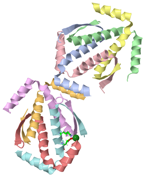
Chain A from PDB  Type:PROTEIN  Length:45
                                                                             
               SCOP domains --------------------------------------------- SCOP domains
               CATH domains --------------------------------------------- CATH domains
               Pfam domains --------------------------------------------- Pfam domains
         Sec.struct. author ...eeeeeeehhhhhhhhhhhhhhhhhhhhhhhhhhhhhhhhhhh Sec.struct. author
                 SAPs(SNPs) --------------------------------------------- SAPs(SNPs)
                    PROSITE --------------------------------------------- PROSITE
                 Transcript --------------------------------------------- Transcript
                 5hob A 351 DEDTYYLQVRGRKNFEILMELKRSLELMELVPQPLVDSYEQQQQL 395
                                   360       370       380       390     

Chain B from PDB  Type:PROTEIN  Length:45
                                                                             
               SCOP domains --------------------------------------------- SCOP domains
               CATH domains --------------------------------------------- CATH domains
               Pfam domains --------------------------------------------- Pfam domains
         Sec.struct. author .eeeeeeehhhhhhhhhhhhhhhhhhh..hhhhhhhhhhhhh... Sec.struct. author
                 SAPs(SNPs) --------------------------------------------- SAPs(SNPs)
                    PROSITE --------------------------------------------- PROSITE
                 Transcript --------------------------------------------- Transcript
                 5hob B 353 DTYYLQVRGRKNFEILMELKRSLELMELVPQPLVDSYEQQQQLLQ 397
                                   362       372       382       392     

Chain C from PDB  Type:PROTEIN  Length:46
                                                                              
               SCOP domains ---------------------------------------------- SCOP domains
               CATH domains ---------------------------------------------- CATH domains
               Pfam domains ---------------------------------------------- Pfam domains
         Sec.struct. author ..eeeeeeehhhhhhhhhhhhhhhhhhh..hhhhhhhhhhhhh... Sec.struct. author
                 SAPs(SNPs) ---------------------------------------------- SAPs(SNPs)
                    PROSITE ---------------------------------------------- PROSITE
                 Transcript ---------------------------------------------- Transcript
                 5hob C 352 EDTYYLQVRGRKNFEILMELKRSLELMELVPQPLVDSYEQQQQLLQ 397
                                   361       371       381       391      

Chain D from PDB  Type:PROTEIN  Length:44
                                                                            
               SCOP domains -------------------------------------------- SCOP domains
               CATH domains -------------------------------------------- CATH domains
               Pfam domains -------------------------------------------- Pfam domains
         Sec.struct. author ..eeeeeeehhhhhhhhhhhhhhhhhhhhhhhhhhhhhhhhhhh Sec.struct. author
                 SAPs(SNPs) -------------------------------------------- SAPs(SNPs)
                    PROSITE -------------------------------------------- PROSITE
                 Transcript -------------------------------------------- Transcript
                 5hob D 352 EDTYYLQVRGRKNFEILMELKRSLELMELVPQPLVDSYEQQQQL 395
                                   361       371       381       391    

Chain E from PDB  Type:PROTEIN  Length:48
                                                                                
               SCOP domains ------------------------------------------------ SCOP domains
               CATH domains ------------------------------------------------ CATH domains
               Pfam domains ------------------------------------------------ Pfam domains
         Sec.struct. author .....eeeeeeehhhhhhhhhhhhhhhhhhhhhhhhhhhhhhhhhhh. Sec.struct. author
                 SAPs(SNPs) ------------------------------------------------ SAPs(SNPs)
                    PROSITE ------------------------------------------------ PROSITE
                 Transcript ------------------------------------------------ Transcript
                 5hob E 349 GSDEDTYYLQVRGRKNFEILMELKRSLELMELVPQPLVDSYEQQQQLL 396
                                   358       368       378       388        

Chain F from PDB  Type:PROTEIN  Length:44
                                                                            
               SCOP domains -------------------------------------------- SCOP domains
               CATH domains -------------------------------------------- CATH domains
               Pfam domains -------------------------------------------- Pfam domains
         Sec.struct. author .eeeeeeehhhhhhhhhhhhhhhhhhh..hhhhhhhhhhhhhhh Sec.struct. author
                 SAPs(SNPs) -------------------------------------------- SAPs(SNPs)
                    PROSITE -------------------------------------------- PROSITE
                 Transcript -------------------------------------------- Transcript
                 5hob F 353 DTYYLQVRGRKNFEILMELKRSLELMELVPQPLVDSYEQQQQLL 396
                                   362       372       382       392    

Chain G from PDB  Type:PROTEIN  Length:47
                                                                               
               SCOP domains ----------------------------------------------- SCOP domains
               CATH domains ----------------------------------------------- CATH domains
               Pfam domains ----------------------------------------------- Pfam domains
         Sec.struct. author ...eeeeeeehhhhhhhhhhhhhhhhhhh..hhhhhhhhhhhhhhh. Sec.struct. author
                 SAPs(SNPs) ----------------------------------------------- SAPs(SNPs)
                    PROSITE ----------------------------------------------- PROSITE
                 Transcript ----------------------------------------------- Transcript
                 5hob G 351 DEDTYYLQVRGRKNFEILMELKRSLELMELVPQPLVDSYEQQQQLLQ 397
                                   360       370       380       390       

Chain H from PDB  Type:PROTEIN  Length:46
                                                                              
               SCOP domains ---------------------------------------------- SCOP domains
               CATH domains ---------------------------------------------- CATH domains
               Pfam domains ---------------------------------------------- Pfam domains
         Sec.struct. author .....eeeeeeehhhhhhhhhhhhhhhhhhhhhhhhhhhhhhhhhh Sec.struct. author
                 SAPs(SNPs) ---------------------------------------------- SAPs(SNPs)
                    PROSITE ---------------------------------------------- PROSITE
                 Transcript ---------------------------------------------- Transcript
                 5hob H 349 GSDEDTYYLQVRGRKNFEILMELKRSLELMELVPQPLVDSYEQQQQ 394
                                   358       368       378       388      

   Legend:   → Mismatch (orange background)
  - → Gap (green background, '-', border residues have a numbering label)
    → Modified Residue (blue background, lower-case, 'x' indicates undefined single-letter code, labelled with number + name)
  x → Chemical Group (purple background, 'x', labelled with number + name, e.g. ACE or NH2)
  extra numbering lines below/above indicate numbering irregularities and modified residue names etc., number ends below/above '|'

 Classification and Annotation

(-) SCOP Domains  (0, 0)

(no "SCOP Domain" information available for 5HOB)

(-) CATH Domains  (0, 0)

(no "CATH Domain" information available for 5HOB)

(-) Pfam Domains  (0, 0)

(no "Pfam Domain" information available for 5HOB)

(-) Gene Ontology  (62, 62)

Asymmetric Unit(hide GO term definitions)

 Visualization

(-) Interactive Views

Asymmetric Unit
  Complete Structure
    Jena3D(integrated viewing of ligand, site, SAP, PROSITE, SCOP information)
    WebMol | AstexViewer[tm]@PDBe
(Java Applets, require no local installation except for Java; loading may be slow)
    STRAP
(Java WebStart application, automatic local installation, requires Java; full application with system access!)
    RasMol
(require local installation)
    Molscript (VRML)
(requires installation of a VRML viewer; select preferred view via VRML and generate a mono or stereo PDF format file)
 
  Ligands, Modified Residues, Ions
    MG  [ RasMol | Jena3D ]  +environment [ RasMol | Jena3D ]
 
  Sites
    AC1  [ RasMol ]  +environment [ RasMol ]
 
  Cis Peptide Bonds
(no "Cis Peptide Bonds" information available for 5hob)
 
Biological Units
  Complete Structure
    Biological Unit 1  [ Jena3D ]
    Biological Unit 2  [ Jena3D ]

(-) Still Images

Jmol
  protein: cartoon or spacefill or dots and stick; nucleic acid: cartoon and stick; ligands: spacefill; active site: stick
Molscript
  protein, nucleic acid: cartoon; ligands: spacefill; active site: ball and stick

 Databases and Analysis Tools

(-) Databases

Access by PDB/NDB ID
  5hob
    Family and Domain InformationProDom | SYSTERS
    General Structural InformationGlycoscienceDB | MMDB | NDB | OCA | PDB | PDBe | PDBj | PDBsum | PDBWiki | PQS | PROTEOPEDIA
    Orientation in MembranesOPM
    Protein SurfaceSURFACE
    Secondary StructureDSSP (structure derived) | HSSP (homology derived)
    Structural GenomicsGeneCensus
    Structural NeighboursCE | VAST
    Structure ClassificationCATH | Dali | SCOP
    Validation and Original DataBMRB Data View | BMRB Restraints Grid | EDS | PROCHECK | RECOORD | WHAT_CHECK
 
Access by UniProt ID/Accession number
  P73_HUMAN | O15350
    Comparative Protein Structure ModelsModBase
    Genomic InformationEnsembl
    Protein-protein InteractionDIP
    Sequence, Family and Domain InformationInterPro | Pfam | SMART | UniProtKB/SwissProt
 
Access by Enzyme Classificator   (EC Number)
  (no 'Enzyme Classificator' available)
    General Enzyme InformationBRENDA | EC-PDB | Enzyme | IntEnz
    PathwayKEGG | MetaCyc
 
Access by Disease Identifier   (MIM ID)
  (no 'MIM ID' available)
    Disease InformationOMIM
 
Access by GenAge ID
  (no 'GenAge ID' available)
    Age Related InformationGenAge

(-) Analysis Tools

Access by PDB/NDB ID
    Domain InformationXDom
    Interatomic Contacts of Structural UnitsCSU
    Ligand-protein ContactsLPC
    Protein CavitiescastP
    Sequence and Secondary StructurePDBCartoon
    Structure AlignmentSTRAP(Java WebStart application, automatic local installation, requires Java; full application with system access!)
    Structure and Sequence BrowserSTING
 
Access by UniProt ID/Accession number
  P73_HUMAN | O15350
    Protein Disorder PredictionDisEMBL | FoldIndex | GLOBPLOT (for more information see DisProt)

 Related Entries

(-) Entries Sharing at Least One Protein Chain (UniProt ID)

UniProtKB/Swiss-Prot
        P73_HUMAN | O153501cok 1dxs 2kby 2mps 2nb1 2wqi 2wqj 2wtt 2xwc 3vd0 3vd1 3vd2 4a63 4g82 4g83 4guo 4guq 5hoc 5kbd

(-) Related Entries Specified in the PDB File

2kby 2KBY CONTAINS THE NATIVE P73 TETRAMERIZATION DOMAIN
2nb1 2NB1 CONTAINS THE P63/P73 HETERO-TETRAMERIZATION DOMAIN THAT CANNOT BE FORMED BY THIS MUTATED P73 TD
4a9z 4A9Z CONTAINS THE NATIVE P63 TETRAMERIZATION DOMAIN (TD) THAT CANNOT FORM HETERO-TETRAMERS WITH THIS MUTATED P73 TD
5hoc 5HOC CONTAINS A LATER AND OPTIMIZED VERSION OF THIS CONSTRUCT, THAT ALSO FORMS TETRAMERS AT LOW CONCENTRATIONS