Show PDB file:   
         Plain Text   HTML   (compressed file size)
QuickSearch:   
by PDB,NDB,UniProt,PROSITE Code or Search Term(s)  
(-)Asymmetric Unit
(-)Asym. Unit - sites
(-)Biological Unit 1
(-)Biol. Unit 1 - sites
collapse expand < >
Image Asymmetric Unit
Asymmetric Unit  (Jmol Viewer)
Image Asym. Unit - sites
Asym. Unit - sites  (Jmol Viewer)
Image Biological Unit 1
Biological Unit 1  (Jmol Viewer)
Image Biol. Unit 1 - sites
Biol. Unit 1 - sites  (Jmol Viewer)

(-) Description

Title :  CRYSTAL STRUCTURE OF P73 DNA-BINDING DOMAIN TETRAMER BOUND TO A FULL RESPONSE-ELEMENT
 
Authors :  A. S. Ethayathulla, H. Viadiu
Date :  20 Jul 12  (Deposition) - 16 Jan 13  (Release) - 20 Feb 13  (Revision)
Method :  X-RAY DIFFRACTION
Resolution :  3.10
Chains :  Asym. Unit :  A,B,E,F
Biol. Unit 1:  A,B,E,F  (2x)
Keywords :  Beta-Immunoglobulin Like Fold, Tumor Suppressor, Dna, Dna Binding Protein-Dna Complex (Keyword Search: [Gene Ontology, PubMed, Web (Google))
 
Reference :  A. S. Ethayathulla, H. Viadiu
Crystal Structure Of P73 Dna-Binding Domain Tetramer Bound To A Full Response-Element
J. Biol. Chem. 2013
PubMed: search

(-) Compounds

Molecule 1 - TUMOR PROTEIN P73
    ChainsA, B
    EngineeredYES
    Expression SystemESCHERICHIA COLI
    Expression System PlasmidPET28
    Expression System StrainBL21(DE3)
    Expression System Taxid469008
    Expression System Vector TypePLASMID
    FragmentUNP RESIDUES 115-312
    GeneHUMAN, P73, TP73
    Organism CommonHUMAN
    Organism ScientificHOMO SAPIENS
    Organism Taxid9606
    SynonymP53-LIKE TRANSCRIPTION FACTOR, P53-RELATED PROTEIN
 
Molecule 2 - DNA (5'-D(P*GP*AP*AP*CP*AP*TP*GP*TP*TP*C)-3')
    ChainsE, F
    EngineeredYES
    SyntheticYES

 Structural Features

(-) Chains, Units

  1234
Asymmetric Unit ABEF
Biological Unit 1 (2x)ABEF

Summary Information (see also Sequences/Alignments below)

(-) Ligands, Modified Residues, Ions  (1, 2)

Asymmetric Unit (1, 2)
No.NameCountTypeFull Name
1ZN2Ligand/IonZINC ION
Biological Unit 1 (0, 0)
No.NameCountTypeFull Name
1ZN-1Ligand/IonZINC ION

(-) Sites  (2, 2)

Asymmetric Unit (2, 2)
No.NameEvidenceResiduesDescription
1AC1SOFTWARECYS A:194 , HIS A:197 , CYS A:258 , CYS A:262BINDING SITE FOR RESIDUE ZN A 401
2AC2SOFTWARECYS B:194 , HIS B:197 , CYS B:258 , CYS B:262BINDING SITE FOR RESIDUE ZN B 401

(-) SS Bonds  (0, 0)

(no "SS Bond" information available for 4G82)

(-) Cis Peptide Bonds  (0, 0)

(no "Cis Peptide Bond" information available for 4G82)

 Sequence-Structure Mapping

(-) SAPs(SNPs)/Variants  (0, 0)

(no "SAP(SNP)/Variant" information available for 4G82)

(-) PROSITE Motifs  (0, 0)

(no "PROSITE Motif" information available for 4G82)

(-) Exons   (0, 0)

(no "Exon" information available for 4G82)

(-) Sequences/Alignments

Asymmetric Unit
   Reformat: Number of residues per line =  ('0' or empty: single-line sequence representation)
  Number of residues per labelling interval =   
  UniProt sequence: complete  aligned part    
   Show mapping: SCOP domains CATH domains Pfam domains Secondary structure (by author)
SAPs(SNPs) PROSITE motifs Exons
(details for a mapped element are shown in a popup box when the mouse pointer rests over it)
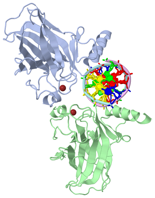
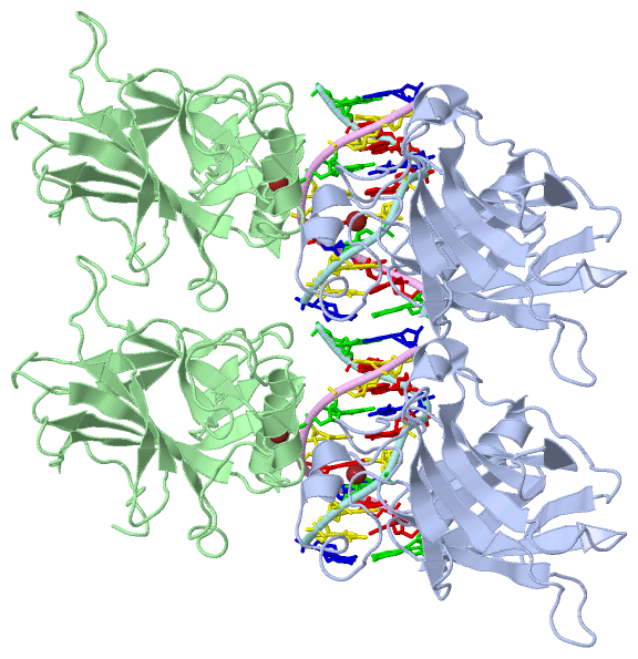
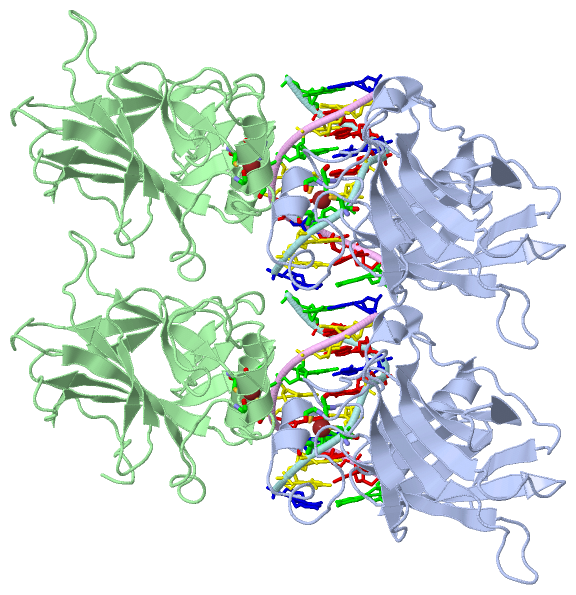
Chain A from PDB  Type:PROTEIN  Length:201
                                                                                                                                                                                                                                         
               SCOP domains d4g82a_ A: automated matches                                                                                                                                                                              SCOP domains
               CATH domains --------------------------------------------------------------------------------------------------------------------------------------------------------------------------------------------------------- CATH domains
               Pfam domains --------------------------------------------------------------------------------------------------------------------------------------------------------------------------------------------------------- Pfam domains
         Sec.struct. author ...............eeee...........eeee....eeee....eeeeeeee........eeeeeeee.hhhhhh.....hhhhhh...............eeee.....eeee......eeeeee..........eeeeeee..............eeeeeeee.....eeeeeeeeeee..hhhhhhhhhhhhhhhh Sec.struct. author
                 SAPs(SNPs) --------------------------------------------------------------------------------------------------------------------------------------------------------------------------------------------------------- SAPs(SNPs)
                    PROSITE --------------------------------------------------------------------------------------------------------------------------------------------------------------------------------------------------------- PROSITE
                 Transcript --------------------------------------------------------------------------------------------------------------------------------------------------------------------------------------------------------- Transcript
                 4g82 A 112 HEFIPSNTDYPGPHHFEVTFQQSSTAKSATWTYSPLLKKLYCQIAKTCPIQIKVSTPPPPGTAIRAMPVYKKAEHVTDVVKRCPNHELGRDFNEGQSAPASHLIRVEGNNLSQYVDDPVTGRQSVVVPYEPPQVGTEFTTILYNFMCNSSCVGGMNRRPILIIITLEMRDGQVLGRRSFEGRICACPGRDRKADEDHYREQ 312
                                   121       131       141       151       161       171       181       191       201       211       221       231       241       251       261       271       281       291       301       311 

Chain B from PDB  Type:PROTEIN  Length:200
                                                                                                                                                                                                                                        
               SCOP domains d4g82b_ B: automated matches                                                                                                                                                                             SCOP domains
               CATH domains -------------------------------------------------------------------------------------------------------------------------------------------------------------------------------------------------------- CATH domains
               Pfam domains -------------------------------------------------------------------------------------------------------------------------------------------------------------------------------------------------------- Pfam domains
         Sec.struct. author ...............eee............eee....eeee.....eeeeee.........eeeeeeee............hhhhhh...............eee......eeee......eeeeee...........eeeeee..............eeeeeeee.....eeeeeeeeeee..hhhhhhhhhhhhhhh. Sec.struct. author
                 SAPs(SNPs) -------------------------------------------------------------------------------------------------------------------------------------------------------------------------------------------------------- SAPs(SNPs)
                    PROSITE -------------------------------------------------------------------------------------------------------------------------------------------------------------------------------------------------------- PROSITE
                 Transcript -------------------------------------------------------------------------------------------------------------------------------------------------------------------------------------------------------- Transcript
                 4g82 B 113 EFIPSNTDYPGPHHFEVTFQQSSTAKSATWTYSPLLKKLYCQIAKTCPIQIKVSTPPPPGTAIRAMPVYKKAEHVTDVVKRCPNHELGRDFNEGQSAPASHLIRVEGNNLSQYVDDPVTGRQSVVVPYEPPQVGTEFTTILYNFMCNSSCVGGMNRRPILIIITLEMRDGQVLGRRSFEGRICACPGRDRKADEDHYREQ 312
                                   122       132       142       152       162       172       182       192       202       212       222       232       242       252       262       272       282       292       302       312

Chain E from PDB  Type:DNA  Length:10
                                          
                 4g82 E 400 GAACATGTTC 409
                                   409

Chain F from PDB  Type:DNA  Length:10
                                          
                 4g82 F 410 GAACATGTTC 419
                                   419

   Legend:   → Mismatch (orange background)
  - → Gap (green background, '-', border residues have a numbering label)
    → Modified Residue (blue background, lower-case, 'x' indicates undefined single-letter code, labelled with number + name)
  x → Chemical Group (purple background, 'x', labelled with number + name, e.g. ACE or NH2)
  extra numbering lines below/above indicate numbering irregularities and modified residue names etc., number ends below/above '|'

 Classification and Annotation

(-) SCOP Domains  (1, 2)

Asymmetric Unit

(-) CATH Domains  (0, 0)

(no "CATH Domain" information available for 4G82)

(-) Pfam Domains  (0, 0)

(no "Pfam Domain" information available for 4G82)

(-) Gene Ontology  (62, 62)

Asymmetric Unit(hide GO term definitions)

 Visualization

(-) Interactive Views

Asymmetric Unit
  Complete Structure
    Jena3D(integrated viewing of ligand, site, SAP, PROSITE, SCOP information)
    WebMol | AstexViewer[tm]@PDBe
(Java Applets, require no local installation except for Java; loading may be slow)
    STRAP
(Java WebStart application, automatic local installation, requires Java; full application with system access!)
    RasMol
(require local installation)
    Molscript (VRML)
(requires installation of a VRML viewer; select preferred view via VRML and generate a mono or stereo PDF format file)
 
  Ligands, Modified Residues, Ions
    ZN  [ RasMol | Jena3D ]  +environment [ RasMol | Jena3D ]
 
  Sites
    AC1  [ RasMol ]  +environment [ RasMol ]
    AC2  [ RasMol ]  +environment [ RasMol ]
 
  Cis Peptide Bonds
(no "Cis Peptide Bonds" information available for 4g82)
 
Biological Unit
  Complete Structure
    Biological Unit 1  [ Jena3D ]

(-) Still Images

Jmol
  protein: cartoon or spacefill or dots and stick; nucleic acid: cartoon and stick; ligands: spacefill; active site: stick
Molscript
  protein, nucleic acid: cartoon; ligands: spacefill; active site: ball and stick

 Databases and Analysis Tools

(-) Databases

Access by PDB/NDB ID
  4g82
    Family and Domain InformationProDom | SYSTERS
    General Structural InformationGlycoscienceDB | MMDB | NDB | OCA | PDB | PDBe | PDBj | PDBsum | PDBWiki | PQS | PROTEOPEDIA
    Orientation in MembranesOPM
    Protein SurfaceSURFACE
    Secondary StructureDSSP (structure derived) | HSSP (homology derived)
    Structural GenomicsGeneCensus
    Structural NeighboursCE | VAST
    Structure ClassificationCATH | Dali | SCOP
    Validation and Original DataBMRB Data View | BMRB Restraints Grid | EDS | PROCHECK | RECOORD | WHAT_CHECK
 
Access by UniProt ID/Accession number
  P73_HUMAN | O15350
    Comparative Protein Structure ModelsModBase
    Genomic InformationEnsembl
    Protein-protein InteractionDIP
    Sequence, Family and Domain InformationInterPro | Pfam | SMART | UniProtKB/SwissProt
 
Access by Enzyme Classificator   (EC Number)
  (no 'Enzyme Classificator' available)
    General Enzyme InformationBRENDA | EC-PDB | Enzyme | IntEnz
    PathwayKEGG | MetaCyc
 
Access by Disease Identifier   (MIM ID)
  (no 'MIM ID' available)
    Disease InformationOMIM
 
Access by GenAge ID
  (no 'GenAge ID' available)
    Age Related InformationGenAge

(-) Analysis Tools

Access by PDB/NDB ID
    Domain InformationXDom
    Interatomic Contacts of Structural UnitsCSU
    Ligand-protein ContactsLPC
    Protein CavitiescastP
    Sequence and Secondary StructurePDBCartoon
    Structure AlignmentSTRAP(Java WebStart application, automatic local installation, requires Java; full application with system access!)
    Structure and Sequence BrowserSTING
 
Access by UniProt ID/Accession number
  P73_HUMAN | O15350
    Protein Disorder PredictionDisEMBL | FoldIndex | GLOBPLOT (for more information see DisProt)

 Related Entries

(-) Entries Sharing at Least One Protein Chain (UniProt ID)

UniProtKB/Swiss-Prot
        P73_HUMAN | O153501cok 1dxs 2kby 2mps 2nb1 2wqi 2wqj 2wtt 2xwc 3vd0 3vd1 3vd2 4a63 4g83 4guo 4guq 5hob 5hoc 5kbd

(-) Related Entries Specified in the PDB File

3vd0 STRUCTURE OF P73 DNA-BINDING DOMAIN TETRAMER MODULATES P73 TRANSACTIVATION.
3vd1 STRUCTURE OF P73 DNA-BINDING DOMAIN TETRAMER MODULATES P73 TRANSACTIVATION.
3vd2 STRUCTURE OF P73 DNA-BINDING DOMAIN TETRAMER MODULATES P73 TRANSACTIVATION.