|
|
|
|
 Description
|
|
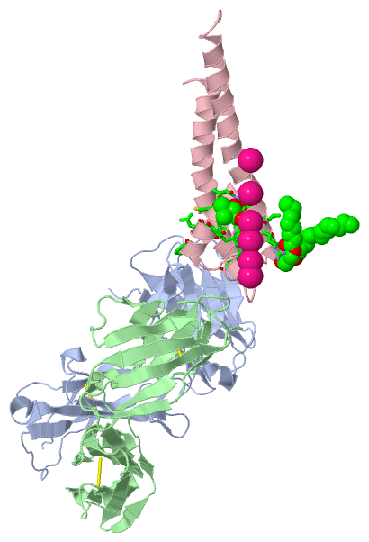
Compounds
|
||||||||||||||||||||||||||||||||||||||||||||||||||||||||||||||||||||||||||||||||||||||||||||||||||||||||||||||||||||||||||||||||
Chains, Units
Summary Information (see also Sequences/Alignments below) |
Ligands, Modified Residues, Ions (4, 9)| Asymmetric Unit (4, 9) Biological Unit 1 (3, 12) |
Sites (8, 8)
Asymmetric Unit (8, 8)
|
SS Bonds (4, 4)
Asymmetric Unit
|
||||||||||||||||||||
Cis Peptide Bonds (6, 6)
Asymmetric Unit
|
||||||||||||||||||||||||||||
SAPs(SNPs)/Variants (0, 0)| (no "SAP(SNP)/Variant" information available for 5EC1) |
PROSITE Motifs (0, 0)| (no "PROSITE Motif" information available for 5EC1) |
Exons (0, 0)| (no "Exon" information available for 5EC1) |
Sequences/Alignments
Asymmetric Unit
Chain A from PDB Type:PROTEIN Length:219
SCOP domains --------------------------------------------------------------------------------------------------------------------------------------------------------------------------------------------------------------------------- SCOP domains
CATH domains --------------------------------------------------------------------------------------------------------------------------------------------------------------------------------------------------------------------------- CATH domains
Pfam domains --------------------------------------------------------------------------------------------------------------------------------------------------------------------------------------------------------------------------- Pfam domains
SAPs(SNPs) --------------------------------------------------------------------------------------------------------------------------------------------------------------------------------------------------------------------------- SAPs(SNPs)
PROSITE --------------------------------------------------------------------------------------------------------------------------------------------------------------------------------------------------------------------------- PROSITE
Transcript --------------------------------------------------------------------------------------------------------------------------------------------------------------------------------------------------------------------------- Transcript
5ec1 A 1 QVQLQQPGAELVKPGASVKLSCKASGYTFTSDWIHWVKQRPGHGLEWIGEIIPSYGRANYNEKIQKKATLTADKSSSTAFMQLSSLTSEDSAVYYCARERGDGYFAVWGAGTTVTVSSAKTTPPSVYPLAPGSAAQTNSMVTLGCLVKGYFPEPVTVTWNSGSLSSGVHTFPAVLQSDLYTLSSSVTVPSSSWPSETVTCNVAHPASSTKVDKKIVPRD 219
10 20 30 40 50 60 70 80 90 100 110 120 130 140 150 160 170 180 190 200 210
Chain B from PDB Type:PROTEIN Length:209
SCOP domains ----------------------------------------------------------------------------------------------------------------------------------------------------------------------------------------------------------------- SCOP domains
CATH domains ----------------------------------------------------------------------------------------------------------------------------------------------------------------------------------------------------------------- CATH domains
Pfam domains ----------------------------------------------------------------------------------------------------------------------------------------------------------------------------------------------------------------- Pfam domains
SAPs(SNPs) ----------------------------------------------------------------------------------------------------------------------------------------------------------------------------------------------------------------- SAPs(SNPs)
PROSITE ----------------------------------------------------------------------------------------------------------------------------------------------------------------------------------------------------------------- PROSITE
Transcript ----------------------------------------------------------------------------------------------------------------------------------------------------------------------------------------------------------------- Transcript
5ec1 B 1 DILLTQSPAILSVSPGERVSFSCRASQSIGTDIHWYQQRTNGSPRLLIKYASESISGIPSRFSGSGSGTDFTLSINSVESEDIANYYCQQSNRWPFTFGSGTKLEIKRADAAPTVSIFPPSSEQLTSGGASVVCFLNNFYPKDINVKWKIDGSERQNGVLNSWTDQDSKDSTYSMSSTLTLTKDEYERHNSYTCEATHKTSTSPIVKSF 209
10 20 30 40 50 60 70 80 90 100 110 120 130 140 150 160 170 180 190 200
Chain C from PDB Type:PROTEIN Length:103
SCOP domains ------------------------------------------------------------------------------------------------------- SCOP domains
CATH domains ------------------------------------------------------------------------------------------------------- CATH domains
Pfam domains ------------------------------------------------------------------------------------------------------- Pfam domains
SAPs(SNPs) ------------------------------------------------------------------------------------------------------- SAPs(SNPs)
PROSITE ------------------------------------------------------------------------------------------------------- PROSITE
Transcript ------------------------------------------------------------------------------------------------------- Transcript
5ec1 C 22 SALHWRAAGAATVLLVIVLLAGSYLAVLAERGAPGAQLITYPRALWWACETATTxGYGDLCPVTLWGRLVAVVVMVAGITSFGLVTAALATWFVGREQERRGH 124
31 41 51 61 71 | 81 91 101 111 121
76-LHV
|
||||||||||||||||||||
SCOP Domains (0, 0)| (no "SCOP Domain" information available for 5EC1) |
CATH Domains (0, 0)| (no "CATH Domain" information available for 5EC1) |
Pfam Domains (0, 0)| (no "Pfam Domain" information available for 5EC1) |
Gene Ontology (10, 10)|
Asymmetric Unit(hide GO term definitions) |
Interactive Views
|
||||||||||||||||||||||||||||||||||||||||||||||||||||||||||||||||||||||||||||||||||||||||||||||||||||||||||||||||||||||||||||||||||||||||||||||||||||||||||||||||||||||||||||||||||||||||||||||||||||||||||||||||||||||||||||||||||||||||||||||||||
Still Images
|
||||||||||||||||
Databases
|
||||||||||||||||||||||||||||||||||||||||||||||||||||||||||||||||||||||||||||||||||||||||||||||||||||||||||||||||||||||||||||||||||||||||||||||||||||||||||||||||
Analysis Tools
|
|||||||||||||||||||||||||||||||||||||||||||||||||||||||||||||
Entries Sharing at Least One Protein Chain (UniProt ID)
Related Entries Specified in the PDB File
|
|