Show PDB file:   
         Plain Text   HTML   (compressed file size)
QuickSearch:   
by PDB,NDB,UniProt,PROSITE Code or Search Term(s)  
(-)Asym./Biol. Unit
(-)Asym./Biol. Unit - sites
collapse expand < >
Image Asym./Biol. Unit
Asym./Biol. Unit  (Jmol Viewer)
Image Asym./Biol. Unit - sites
Asym./Biol. Unit - sites  (Jmol Viewer)

(-) Description

Title :  ANTI-TAU AT8 FAB WITH DOUBLY PHOSPHORYLATED TAU PEPTIDE
 
Authors :  T. Malia, A. Teplyakov
Date :  01 Oct 15  (Deposition) - 24 Feb 16  (Release) - 30 Mar 16  (Revision)
Method :  X-RAY DIFFRACTION
Resolution :  1.64
Chains :  Asym./Biol. Unit :  H,L,P
Keywords :  Immune System (Keyword Search: [Gene Ontology, PubMed, Web (Google))
 
Reference :  T. J. Malia, A. Teplyakov, R. Ernst, S. J. Wu, E. R. Lacy, X. Liu, M. Vandermeeren, M. Mercken, J. Luo, R. W. Sweet, G. L. Gilliland
Epitope Mapping And Structural Basis For The Recognition Of Phosphorylated Tau By The Anti-Tau Antibody At8.
Proteins V. 84 427 2016
PubMed-ID: 26800003  |  Reference-DOI: 10.1002/PROT.24988

(-) Compounds

Molecule 1 - AT8 LIGHT CHAIN
    ChainsL
    EngineeredYES
    Expression SystemHOMO SAPIENS
    Expression System Taxid9606
    Organism ScientificMUS MUSCULUS
    Organism Taxid10090
 
Molecule 2 - AT8 HEAVY CHAIN
    ChainsH
    EngineeredYES
    Expression SystemHOMO SAPIENS
    Expression System Taxid9606
    Organism ScientificMUS MUSCULUS
    Organism Taxid10090
 
Molecule 3 - TAU PHOSPHOPEPTIDE
    ChainsP
    EngineeredYES
    Organism ScientificHOMO SAPIENS
    Organism Taxid9606
    SyntheticYES

 Structural Features

(-) Chains, Units

  123
Asymmetric/Biological Unit HLP

Summary Information (see also Sequences/Alignments below)

(-) Ligands, Modified Residues, Ions  (3, 4)

Asymmetric/Biological Unit (3, 4)
No.NameCountTypeFull Name
1GOL2Ligand/IonGLYCEROL
2SEP1Mod. Amino AcidPHOSPHOSERINE
3TPO1Mod. Amino AcidPHOSPHOTHREONINE

(-) Sites  (2, 2)

Asymmetric Unit (2, 2)
No.NameEvidenceResiduesDescription
1AC1SOFTWARESER L:72 , GLY L:73 , THR L:74 , GLY L:133 , LYS L:188 , ALA L:189 , HOH L:490 , HOH L:590binding site for residue GOL L 301
2AC2SOFTWARETYR L:39 , HIS L:54 , ARG L:55 , TPO P:205 , HOH P:1102 , HOH P:1103 , HOH P:1108binding site for residue GOL P 1001

(-) SS Bonds  (4, 4)

Asymmetric/Biological Unit
No.Residues
1H:22 -H:96
2H:140 -H:196
3L:23 -L:93
4L:139 -L:199

(-) Cis Peptide Bonds  (4, 4)

Asymmetric/Biological Unit
No.Residues
1Tyr L:99 -Pro L:100
2Tyr L:145 -Pro L:146
3Phe H:146 -Pro H:147
4Glu H:148 -Pro H:149

 Sequence-Structure Mapping

(-) SAPs(SNPs)/Variants  (0, 0)

(no "SAP(SNP)/Variant" information available for 5E2V)

(-) PROSITE Motifs  (0, 0)

(no "PROSITE Motif" information available for 5E2V)

(-) Exons   (0, 0)

(no "Exon" information available for 5E2V)

(-) Sequences/Alignments

Asymmetric/Biological Unit
   Reformat: Number of residues per line =  ('0' or empty: single-line sequence representation)
  Number of residues per labelling interval =   
  UniProt sequence: complete  aligned part    
   Show mapping: SCOP domains CATH domains Pfam domains Secondary structure (by author)
SAPs(SNPs) PROSITE motifs Exons
(details for a mapped element are shown in a popup box when the mouse pointer rests over it)
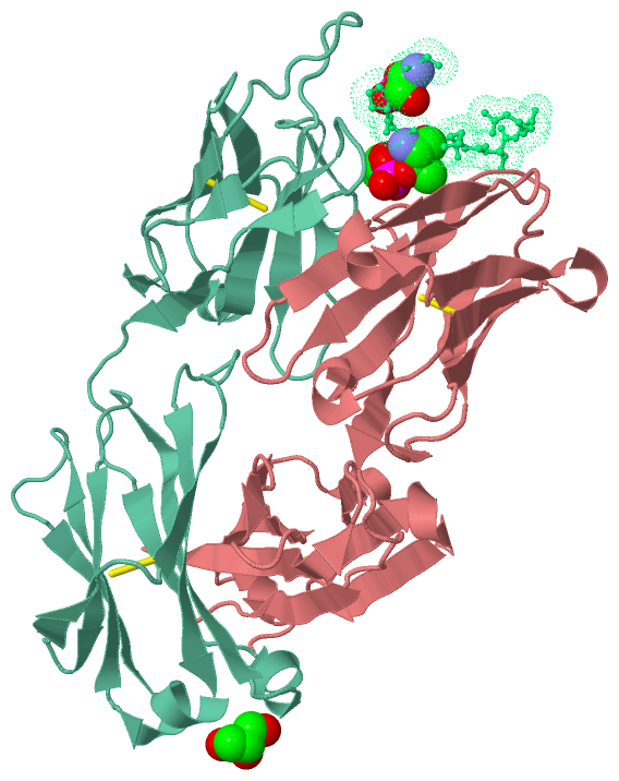
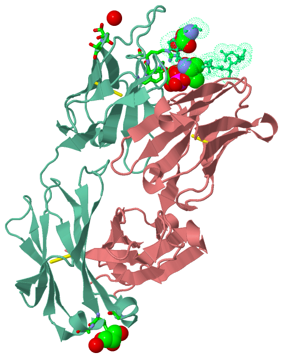
Chain H from PDB  Type:PROTEIN  Length:208
                                                                                                                                                                                                                                                
               SCOP domains ---------------------------------------------------------------------------------------------------------------------------------------------------------------------------------------------------------------- SCOP domains
               CATH domains ---------------------------------------------------------------------------------------------------------------------------------------------------------------------------------------------------------------- CATH domains
               Pfam domains ---------------------------------------------------------------------------------------------------------------------------------------------------------------------------------------------------------------- Pfam domains
         Sec.struct. author ..eeeee...ee.....eeeeeeee........eeeeeee.....eeeeeeee....eee.hhhh..eeeeeehhh.eeeeee...hhhhheeeeeee...ee...eeeee........eeeee.....eeeeeeeeeee.....eeee.hhh....eee...ee.....eeeeeeeeee.hhh.....eeeeeehhhheeeeee... Sec.struct. author
                 SAPs(SNPs) ---------------------------------------------------------------------------------------------------------------------------------------------------------------------------------------------------------------- SAPs(SNPs)
                    PROSITE ---------------------------------------------------------------------------------------------------------------------------------------------------------------------------------------------------------------- PROSITE
                 Transcript ---------------------------------------------------------------------------------------------------------------------------------------------------------------------------------------------------------------- Transcript
                 5e2v H   1 DVQLQESGPGLVKPSQSLSLTCSVTDYSITSGYYWNWIRQFPGNKLEWMGYISYDGSNNYNPSLKNRISITRDPSKDQFFLNLNSVTTEDTATYYCTRGSLVWGQGTLVTVSAASTKGPSVFPLAPSGGTAALGCLVKDYFPEPVTVSWNSGALTSGVHTFPAVLQSSGLYSLSSVVTVPSSSLGTQTYICNVNHKPSNTKVDKKVEP 213
                                    10        20        30        40        50        60        70        80        90       100       110       120      |135       145       155       165       175       185       195       205        
                                                                                                                                                        127|                                                                                
                                                                                                                                                         133                                                                                

Chain L from PDB  Type:PROTEIN  Length:216
                                                                                                                                                                                                                                                        
               SCOP domains ------------------------------------------------------------------------------------------------------------------------------------------------------------------------------------------------------------------------ SCOP domains
               CATH domains ------------------------------------------------------------------------------------------------------------------------------------------------------------------------------------------------------------------------ CATH domains
               Pfam domains ------------------------------------------------------------------------------------------------------------------------------------------------------------------------------------------------------------------------ Pfam domains
         Sec.struct. author ...ee....eeee.....eeeeeee............eeeeee......eeeee...ee.......eeeeee..eeeeee...hhhhheeeeeee......ee...eeeee.......eeeee..hhhhhh..eeeeeeeeeee.....eeeeee..ee....eeeee.........eeeeeeeeeehhhhhh..eeeeeee.......eeeeee. Sec.struct. author
                 SAPs(SNPs) ------------------------------------------------------------------------------------------------------------------------------------------------------------------------------------------------------------------------ SAPs(SNPs)
                    PROSITE ------------------------------------------------------------------------------------------------------------------------------------------------------------------------------------------------------------------------ PROSITE
                 Transcript ------------------------------------------------------------------------------------------------------------------------------------------------------------------------------------------------------------------------ Transcript
                 5e2v L   1 DIVMTQAAPSVPVTPGESVSISCRSSKSLLHSNGNTYLYWFLQRPGQSPQLLIHRMSNLASGVPDRFSGSGSGTAFTLRISRVEAEDVGVYYCMQHLEYPYTFGGGTRLEVKRTVAAPSVFIFPPSDEQLKSGTASVVCLLNNFYPREAKVQWKVDNALQSGNSQESVTEQDSKDSTYSLSSTLTLSKADYEKHKVYACEVTHQGLSSPVTKSFNR 216
                                    10        20        30        40        50        60        70        80        90       100       110       120       130       140       150       160       170       180       190       200       210      

Chain P from PDB  Type:PROTEIN  Length:9
                                         
               SCOP domains --------- SCOP domains
               CATH domains --------- CATH domains
               Pfam domains --------- Pfam domains
         Sec.struct. author ......... Sec.struct. author
                 SAPs(SNPs) --------- SAPs(SNPs)
                    PROSITE --------- PROSITE
                 Transcript --------- Transcript
                 5e2v P 201 GsPGtPGSR 209
                             |  |    
                           202-SEP   
                              205-TPO

   Legend:   → Mismatch (orange background)
  - → Gap (green background, '-', border residues have a numbering label)
    → Modified Residue (blue background, lower-case, 'x' indicates undefined single-letter code, labelled with number + name)
  x → Chemical Group (purple background, 'x', labelled with number + name, e.g. ACE or NH2)
  extra numbering lines below/above indicate numbering irregularities and modified residue names etc., number ends below/above '|'

 Classification and Annotation

(-) SCOP Domains  (0, 0)

(no "SCOP Domain" information available for 5E2V)

(-) CATH Domains  (0, 0)

(no "CATH Domain" information available for 5E2V)

(-) Pfam Domains  (0, 0)

(no "Pfam Domain" information available for 5E2V)

(-) Gene Ontology  (53, 53)

Asymmetric/Biological Unit(hide GO term definitions)

 Visualization

(-) Interactive Views

Asymmetric/Biological Unit
  Complete Structure
    Jena3D(integrated viewing of ligand, site, SAP, PROSITE, SCOP information)
    WebMol | AstexViewer[tm]@PDBe
(Java Applets, require no local installation except for Java; loading may be slow)
    STRAP
(Java WebStart application, automatic local installation, requires Java; full application with system access!)
    RasMol
(require local installation)
    Molscript (VRML)
(requires installation of a VRML viewer; select preferred view via VRML and generate a mono or stereo PDF format file)
 
  Ligands, Modified Residues, Ions
    GOL  [ RasMol | Jena3D ]  +environment [ RasMol | Jena3D ]
    SEP  [ RasMol | Jena3D ]  +environment [ RasMol | Jena3D ]
    TPO  [ RasMol | Jena3D ]  +environment [ RasMol | Jena3D ]
 
  Sites
    AC1  [ RasMol ]  +environment [ RasMol ]
    AC2  [ RasMol ]  +environment [ RasMol ]
 
  Cis Peptide Bonds
    Glu H:148 - Pro H:149   [ RasMol ]  
    Phe H:146 - Pro H:147   [ RasMol ]  
    Tyr L:145 - Pro L:146   [ RasMol ]  
    Tyr L:99 - Pro L:100   [ RasMol ]  
 

(-) Still Images

Jmol
  protein: cartoon or spacefill or dots and stick; nucleic acid: cartoon and stick; ligands: spacefill; active site: stick
Molscript
  protein, nucleic acid: cartoon; ligands: spacefill; active site: ball and stick

 Databases and Analysis Tools

(-) Databases

Access by PDB/NDB ID
  5e2v
    Family and Domain InformationProDom | SYSTERS
    General Structural InformationGlycoscienceDB | MMDB | NDB | OCA | PDB | PDBe | PDBj | PDBsum | PDBWiki | PQS | PROTEOPEDIA
    Orientation in MembranesOPM
    Protein SurfaceSURFACE
    Secondary StructureDSSP (structure derived) | HSSP (homology derived)
    Structural GenomicsGeneCensus
    Structural NeighboursCE | VAST
    Structure ClassificationCATH | Dali | SCOP
    Validation and Original DataBMRB Data View | BMRB Restraints Grid | EDS | PROCHECK | RECOORD | WHAT_CHECK
 
Access by UniProt ID/Accession number
  TAU_HUMAN | P10636
    Comparative Protein Structure ModelsModBase
    Genomic InformationEnsembl
    Protein-protein InteractionDIP
    Sequence, Family and Domain InformationInterPro | Pfam | SMART | UniProtKB/SwissProt
 
Access by Enzyme Classificator   (EC Number)
  (no 'Enzyme Classificator' available)
    General Enzyme InformationBRENDA | EC-PDB | Enzyme | IntEnz
    PathwayKEGG | MetaCyc
 
Access by Disease Identifier   (MIM ID)
  (no 'MIM ID' available)
    Disease InformationOMIM
 
Access by GenAge ID
  (no 'GenAge ID' available)
    Age Related InformationGenAge

(-) Analysis Tools

Access by PDB/NDB ID
    Domain InformationXDom
    Interatomic Contacts of Structural UnitsCSU
    Ligand-protein ContactsLPC
    Protein CavitiescastP
    Sequence and Secondary StructurePDBCartoon
    Structure AlignmentSTRAP(Java WebStart application, automatic local installation, requires Java; full application with system access!)
    Structure and Sequence BrowserSTING
 
Access by UniProt ID/Accession number
  TAU_HUMAN | P10636
    Protein Disorder PredictionDisEMBL | FoldIndex | GLOBPLOT (for more information see DisProt)

 Related Entries

(-) Entries Sharing at Least One Protein Chain (UniProt ID)

UniProtKB/Swiss-Prot
        TAU_HUMAN | P106361i8h 2mz7 2on9 3ovl 4e0m 4e0n 4e0o 4fl5 4glr 4np8 4tqe 4y32 4y3b 4y5i 5dmg 5e2w 5hf3 5k7n

(-) Related Entries Specified in the PDB File

5e2t 5e2u 5e2w