Show PDB file:   
         Plain Text   HTML   (compressed file size)
QuickSearch:   
by PDB,NDB,UniProt,PROSITE Code or Search Term(s)  
(-)Asym./Biol. Unit
(-)Asym./Biol. Unit - sites
collapse expand < >
Image Asym./Biol. Unit
Asym./Biol. Unit  (Jmol Viewer)
Image Asym./Biol. Unit - sites
Asym./Biol. Unit - sites  (Jmol Viewer)

(-) Description

Title :  GTB MUTANT WITH MERCURY - E303C
 
Authors :  S. M. L. Gagnon, R. J. Blackler
Date :  22 Jul 15  (Deposition) - 26 Oct 16  (Release) - 19 Apr 17  (Revision)
Method :  X-RAY DIFFRACTION
Resolution :  1.65
Chains :  Asym./Biol. Unit :  X
Keywords :  Human Abo(H) Blood Group System, Glycosyltransferase, Double Turn Motif, Catalytic Domain, Transferase (Keyword Search: [Gene Ontology, PubMed, Web (Google))
 
Reference :  R. J. Blackler, S. M. Gagnon, R. Polakowski, N. L. Rose, R. B. Zheng, J. A. Letts, A. R. Johal, B. Schuman, S. N. Borisova, M. M. Palcic, S. V. Evans
Glycosyltransfer In Mutants Of Putative Catalytic Residue Glu303 Of The Human Abo(H) A And B Blood Group Glycosyltransferases Gta And Gtb Proceeds Through A Labile Active Site.
Glycobiology V. 27 370 2017
PubMed-ID: 27979997  |  Reference-DOI: 10.1093/GLYCOB/CWW117

(-) Compounds

Molecule 1 - HISTO-BLOOD GROUP ABO SYSTEM TRANSFERASE
    ChainsX
    EC Number2.4.1.40, 2.4.1.37
    EngineeredYES
    Expression SystemESCHERICHIA COLI
    Expression System Taxid562
    FragmentCATALYTIC DOMAIN (UNP RESIDUES 64-354)
    GeneABO
    MutationYES
    Organism CommonHUMAN
    Organism ScientificHOMO SAPIENS
    Organism Taxid9606
    SynonymFUCOSYLGLYCOPROTEIN 3-ALPHA-GALACTOSYLTRANSFERASE, FUCOSYLGLYCOPROTEIN ALPHA-N-ACETYLGALACTOSAMINYLTRANSFERASE, GLYCOPROTEIN-FUCOSYLGALACTOSIDE ALPHA-N- ACETYLGALACTOSAMINYLTRANSFERASE,GLYCOPROTEIN-FUCOSYLGALACTOSIDE ALPHA-GALACTOSYLTRANSFERASE,HISTO-BLOOD GROUP A TRANSFERASE,A TRANSFERASE,HISTO-BLOOD GROUP B TRANSFERASE,B TRANSFERASE,NAGAT

 Structural Features

(-) Chains, Units

  1
Asymmetric/Biological Unit X

Summary Information (see also Sequences/Alignments below)

(-) Ligands, Modified Residues, Ions  (1, 5)

Asymmetric/Biological Unit (1, 5)
No.NameCountTypeFull Name
1HG5Ligand/IonMERCURY (II) ION

(-) Sites  (5, 5)

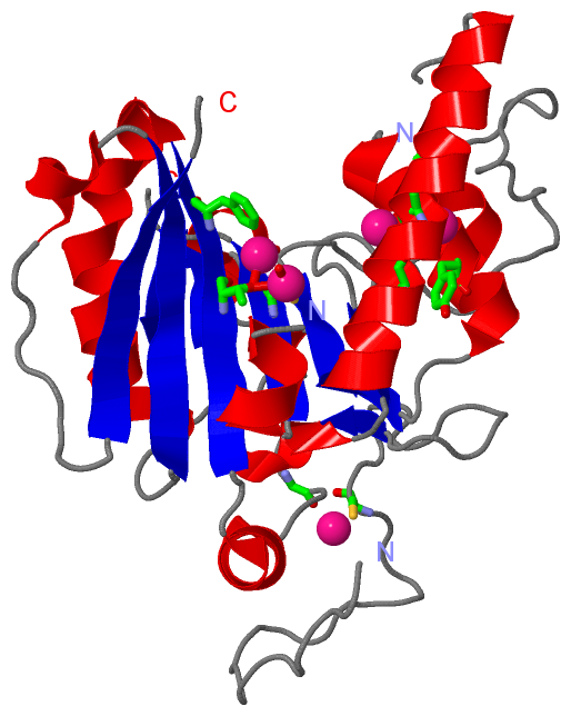
Asymmetric Unit (5, 5)
No.NameEvidenceResiduesDescription
1AC1SOFTWARETHR X:119 , CYS X:209 , HG X:405 , HOH X:680binding site for residue HG X 401
2AC2SOFTWARELEU X:280 , CYS X:284 , HOH X:676binding site for residue HG X 402
3AC3SOFTWARECYS X:284 , HIS X:305 , TYR X:309 , HOH X:694binding site for residue HG X 403
4AC4SOFTWARECYS X:80 , GLY X:98binding site for residue HG X 404
5AC5SOFTWARETHR X:119 , PHE X:121 , HG X:401 , HOH X:684binding site for residue HG X 405

(-) SS Bonds  (0, 0)

(no "SS Bond" information available for 5CQM)

(-) Cis Peptide Bonds  (0, 0)

(no "Cis Peptide Bond" information available for 5CQM)

 Sequence-Structure Mapping

(-) SAPs(SNPs)/Variants  (0, 0)

(no "SAP(SNP)/Variant" information available for 5CQM)

(-) PROSITE Motifs  (0, 0)

(no "PROSITE Motif" information available for 5CQM)

(-) Exons   (0, 0)

(no "Exon" information available for 5CQM)

(-) Sequences/Alignments

Asymmetric/Biological Unit
   Reformat: Number of residues per line =  ('0' or empty: single-line sequence representation)
  Number of residues per labelling interval =   
  UniProt sequence: complete  aligned part    
   Show mapping: SCOP domains CATH domains Pfam domains Secondary structure (by author)
SAPs(SNPs) PROSITE motifs Exons
(details for a mapped element are shown in a popup box when the mouse pointer rests over it)
Chain X from PDB  Type:PROTEIN  Length:262
                                                                                                                                                                                                                                                                                                      
               SCOP domains ---------------------------------------------------------------------------------------------------------------------------------------------------------------------------------------------------------------------------------------------------------------------- SCOP domains
               CATH domains ---------------------------------------------------------------------------------------------------------------------------------------------------------------------------------------------------------------------------------------------------------------------- CATH domains
               Pfam domains ---------------------------------------------------------------------------------------------------------------------------------------------------------------------------------------------------------------------------------------------------------------------- Pfam domains
         Sec.struct. author .................................ee.....hhhhhhhhhhhh..eeeeeeeehhhhhhhhhhhhhhhhhhh....eeeeeeee.hhhhh........eeeeeee...hhhhhh.eeeee...eee....hhhhh..eeee........hhhhh........................eeeeehhhhhhhhhhhhhhhhhhhhh....hhhhhhhhhhhhh...eee.hhhh.hhhhhh.........eee.. Sec.struct. author
                 SAPs(SNPs) ---------------------------------------------------------------------------------------------------------------------------------------------------------------------------------------------------------------------------------------------------------------------- SAPs(SNPs)
                    PROSITE ---------------------------------------------------------------------------------------------------------------------------------------------------------------------------------------------------------------------------------------------------------------------- PROSITE
                 Transcript ---------------------------------------------------------------------------------------------------------------------------------------------------------------------------------------------------------------------------------------------------------------------- Transcript
                 5cqm X  61 EFMVSLPRMVYPQPKVLTPCRKDVLVVTPWLAPIVWEGTFNIDILNEQFRLQNTTIGLTVFAIKKYVAFLKLFLETAEKHFMVGHRVHYYVFTDQPAAVPRVTLGTGRQLSVLEVGRRFLSEVDYLVCVDVDMEFRDHVGVEILTPLFGTLHPSFYGSSREAFTYERRPQSQAYIPKDEGDFYYMGAFFGGSVQEVQRLTRACHQAMMVDQANGIEWHDCSHLNKYLLRHKPTKVLSPEYLWDQQLLGWPAVLRKLRFTAVP 345
                                    70        80        90       100       110       120       130       140       150       160       170     ||201       211       221       231       241       251       261       271       281       291     ||303       313       323       333       343  
                                                                                                                                             176|                                                                                                297|                                             
                                                                                                                                              198                                                                                                 300                                             

   Legend:   → Mismatch (orange background)
  - → Gap (green background, '-', border residues have a numbering label)
    → Modified Residue (blue background, lower-case, 'x' indicates undefined single-letter code, labelled with number + name)
  x → Chemical Group (purple background, 'x', labelled with number + name, e.g. ACE or NH2)
  extra numbering lines below/above indicate numbering irregularities and modified residue names etc., number ends below/above '|'

 Classification and Annotation

(-) SCOP Domains  (0, 0)

(no "SCOP Domain" information available for 5CQM)

(-) CATH Domains  (0, 0)

(no "CATH Domain" information available for 5CQM)

(-) Pfam Domains  (0, 0)

(no "Pfam Domain" information available for 5CQM)

(-) Gene Ontology  (13, 13)

Asymmetric/Biological Unit(hide GO term definitions)

 Visualization

(-) Interactive Views

Asymmetric/Biological Unit
  Complete Structure
    Jena3D(integrated viewing of ligand, site, SAP, PROSITE, SCOP information)
    WebMol | AstexViewer[tm]@PDBe
(Java Applets, require no local installation except for Java; loading may be slow)
    STRAP
(Java WebStart application, automatic local installation, requires Java; full application with system access!)
    RasMol
(require local installation)
    Molscript (VRML)
(requires installation of a VRML viewer; select preferred view via VRML and generate a mono or stereo PDF format file)
 
  Ligands, Modified Residues, Ions
    HG  [ RasMol | Jena3D ]  +environment [ RasMol | Jena3D ]
 
  Sites
    AC1  [ RasMol ]  +environment [ RasMol ]
    AC2  [ RasMol ]  +environment [ RasMol ]
    AC3  [ RasMol ]  +environment [ RasMol ]
    AC4  [ RasMol ]  +environment [ RasMol ]
    AC5  [ RasMol ]  +environment [ RasMol ]
 
  Cis Peptide Bonds
(no "Cis Peptide Bonds" information available for 5cqm)
 

(-) Still Images

Jmol
  protein: cartoon or spacefill or dots and stick; nucleic acid: cartoon and stick; ligands: spacefill; active site: stick
Molscript
  protein, nucleic acid: cartoon; ligands: spacefill; active site: ball and stick

 Databases and Analysis Tools

(-) Databases

Access by PDB/NDB ID
  5cqm
    Family and Domain InformationProDom | SYSTERS
    General Structural InformationGlycoscienceDB | MMDB | NDB | OCA | PDB | PDBe | PDBj | PDBsum | PDBWiki | PQS | PROTEOPEDIA
    Orientation in MembranesOPM
    Protein SurfaceSURFACE
    Secondary StructureDSSP (structure derived) | HSSP (homology derived)
    Structural GenomicsGeneCensus
    Structural NeighboursCE | VAST
    Structure ClassificationCATH | Dali | SCOP
    Validation and Original DataBMRB Data View | BMRB Restraints Grid | EDS | PROCHECK | RECOORD | WHAT_CHECK
 
Access by UniProt ID/Accession number
  BGAT_HUMAN | P16442
    Comparative Protein Structure ModelsModBase
    Genomic InformationEnsembl
    Protein-protein InteractionDIP
    Sequence, Family and Domain InformationInterPro | Pfam | SMART | UniProtKB/SwissProt
 
Access by Enzyme Classificator   (EC Number)
  2.4.1.37
    General Enzyme InformationBRENDA | EC-PDB | Enzyme | IntEnz
    PathwayKEGG | MetaCyc
  2.4.1.40
    General Enzyme InformationBRENDA | EC-PDB | Enzyme | IntEnz
    PathwayKEGG | MetaCyc
 
Access by Disease Identifier   (MIM ID)
  (no 'MIM ID' available)
    Disease InformationOMIM
 
Access by GenAge ID
  (no 'GenAge ID' available)
    Age Related InformationGenAge

(-) Analysis Tools

Access by PDB/NDB ID
    Domain InformationXDom
    Interatomic Contacts of Structural UnitsCSU
    Ligand-protein ContactsLPC
    Protein CavitiescastP
    Sequence and Secondary StructurePDBCartoon
    Structure AlignmentSTRAP(Java WebStart application, automatic local installation, requires Java; full application with system access!)
    Structure and Sequence BrowserSTING
 
Access by UniProt ID/Accession number
  BGAT_HUMAN | P16442
    Protein Disorder PredictionDisEMBL | FoldIndex | GLOBPLOT (for more information see DisProt)

 Related Entries

(-) Entries Sharing at Least One Protein Chain (UniProt ID)

UniProtKB/Swiss-Prot
        BGAT_HUMAN | P164421lz0 1lz7 1lzi 1lzj 1r7t 1r7u 1r7v 1r7x 1r7y 1r80 1r81 1r82 1wsz 1wt0 1wt1 1wt2 1wt3 1xz6 1zhj 1zi1 1zi3 1zi4 1zi5 1ziz 1zj0 1zj1 1zj2 1zj3 1zjo 1zjp 2a8u 2a8w 2i7b 2o1f 2o1g 2o1h 2pgv 2pgy 2rit 2rix 2riy 2riz 2rj0 2rj1 2rj4 2rj5 2rj6 2rj7 2rj8 2rj9 2y7a 3i0c 3i0d 3i0e 3i0f 3i0g 3i0h 3i0i 3i0j 3i0k 3i0l 3ioh 3ioi 3ioj 3sx3 3sx5 3sx7 3sx8 3sxa 3sxb 3sxc 3sxd 3sxe 3sxg 3u0x 3u0y 3v0l 3v0m 3v0n 3v0o 3v0p 3v0q 3zgf 3zgg 4c2s 4fqw 4fra 4frb 4frd 4fre 4frh 4frl 4frm 4fro 4frp 4frq 4gbp 4kc1 4kc2 4kc4 4kxo 4y62 4y63 4y64 5bxc 5c1g 5c1h 5c1l 5c36 5c38 5c3a 5c3b 5c3d 5c47 5c48 5c49 5c4b 5c4c 5c4d 5c4e 5c4f 5c8r 5cmf 5cmg 5cmh 5cmi 5cmj 5cql 5cqn 5cqo 5cqp 5tjk 5tjl 5tjn 5tjo

(-) Related Entries Specified in the PDB File

5cmf 5cmg 5cmh 5cmi 5cmj 5cql 5cqn 5cqo 5cqp