Show PDB file:   
         Plain Text   HTML   (compressed file size)
QuickSearch:   
by PDB,NDB,UniProt,PROSITE Code or Search Term(s)  
(-)Asymmetric Unit
(-)Asym. Unit - sites
(-)Biological Unit 1
(-)Biol. Unit 1 - sites
(-)Biological Unit 2
collapse expand < >
Image Asymmetric Unit
Asymmetric Unit  (Jmol Viewer)
Image Asym. Unit - sites
Asym. Unit - sites  (Jmol Viewer)
Image Biological Unit 1
Biological Unit 1  (Jmol Viewer)
Image Biol. Unit 1 - sites
Biol. Unit 1 - sites  (Jmol Viewer)
Image Biological Unit 2
Biological Unit 2  (Jmol Viewer)

(-) Description

Title :  CRYSTAL STRUCTURE OF SCHIZOSACCHAROMYCES POMBE AMSH-LIKE PROTEIN SST2 T319I MUTANT
 
Authors :  R. K. Shrestha, J. A. Ronau, C. Das
Date :  18 Sep 13  (Deposition) - 18 Jun 14  (Release) - 18 Jun 14  (Revision)
Method :  X-RAY DIFFRACTION
Resolution :  1.90
Chains :  Asym. Unit :  A,B
Biol. Unit 1:  A  (1x)
Biol. Unit 2:  B  (1x)
Keywords :  Helix-Beta-Helix Sandwich, Ubiquitin, Deubiquitination, Zinc Metalloprotease, Amsh, Lysine 63-Linked Polyubiquitin, Cytosol, Hydrolase (Keyword Search: [Gene Ontology, PubMed, Web (Google))
 
Reference :  R. K. Shrestha, J. A. Ronau, C. W. Davies, R. G. Guenette, E. R. Strieter L. N. Paul, C. Das
Insights Into The Mechanism Of Deubiquitination By Jamm Deubiquitinases From Cocrystal Structures Of The Enzyme Wit The Substrate And Product.
Biochemistry V. 53 3199 2014
PubMed-ID: 24787148  |  Reference-DOI: 10.1021/BI5003162

(-) Compounds

Molecule 1 - AMSH-LIKE PROTEASE SST2
    ChainsA, B
    EC Number3.4.19.-
    EngineeredYES
    Expression SystemESCHERICHIA COLI
    Expression System PlasmidPGEX-6P-1
    Expression System StrainROSETTA
    Expression System Taxid511693
    Expression System Vector TypePLASMID
    FragmentCATALYTIC DOMAIN, UNP RESIDUES 245-435
    GeneSST2, SPAC19B12.10
    MutationYES
    Organism CommonFISSION YEAST
    Organism ScientificSCHIZOSACCHAROMYCES POMBE
    Organism Taxid284812
    Strain972/ATCC 24843
    SynonymSUPPRESSOR OF STE12 DELETION PROTEIN 2

 Structural Features

(-) Chains, Units

  12
Asymmetric Unit AB
Biological Unit 1 (1x)A 
Biological Unit 2 (1x) B

Summary Information (see also Sequences/Alignments below)

(-) Ligands, Modified Residues, Ions  (3, 19)

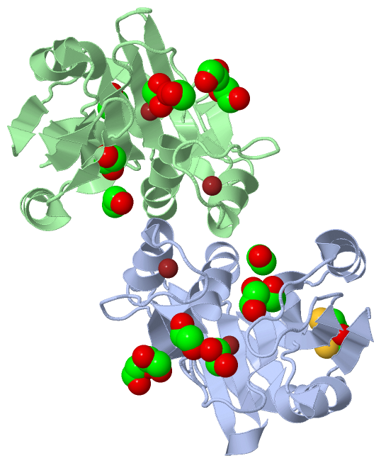
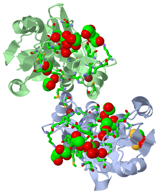
Asymmetric Unit (3, 19)
No.NameCountTypeFull Name
1DTT1Ligand/Ion2,3-DIHYDROXY-1,4-DITHIOBUTANE
2EDO14Ligand/Ion1,2-ETHANEDIOL
3ZN4Ligand/IonZINC ION
Biological Unit 1 (2, 8)
No.NameCountTypeFull Name
1DTT1Ligand/Ion2,3-DIHYDROXY-1,4-DITHIOBUTANE
2EDO7Ligand/Ion1,2-ETHANEDIOL
3ZN-1Ligand/IonZINC ION
Biological Unit 2 (1, 7)
No.NameCountTypeFull Name
1DTT-1Ligand/Ion2,3-DIHYDROXY-1,4-DITHIOBUTANE
2EDO7Ligand/Ion1,2-ETHANEDIOL
3ZN-1Ligand/IonZINC ION

(-) Sites  (18, 18)

Asymmetric Unit (18, 18)
No.NameEvidenceResiduesDescription
01AC1SOFTWAREHIS A:341 , HIS A:343 , ASP A:354 , HOH A:621BINDING SITE FOR RESIDUE ZN A 501
02AC2SOFTWAREHIS A:356 , CYS A:397 , HIS A:404 , HIS A:406BINDING SITE FOR RESIDUE ZN A 502
03AC3SOFTWARECYS A:317 , GLY A:318 , ILE A:319 , THR A:357 , TYR A:361 , EDO A:509BINDING SITE FOR RESIDUE EDO A 503
04AC4SOFTWARELYS A:409 , TYR A:411 , MET A:413 , EDO A:508BINDING SITE FOR RESIDUE EDO A 504
05AC5SOFTWAREGLN A:346 , EDO A:507 , HOH A:603 , HOH A:635BINDING SITE FOR RESIDUE EDO A 505
06AC6SOFTWARESER A:360 , HOH A:647BINDING SITE FOR RESIDUE EDO A 506
07AC7SOFTWAREPHE A:349 , SER A:351 , SER A:352 , PHE A:403 , EDO A:505BINDING SITE FOR RESIDUE EDO A 507
08AC8SOFTWARELYS A:409 , THR A:412 , MET A:413 , EDO A:504BINDING SITE FOR RESIDUE EDO A 508
09AC9SOFTWARETHR A:316 , CYS A:317 , VAL A:353 , THR A:357 , EDO A:503 , HOH A:637BINDING SITE FOR RESIDUE EDO A 509
10BC1SOFTWAREARG A:296 , HOH A:632BINDING SITE FOR RESIDUE DTT A 510
11BC2SOFTWAREHIS B:341 , HIS B:343 , ASP B:354 , HOH B:624BINDING SITE FOR RESIDUE ZN B 501
12BC3SOFTWAREHIS B:356 , CYS B:397 , HIS B:404 , HIS B:406BINDING SITE FOR RESIDUE ZN B 502
13BC4SOFTWARELYS B:409 , TYR B:411BINDING SITE FOR RESIDUE EDO B 503
14BC5SOFTWAREASP B:315 , HIS B:343 , GLN B:346 , PHE B:349 , SER B:351 , HOH B:603 , HOH B:604 , HOH B:642BINDING SITE FOR RESIDUE EDO B 504
15BC6SOFTWARECYS B:288 , CYS B:317 , GLY B:318 , THR B:357 , TYR B:361BINDING SITE FOR RESIDUE EDO B 505
16BC7SOFTWAREPHE B:349BINDING SITE FOR RESIDUE EDO B 506
17BC8SOFTWAREMET B:364 , HOH B:611BINDING SITE FOR RESIDUE EDO B 508
18BC9SOFTWAREASP B:315BINDING SITE FOR RESIDUE EDO B 509

(-) SS Bonds  (0, 0)

(no "SS Bond" information available for 4MSD)

(-) Cis Peptide Bonds  (5, 5)

Asymmetric Unit
No.Residues
1Asp A:387 -Pro A:388
2Gln A:416 -Pro A:417
3Asp B:387 -Pro B:388
4Gln B:416 -Pro B:417
5Pro B:417 -Gly B:418

 Sequence-Structure Mapping

(-) SAPs(SNPs)/Variants  (0, 0)

(no "SAP(SNP)/Variant" information available for 4MSD)

(-) PROSITE Motifs  (0, 0)

(no "PROSITE Motif" information available for 4MSD)

(-) Exons   (0, 0)

(no "Exon" information available for 4MSD)

(-) Sequences/Alignments

Asymmetric Unit
   Reformat: Number of residues per line =  ('0' or empty: single-line sequence representation)
  Number of residues per labelling interval =   
  UniProt sequence: complete  aligned part    
   Show mapping: SCOP domains CATH domains Pfam domains Secondary structure (by author)
SAPs(SNPs) PROSITE motifs Exons
(details for a mapped element are shown in a popup box when the mouse pointer rests over it)
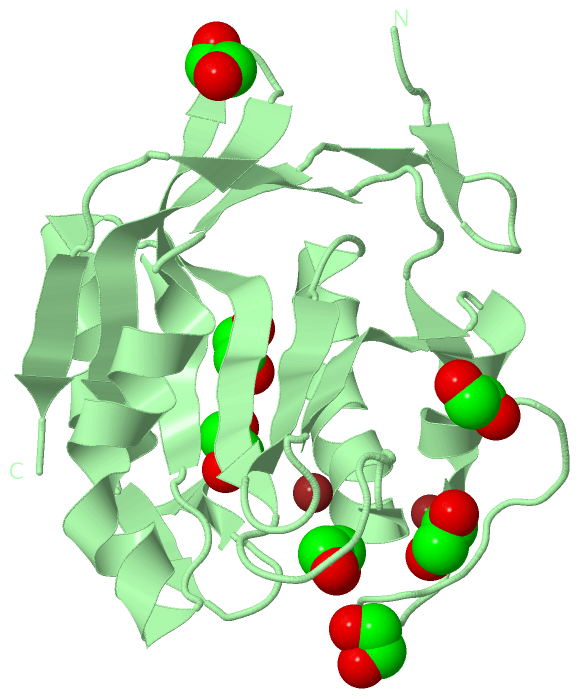
Chain A from PDB  Type:PROTEIN  Length:185
                                                                                                                                                                                                                         
               SCOP domains ----------------------------------------------------------------------------------------------------------------------------------------------------------------------------------------- SCOP domains
               CATH domains ----------------------------------------------------------------------------------------------------------------------------------------------------------------------------------------- CATH domains
               Pfam domains ----------------------------------------------------------------------------------------------------------------------------------------------------------------------------------------- Pfam domains
         Sec.struct. author ...ee.....ee..eeee.hhhhhhhhhhhhhhhh....eeeeeeeee..eeeeeeeee.............hhhhhhhhhhhh..eeeeeeee........hhhhhhhhhhhhhhh...eeeeeehhhheeeeeee..hhhhhhhhhh.............eee......eeee....eeee.. Sec.struct. author
                 SAPs(SNPs) ----------------------------------------------------------------------------------------------------------------------------------------------------------------------------------------- SAPs(SNPs)
                    PROSITE ----------------------------------------------------------------------------------------------------------------------------------------------------------------------------------------- PROSITE
                 Transcript ----------------------------------------------------------------------------------------------------------------------------------------------------------------------------------------- Transcript
                 4msd A 249 KIHAYTEGGKPLRTIYLPKLLKKVFLDVVKPNTKKNLETCGILCGKLRQNAFFITHLVIPLQEATSDTCGITDEASLFEFQDKHNLLTLGWIHTHPTQTCFMSSVDLHTHCSYQLMLPEAIAIVMAPSKNTSGIFRLLDPEGLQTIVKCRKPGLFHPHEGKVYTMVAQPGHVREINSKLQVVDLR 433
                                   258       268       278       288       298       308       318       328       338       348       358       368       378       388       398       408       418       428     

Chain B from PDB  Type:PROTEIN  Length:185
                                                                                                                                                                                                                         
               SCOP domains ----------------------------------------------------------------------------------------------------------------------------------------------------------------------------------------- SCOP domains
               CATH domains ----------------------------------------------------------------------------------------------------------------------------------------------------------------------------------------- CATH domains
               Pfam domains ----------------------------------------------------------------------------------------------------------------------------------------------------------------------------------------- Pfam domains
         Sec.struct. author ...ee.....ee..eeee.hhhhhhhhhhhhhhhh....eeeeeeeee..eeeeeeeee..eee....eee.hhhhhhhhhhhh..eeeeeeee........hhhhhhhhhhhhhhh...eeeeee....eeeeeee..hhhhhhhhhh.............eee......eeee....eeee.. Sec.struct. author
                 SAPs(SNPs) ----------------------------------------------------------------------------------------------------------------------------------------------------------------------------------------- SAPs(SNPs)
                    PROSITE ----------------------------------------------------------------------------------------------------------------------------------------------------------------------------------------- PROSITE
                 Transcript ----------------------------------------------------------------------------------------------------------------------------------------------------------------------------------------- Transcript
                 4msd B 249 KIHAYTEGGKPLRTIYLPKLLKKVFLDVVKPNTKKNLETCGILCGKLRQNAFFITHLVIPLQEATSDTCGITDEASLFEFQDKHNLLTLGWIHTHPTQTCFMSSVDLHTHCSYQLMLPEAIAIVMAPSKNTSGIFRLLDPEGLQTIVKCRKPGLFHPHEGKVYTMVAQPGHVREINSKLQVVDLR 433
                                   258       268       278       288       298       308       318       328       338       348       358       368       378       388       398       408       418       428     

   Legend:   → Mismatch (orange background)
  - → Gap (green background, '-', border residues have a numbering label)
    → Modified Residue (blue background, lower-case, 'x' indicates undefined single-letter code, labelled with number + name)
  x → Chemical Group (purple background, 'x', labelled with number + name, e.g. ACE or NH2)
  extra numbering lines below/above indicate numbering irregularities and modified residue names etc., number ends below/above '|'

 Classification and Annotation

(-) SCOP Domains  (0, 0)

(no "SCOP Domain" information available for 4MSD)

(-) CATH Domains  (0, 0)

(no "CATH Domain" information available for 4MSD)

(-) Pfam Domains  (0, 0)

(no "Pfam Domain" information available for 4MSD)

(-) Gene Ontology  (14, 14)

Asymmetric Unit(hide GO term definitions)

 Visualization

(-) Interactive Views

Asymmetric Unit
  Complete Structure
    Jena3D(integrated viewing of ligand, site, SAP, PROSITE, SCOP information)
    WebMol | AstexViewer[tm]@PDBe
(Java Applets, require no local installation except for Java; loading may be slow)
    STRAP
(Java WebStart application, automatic local installation, requires Java; full application with system access!)
    RasMol
(require local installation)
    Molscript (VRML)
(requires installation of a VRML viewer; select preferred view via VRML and generate a mono or stereo PDF format file)
 
  Ligands, Modified Residues, Ions
    DTT  [ RasMol | Jena3D ]  +environment [ RasMol | Jena3D ]
    EDO  [ RasMol | Jena3D ]  +environment [ RasMol | Jena3D ]
    ZN  [ RasMol | Jena3D ]  +environment [ RasMol | Jena3D ]
 
  Sites
    AC1  [ RasMol ]  +environment [ RasMol ]
    AC2  [ RasMol ]  +environment [ RasMol ]
    AC3  [ RasMol ]  +environment [ RasMol ]
    AC4  [ RasMol ]  +environment [ RasMol ]
    AC5  [ RasMol ]  +environment [ RasMol ]
    AC6  [ RasMol ]  +environment [ RasMol ]
    AC7  [ RasMol ]  +environment [ RasMol ]
    AC8  [ RasMol ]  +environment [ RasMol ]
    AC9  [ RasMol ]  +environment [ RasMol ]
    BC1  [ RasMol ]  +environment [ RasMol ]
    BC2  [ RasMol ]  +environment [ RasMol ]
    BC3  [ RasMol ]  +environment [ RasMol ]
    BC4  [ RasMol ]  +environment [ RasMol ]
    BC5  [ RasMol ]  +environment [ RasMol ]
    BC6  [ RasMol ]  +environment [ RasMol ]
    BC7  [ RasMol ]  +environment [ RasMol ]
    BC8  [ RasMol ]  +environment [ RasMol ]
    BC9  [ RasMol ]  +environment [ RasMol ]
 
  Cis Peptide Bonds
    Asp A:387 - Pro A:388   [ RasMol ]  
    Asp B:387 - Pro B:388   [ RasMol ]  
    Gln A:416 - Pro A:417   [ RasMol ]  
    Gln B:416 - Pro B:417   [ RasMol ]  
    Pro B:417 - Gly B:418   [ RasMol ]  
 
Biological Units
  Complete Structure
    Biological Unit 1  [ Jena3D ]
    Biological Unit 2  [ Jena3D ]

(-) Still Images

Jmol
  protein: cartoon or spacefill or dots and stick; nucleic acid: cartoon and stick; ligands: spacefill; active site: stick
Molscript
  protein, nucleic acid: cartoon; ligands: spacefill; active site: ball and stick

 Databases and Analysis Tools

(-) Databases

Access by PDB/NDB ID
  4msd
    Family and Domain InformationProDom | SYSTERS
    General Structural InformationGlycoscienceDB | MMDB | NDB | OCA | PDB | PDBe | PDBj | PDBsum | PDBWiki | PQS | PROTEOPEDIA
    Orientation in MembranesOPM
    Protein SurfaceSURFACE
    Secondary StructureDSSP (structure derived) | HSSP (homology derived)
    Structural GenomicsGeneCensus
    Structural NeighboursCE | VAST
    Structure ClassificationCATH | Dali | SCOP
    Validation and Original DataBMRB Data View | BMRB Restraints Grid | EDS | PROCHECK | RECOORD | WHAT_CHECK
 
Access by UniProt ID/Accession number
  SST2_SCHPO | Q9P371
    Comparative Protein Structure ModelsModBase
    Genomic InformationEnsembl
    Protein-protein InteractionDIP
    Sequence, Family and Domain InformationInterPro | Pfam | SMART | UniProtKB/SwissProt
 
Access by Enzyme Classificator   (EC Number)
  3.4.19.-
    General Enzyme InformationBRENDA | EC-PDB | Enzyme | IntEnz
    PathwayKEGG | MetaCyc
 
Access by Disease Identifier   (MIM ID)
  (no 'MIM ID' available)
    Disease InformationOMIM
 
Access by GenAge ID
  (no 'GenAge ID' available)
    Age Related InformationGenAge

(-) Analysis Tools

Access by PDB/NDB ID
    Domain InformationXDom
    Interatomic Contacts of Structural UnitsCSU
    Ligand-protein ContactsLPC
    Protein CavitiescastP
    Sequence and Secondary StructurePDBCartoon
    Structure AlignmentSTRAP(Java WebStart application, automatic local installation, requires Java; full application with system access!)
    Structure and Sequence BrowserSTING
 
Access by UniProt ID/Accession number
  SST2_SCHPO | Q9P371
    Protein Disorder PredictionDisEMBL | FoldIndex | GLOBPLOT (for more information see DisProt)

 Related Entries

(-) Entries Sharing at Least One Protein Chain (UniProt ID)

UniProtKB/Swiss-Prot
        SST2_SCHPO | Q9P3714jxe 4k1r 4ms7 4msj 4msm 4msq 4nql 4pqt 4zd4 4zd5 4zfr 4zft

(-) Related Entries Specified in the PDB File

3rzu STRUCTURE OF CATALYTIC DOMAIN, HUMAN PROTEIN AMSH
4jxe STRUCTURE OF CATALYTIC DOMAIN, S.POMBE SSTE
4ms7 CRYSTAL STRUCTURE OF SCHIZOSACCHAROMYCES POMBE SST2 CATALYTIC DOMAIN
4msj CRYSTAL STRUCTURE OF S. POMBE AMSH-LIKE PROTEASE SST2 CATALYTIC DOMAIN FROM P212121 SPACE GROUP
4msm CRYSTAL STRUCTURE OF SCHIZOSACCHAROMYCES POMBE AMSH-LIKE PROTEASE SST2 E286A MUTANT BOUND TO UBIQUITIN
4msq CRYSTAL STRUCTURE OF SCHIZOSACCHAROMYCES POMBE AMSH-LIKE PROTEASE SST2 CATALYTIC DOMAIN BOUND TO UBIQUITIN