|
|
|
|
 Description
|
|
Compounds
|
||||||||||||||||||||||||||||||||||||||||||||||||||||||||||||||||||||||||||||||||||||||||||||||||||||||||||||||||||||||||||
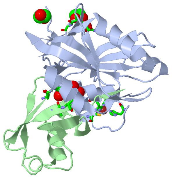
Chains, Units
Summary Information (see also Sequences/Alignments below) |
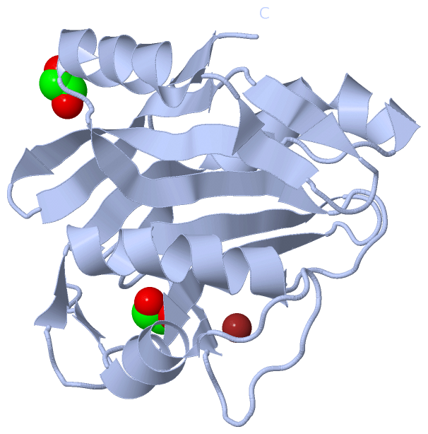
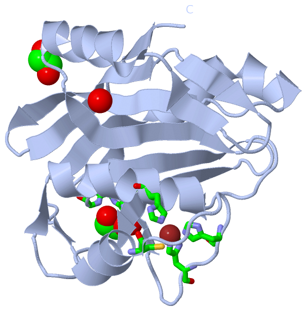
Ligands, Modified Residues, Ions (2, 4)| Asymmetric Unit (2, 4) Biological Unit 1 (1, 2) Biological Unit 2 (1, 1) Biological Unit 3 (1, 3) |
Sites (3, 3)
Asymmetric Unit (3, 3)
|
SS Bonds (0, 0)| (no "SS Bond" information available for 4PQT) |
Cis Peptide Bonds (2, 2)
Asymmetric Unit
|
||||||||||||
SAPs(SNPs)/Variants (0, 0)| (no "SAP(SNP)/Variant" information available for 4PQT) |
PROSITE Motifs (0, 0)| (no "PROSITE Motif" information available for 4PQT) |
Exons (0, 0)| (no "Exon" information available for 4PQT) |
Sequences/Alignments
Asymmetric Unit
Chain A from PDB Type:PROTEIN Length:185
SCOP domains ----------------------------------------------------------------------------------------------------------------------------------------------------------------------------------------- SCOP domains
CATH domains ----------------------------------------------------------------------------------------------------------------------------------------------------------------------------------------- CATH domains
Pfam domains ----------------------------------------------------------------------------------------------------------------------------------------------------------------------------------------- Pfam domains
SAPs(SNPs) ----------------------------------------------------------------------------------------------------------------------------------------------------------------------------------------- SAPs(SNPs)
PROSITE ----------------------------------------------------------------------------------------------------------------------------------------------------------------------------------------- PROSITE
Transcript ----------------------------------------------------------------------------------------------------------------------------------------------------------------------------------------- Transcript
4pqt A 249 KIHAYTEGGKPLRTIYLPKLLKKVFLDVVKPNTKKNLETCGILCGKLRQNAFFITHLVIPLQEATSDTCGTTDEASLFEFQDKHNLLTLGWIHTHPTQTCFMSSVALHTHCSYQLMLPEAIAIVMAPSKNTSGIFRLLDPEGLQTIVKCRKPGLFHPHEGKVYTMVAQPGHVREINSKLQVVDLR 433
258 268 278 288 298 308 318 328 338 348 358 368 378 388 398 408 418 428
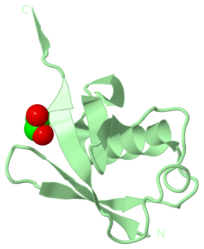
Chain B from PDB Type:PROTEIN Length:76
SCOP domains ---------------------------------------------------------------------------- SCOP domains
CATH domains ---------------------------------------------------------------------------- CATH domains
Pfam domains ---------------------------------------------------------------------------- Pfam domains
SAPs(SNPs) ---------------------------------------------------------------------------- SAPs(SNPs)
PROSITE ---------------------------------------------------------------------------- PROSITE
Transcript ---------------------------------------------------------------------------- Transcript
4pqt B 1 MQIFVKTLTGKTITLEVEPSDTIENVKAKIQDKEGIPPDQQRLIFAGKQLEDGRTLSDYNIQKESTLHLVLRLRGG 76
10 20 30 40 50 60 70
|
||||||||||||||||||||
SCOP Domains (0, 0)| (no "SCOP Domain" information available for 4PQT) |
CATH Domains (0, 0)| (no "CATH Domain" information available for 4PQT) |
Pfam Domains (0, 0)| (no "Pfam Domain" information available for 4PQT) |
Gene Ontology (14, 14)|
Asymmetric Unit(hide GO term definitions) |
Interactive Views
|
|||||||||||||||||||||||||||||||||||||||||||||||||||||||||||||||||||||||||||||||||||||||||||||||||||||||||||||||||||||||||||||||||||||||||||||||||||||||||||||||||||||||||||||||
Still Images
|
||||||||||||||||
Databases
|
||||||||||||||||||||||||||||||||||||||||||||||||||||||||||||||||||||||||||||||||||||||||||||||||||||||||||||||||||||||||||||||||||||||||||||||||||||||||||||||||
Analysis Tools
|
|||||||||||||||||||||||||||||||||||||||||||||||||||||||||||||
Entries Sharing at Least One Protein Chain (UniProt ID)
Related Entries Specified in the PDB File
|
|