| No. | Name | Evidence | Residues | Description |
|---|
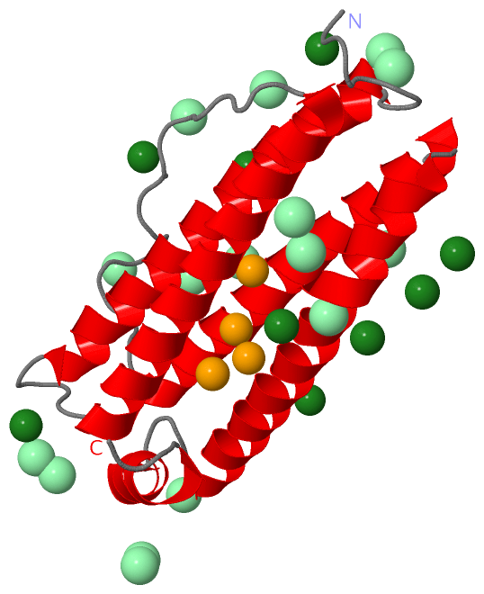
| 01 | AC1 | SOFTWARE | GLU A:23 , GLU A:58 , HIS A:61 , HOH A:663 , HOH A:673 | BINDING SITE FOR RESIDUE FE2 A 201 |
| 02 | AC2 | SOFTWARE | GLU A:57 , GLU A:136 , ASP A:140 , ARG A:143 , HOH A:484 , HOH A:669 | BINDING SITE FOR RESIDUE FE2 A 202 |
| 03 | AC3 | SOFTWARE | GLU A:53 , GLU A:57 , HOH A:437 , HOH A:484 , HOH A:489 , HOH A:546 | BINDING SITE FOR RESIDUE FE2 A 203 |
| 04 | AC4 | SOFTWARE | GLN A:54 , GLU A:103 , ASP A:140 , HOH A:589 , HOH A:649 , HOH A:669 | BINDING SITE FOR RESIDUE FE2 A 204 |
| 05 | AC5 | SOFTWARE | SER A:10 , HOH A:362 , HOH A:441 , HOH A:529 , HOH A:530 , HOH A:591 | BINDING SITE FOR RESIDUE MG A 205 |
| 06 | AC6 | SOFTWARE | HOH A:312 , HOH A:337 | BINDING SITE FOR RESIDUE MG A 206 |
| 07 | AC7 | SOFTWARE | ASP A:127 , HOH A:378 , HOH A:674 | BINDING SITE FOR RESIDUE MG A 207 |
| 08 | AC8 | SOFTWARE | HOH A:471 , HOH A:475 , HOH A:590 , HOH A:593 , HOH A:664 , HOH A:676 | BINDING SITE FOR RESIDUE MG A 208 |
| 09 | AC9 | SOFTWARE | HOH A:396 , HOH A:456 , HOH A:680 , HOH A:681 | BINDING SITE FOR RESIDUE MG A 209 |
| 10 | BC1 | SOFTWARE | HOH A:399 , HOH A:438 , HOH A:460 , HOH A:644 , HOH A:665 , HOH A:679 | BINDING SITE FOR RESIDUE MG A 210 |
| 11 | BC2 | SOFTWARE | HOH A:365 , HOH A:447 , HOH A:533 , HOH A:555 , HOH A:561 , HOH A:601 | BINDING SITE FOR RESIDUE MG A 211 |
| 12 | BC3 | SOFTWARE | HOH A:490 , HOH A:567 , HOH A:578 , HOH A:587 | BINDING SITE FOR RESIDUE MG A 212 |
| 13 | BC4 | SOFTWARE | HOH A:348 , HOH A:429 , HOH A:553 , HOH A:600 , HOH A:623 , HOH A:650 | BINDING SITE FOR RESIDUE MG A 213 |
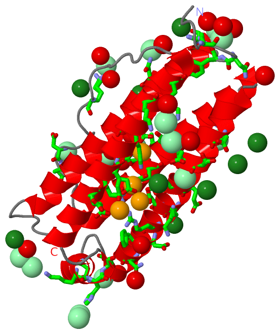
| 14 | BC5 | SOFTWARE | SER A:131 , GLU A:132 , TYR A:133 , GLU A:135 , GLU A:136 | BINDING SITE FOR RESIDUE CL A 214 |
| 15 | BC6 | SOFTWARE | ASP A:87 , GLU A:88 , HOH A:519 | BINDING SITE FOR RESIDUE CL A 215 |
| 16 | BC7 | SOFTWARE | LYS A:82 , HOH A:307 , HOH A:394 | BINDING SITE FOR RESIDUE CL A 216 |
| 17 | BC8 | SOFTWARE | ASN A:7 , GLN A:108 , LYS A:115 , HOH A:371 , HOH A:418 | BINDING SITE FOR RESIDUE CL A 217 |
| 18 | BC9 | SOFTWARE | HOH A:373 , HOH A:402 , HOH A:436 | BINDING SITE FOR RESIDUE CL A 218 |
| 19 | CC1 | SOFTWARE | GLN A:101 , LEU A:102 , THR A:105 , HOH A:500 | BINDING SITE FOR RESIDUE CL A 219 |
| 20 | CC2 | SOFTWARE | HIS A:169 | BINDING SITE FOR RESIDUE CL A 220 |
| 21 | CC3 | SOFTWARE | HIS A:169 | BINDING SITE FOR RESIDUE CL A 221 |
| 22 | CC4 | SOFTWARE | ARG A:5 , ASN A:7 , TYR A:8 , HOH A:543 | BINDING SITE FOR RESIDUE CL A 222 |
| 23 | CC5 | SOFTWARE | ASN A:17 , HOH A:361 | BINDING SITE FOR RESIDUE CL A 223 |
| 24 | CC6 | SOFTWARE | SER A:10 , HOH A:544 , HOH A:682 | BINDING SITE FOR RESIDUE CL A 224 |
| 25 | CC7 | SOFTWARE | ASN A:150 , LYS A:168 , SER A:170 , CL A:226 , HOH A:313 | BINDING SITE FOR RESIDUE CL A 225 |
| 26 | CC8 | SOFTWARE | ASP A:146 , PHE A:147 , ASN A:150 , CL A:225 , HOH A:331 , HOH A:403 | BINDING SITE FOR RESIDUE CL A 226 |
| 27 | CC9 | SOFTWARE | GLY A:155 , ASN A:159 , HOH A:656 | BINDING SITE FOR RESIDUE CL A 227 |
| 28 | DC1 | SOFTWARE | HOH A:570 | BINDING SITE FOR RESIDUE CL A 228 |
| 29 | DC2 | SOFTWARE | LYS A:67 , LYS A:71 , LYS A:139 , HOH A:381 | BINDING SITE FOR RESIDUE CL A 229 |
 Description