Show PDB file:   
         Plain Text   HTML   (compressed file size)
QuickSearch:   
by PDB,NDB,UniProt,PROSITE Code or Search Term(s)  
(-)Asymmetric Unit
(-)Asym. Unit - sites
(-)Biological Unit 1
(-)Biol. Unit 1 - sites
(-)Biological Unit 2
collapse expand < >
Image Asymmetric Unit
Asymmetric Unit  (Jmol Viewer)
Image Asym. Unit - sites
Asym. Unit - sites  (Jmol Viewer)
Image Biological Unit 1
Biological Unit 1  (Jmol Viewer)
Image Biol. Unit 1 - sites
Biol. Unit 1 - sites  (Jmol Viewer)
Image Biological Unit 2
Biological Unit 2  (Jmol Viewer)

(-) Description

Title :  STRUCTURE OF MNTR H77A MUTANT IN APO- AND MN-BOUND FORMS
 
Authors :  A. Glasfeld, K. Grauer-Gray, A. Mcguire
Date :  05 Nov 12  (Deposition) - 21 Nov 12  (Release) - 17 Apr 13  (Revision)
Method :  X-RAY DIFFRACTION
Resolution :  2.30
Chains :  Asym. Unit :  A,B
Biol. Unit 1:  A  (2x)
Biol. Unit 2:  B  (2x)
Keywords :  Winged Helix-Turn-Helix, Transcription Regulator, Metalloregulator, Transcription (Keyword Search: [Gene Ontology, PubMed, Web (Google))
 
Reference :  A. M. Mcguire, B. J. Cuthbert, Z. Ma, K. D. Grauer-Gray, M. Brunjes Brophy, K. A. Spear, S. Soonsanga, J. I. Kliegman, S. L. Griner, J. D. Helmann, A. Glasfeld
Roles Of The A And C Sites In The Manganese-Specific Activation Of Mntr.
Biochemistry V. 52 701 2013
PubMed-ID: 23298157  |  Reference-DOI: 10.1021/BI301550T

(-) Compounds

Molecule 1 - TRANSCRIPTIONAL REGULATOR MNTR
    ChainsA, B
    EngineeredYES
    Expression SystemESCHERICHIA COLI
    Expression System Taxid562
    FragmentMNTR (UNP RESIDUES 2-142)
    GeneBSU24520, MNTR, YQHN
    MutationYES
    Organism ScientificBACILLUS SUBTILIS SUBSP. SUBTILIS
    Organism Taxid224308
    Strain168
    SynonymMANGANESE TRANSPORT REGULATOR

 Structural Features

(-) Chains, Units

  12
Asymmetric Unit AB
Biological Unit 1 (2x)A 
Biological Unit 2 (2x) B

Summary Information (see also Sequences/Alignments below)
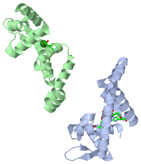
(-) Ligands, Modified Residues, Ions  (2, 3)

Asymmetric Unit (2, 3)
No.NameCountTypeFull Name
1MG1Ligand/IonMAGNESIUM ION
2MN2Ligand/IonMANGANESE (II) ION
Biological Unit 1 (0, 0)
No.NameCountTypeFull Name
1MG-1Ligand/IonMAGNESIUM ION
2MN-1Ligand/IonMANGANESE (II) ION
Biological Unit 2 (0, 0)
No.NameCountTypeFull Name
1MG-1Ligand/IonMAGNESIUM ION
2MN-1Ligand/IonMANGANESE (II) ION

(-) Sites  (3, 3)

Asymmetric Unit (3, 3)
No.NameEvidenceResiduesDescription
1AC1SOFTWAREASP A:8 , GLU A:99 , GLU A:102 , HIS A:103 , HOH A:301BINDING SITE FOR RESIDUE MN A 201
2AC2SOFTWAREASP A:8 , GLU A:11 , GLU A:99 , GLU A:102 , HOH A:306BINDING SITE FOR RESIDUE MN A 202
3AC3SOFTWAREGLU B:99 , GLU B:102 , HOH B:306 , HOH B:307BINDING SITE FOR RESIDUE MG B 201

(-) SS Bonds  (0, 0)

(no "SS Bond" information available for 4HV6)

(-) Cis Peptide Bonds  (0, 0)

(no "Cis Peptide Bond" information available for 4HV6)

 Sequence-Structure Mapping

(-) SAPs(SNPs)/Variants  (0, 0)

(no "SAP(SNP)/Variant" information available for 4HV6)

(-) PROSITE Motifs  (0, 0)

(no "PROSITE Motif" information available for 4HV6)

(-) Exons   (0, 0)

(no "Exon" information available for 4HV6)

(-) Sequences/Alignments

Asymmetric Unit
   Reformat: Number of residues per line =  ('0' or empty: single-line sequence representation)
  Number of residues per labelling interval =   
  UniProt sequence: complete  aligned part    
   Show mapping: SCOP domains CATH domains Pfam domains Secondary structure (by author)
SAPs(SNPs) PROSITE motifs Exons
(details for a mapped element are shown in a popup box when the mouse pointer rests over it)
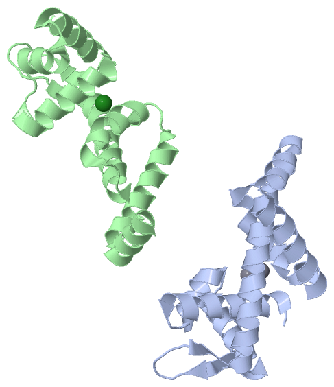
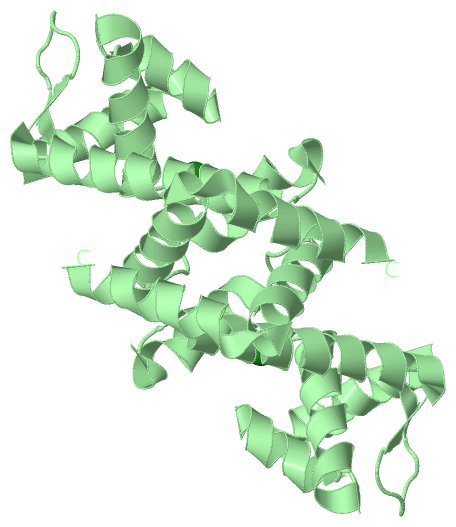
Chain A from PDB  Type:PROTEIN  Length:133
                                                                                                                                                                     
               SCOP domains d4hv6a1 A:2-62 automated matches                             d4hv6a2 A:63-134 automated matches                                       SCOP domains
               CATH domains ------------------------------------------------------------------------------------------------------------------------------------- CATH domains
               Pfam domains ------------------------------------------------------------------------------------------------------------------------------------- Pfam domains
         Sec.struct. author .hhhhhhhhhhhhhhhhhh...hhhhhhhhhh.hhhhhhhhhhhhhhh..eee.....eeehhhhhhhhhhhhhhhhhhhhhhhhhh.hhhhhhhhhhhhhhhhhhhhhhhhhhhhhhhhhhhhhhhhhhhhh Sec.struct. author
                 SAPs(SNPs) ------------------------------------------------------------------------------------------------------------------------------------- SAPs(SNPs)
                    PROSITE ------------------------------------------------------------------------------------------------------------------------------------- PROSITE
                 Transcript ------------------------------------------------------------------------------------------------------------------------------------- Transcript
                 4hv6 A   2 TTPSMEDYIEQIYMLIEEKGYARVSDIAEALAVHPSSVTKMVQKLDKDEYLIYEKYRGLVLTSKGKKIGKRLVYRAELLEQFLRIIGVDEEKIYNDVEGIEHHLSWNSIDRIGDLVQYFEEDDARKKDLKSIQ 134
                                    11        21        31        41        51        61        71        81        91       101       111       121       131   

Chain B from PDB  Type:PROTEIN  Length:133
                                                                                                                                                                     
               SCOP domains d4hv6b1 B:4-62 automated matches                           d4hv6b2 B:63-136 automated matches                                         SCOP domains
               CATH domains ------------------------------------------------------------------------------------------------------------------------------------- CATH domains
               Pfam domains ------------------------------------------------------------------------------------------------------------------------------------- Pfam domains
         Sec.struct. author .hhhhhhhhhhhhhhhhh..hhhhhhhhhh.hhhhhhhhhhhhhhh..ee.......eehhhhhhhhhhhhhhhhhhhhhhhhh..hhhhhhhhhhhhh...hhhhhhhhhhhhhhhhhhhhhhhhhhhhhhh Sec.struct. author
                 SAPs(SNPs) ------------------------------------------------------------------------------------------------------------------------------------- SAPs(SNPs)
                    PROSITE ------------------------------------------------------------------------------------------------------------------------------------- PROSITE
                 Transcript ------------------------------------------------------------------------------------------------------------------------------------- Transcript
                 4hv6 B   4 PSMEDYIEQIYMLIEEKGYARVSDIAEALAVHPSSVTKMVQKLDKDEYLIYEKYRGLVLTSKGKKIGKRLVYRAELLEQFLRIIGVDEEKIYNDVEGIEHHLSWNSIDRIGDLVQYFEEDDARKKDLKSIQKK 136
                                    13        23        33        43        53        63        73        83        93       103       113       123       133   

   Legend:   → Mismatch (orange background)
  - → Gap (green background, '-', border residues have a numbering label)
    → Modified Residue (blue background, lower-case, 'x' indicates undefined single-letter code, labelled with number + name)
  x → Chemical Group (purple background, 'x', labelled with number + name, e.g. ACE or NH2)
  extra numbering lines below/above indicate numbering irregularities and modified residue names etc., number ends below/above '|'

 Classification and Annotation

(-) SCOP Domains  (2, 4)

Asymmetric Unit

(-) CATH Domains  (0, 0)

(no "CATH Domain" information available for 4HV6)

(-) Pfam Domains  (0, 0)

(no "Pfam Domain" information available for 4HV6)

(-) Gene Ontology  (10, 10)

Asymmetric Unit(hide GO term definitions)

 Visualization

(-) Interactive Views

Asymmetric Unit
  Complete Structure
    Jena3D(integrated viewing of ligand, site, SAP, PROSITE, SCOP information)
    WebMol | AstexViewer[tm]@PDBe
(Java Applets, require no local installation except for Java; loading may be slow)
    STRAP
(Java WebStart application, automatic local installation, requires Java; full application with system access!)
    RasMol
(require local installation)
    Molscript (VRML)
(requires installation of a VRML viewer; select preferred view via VRML and generate a mono or stereo PDF format file)
 
  Ligands, Modified Residues, Ions
    MG  [ RasMol | Jena3D ]  +environment [ RasMol | Jena3D ]
    MN  [ RasMol | Jena3D ]  +environment [ RasMol | Jena3D ]
 
  Sites
    AC1  [ RasMol ]  +environment [ RasMol ]
    AC2  [ RasMol ]  +environment [ RasMol ]
    AC3  [ RasMol ]  +environment [ RasMol ]
 
  Cis Peptide Bonds
(no "Cis Peptide Bonds" information available for 4hv6)
 
Biological Units
  Complete Structure
    Biological Unit 1  [ Jena3D ]
    Biological Unit 2  [ Jena3D ]

(-) Still Images

Jmol
  protein: cartoon or spacefill or dots and stick; nucleic acid: cartoon and stick; ligands: spacefill; active site: stick
Molscript
  protein, nucleic acid: cartoon; ligands: spacefill; active site: ball and stick

 Databases and Analysis Tools

(-) Databases

Access by PDB/NDB ID
  4hv6
    Family and Domain InformationProDom | SYSTERS
    General Structural InformationGlycoscienceDB | MMDB | NDB | OCA | PDB | PDBe | PDBj | PDBsum | PDBWiki | PQS | PROTEOPEDIA
    Orientation in MembranesOPM
    Protein SurfaceSURFACE
    Secondary StructureDSSP (structure derived) | HSSP (homology derived)
    Structural GenomicsGeneCensus
    Structural NeighboursCE | VAST
    Structure ClassificationCATH | Dali | SCOP
    Validation and Original DataBMRB Data View | BMRB Restraints Grid | EDS | PROCHECK | RECOORD | WHAT_CHECK
 
Access by UniProt ID/Accession number
  MNTR_BACSU | P54512
    Comparative Protein Structure ModelsModBase
    Genomic InformationEnsembl
    Protein-protein InteractionDIP
    Sequence, Family and Domain InformationInterPro | Pfam | SMART | UniProtKB/SwissProt
 
Access by Enzyme Classificator   (EC Number)
  (no 'Enzyme Classificator' available)
    General Enzyme InformationBRENDA | EC-PDB | Enzyme | IntEnz
    PathwayKEGG | MetaCyc
 
Access by Disease Identifier   (MIM ID)
  (no 'MIM ID' available)
    Disease InformationOMIM
 
Access by GenAge ID
  (no 'GenAge ID' available)
    Age Related InformationGenAge

(-) Analysis Tools

Access by PDB/NDB ID
    Domain InformationXDom
    Interatomic Contacts of Structural UnitsCSU
    Ligand-protein ContactsLPC
    Protein CavitiescastP
    Sequence and Secondary StructurePDBCartoon
    Structure AlignmentSTRAP(Java WebStart application, automatic local installation, requires Java; full application with system access!)
    Structure and Sequence BrowserSTING
 
Access by UniProt ID/Accession number
  MNTR_BACSU | P54512
    Protein Disorder PredictionDisEMBL | FoldIndex | GLOBPLOT (for more information see DisProt)

 Related Entries

(-) Entries Sharing at Least One Protein Chain (UniProt ID)

UniProtKB/Swiss-Prot
        MNTR_BACSU | P545121on1 1on2 2ev0 2ev5 2ev6 2f5c 2f5d 2f5e 2f5f 2hyf 2hyg 3r60 3r61 4hv5 4hx4 4hx7 4hx8

(-) Related Entries Specified in the PDB File

4hv5