|
|
|
|
 Description
|
|
Compounds
|
||||||||||||||||||||||||||||||||||||||||||||||||||||||||||||||||||||||||||||||||||||||||||||||||||||||||||||||||||
Chains, Units
Summary Information (see also Sequences/Alignments below) |
Ligands, Modified Residues, Ions (1, 1)
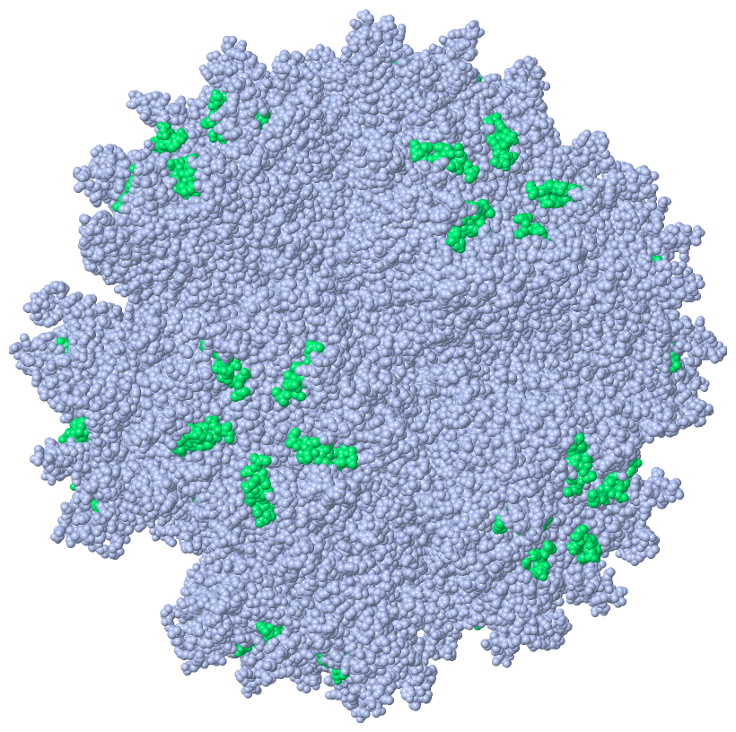
Asymmetric Unit (1, 1)
|
Sites (0, 0)| (no "Site" information available for 4AR2) |
SS Bonds (0, 0)| (no "SS Bond" information available for 4AR2) |
Cis Peptide Bonds (0, 0)| (no "Cis Peptide Bond" information available for 4AR2) |
SAPs(SNPs)/Variants (0, 0)| (no "SAP(SNP)/Variant" information available for 4AR2) |
PROSITE Motifs (0, 0)| (no "PROSITE Motif" information available for 4AR2) |
Exons (0, 0)| (no "Exon" information available for 4AR2) |
Sequences/Alignments
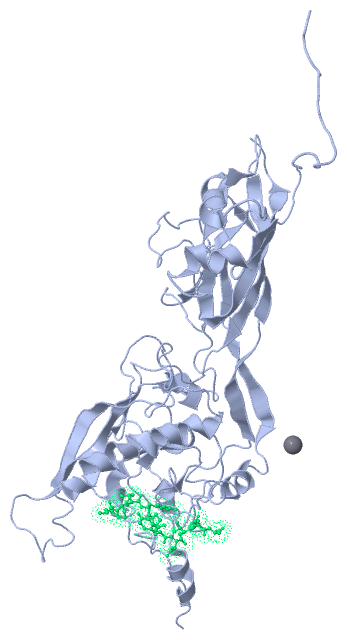
Asymmetric UnitChain A from PDB Type:PROTEIN Length:455 aligned with Q2Y0H9_ADE03 | Q2Y0H9 from UniProtKB/TrEMBL Length:544 Alignment length:495 57 67 77 87 97 107 117 127 137 147 157 167 177 187 197 207 217 227 237 247 257 267 277 287 297 307 317 327 337 347 357 367 377 387 397 407 417 427 437 447 457 467 477 487 497 507 517 527 537 Q2Y0H9_ADE03 48 PTEGRNSIRYSELSPLYDTTKLYLVDNKSADIASLNYQNDHSNFLTTVVQNNDFTPTEASTQTINFDERSRWGGQLKTIMHTNMPNVNEYMFSNKFKARVMVSRKAPEGVTVNDTYDHKEDILKYEWFEFILPEGNFSATMTIDLMNNAIIDNYLEIGRQNGVLESDIGVKFDTRNFRLGWDPETKLIMPGVYTYEAFHPDIVLLPGCGVDFTESRLSNLLGIRKRHPFQEGFKIMYEDLEGGNIPALLDVTAYEESKKDTTTETTTLAVAEETSEDDDITRGDTYITEKQKREAAAAEVKKELKIQPLEKDSKSRSYNVLEDKINTAYRSWYLSYNYGNPEKGIRSWTLLTTSDVTCGAEQVYWSLPDMMQDPVTFRSTRQVNNYPVVGAELMPVFSKSFYNEQAVYSQQLRQATSLTHVFNRFPENQILIRPPAPTITTVSENVPALTDHGTLPLRSSIRGVQRVTVTDARRRTCPYVYKALGIVAPRVLSSR 542 SCOP domains --------------------------------------------------------------------------------------------------------------------------------------------------------------------------------------------------------------------------------------------------------------------------------------------------------------------------------------------------------------------------------------------------------------------------------------------------------------------------------------------------------------- SCOP domains CATH domains --------------------------------------------------------------------------------------------------------------------------------------------------------------------------------------------------------------------------------------------------------------------------------------------------------------------------------------------------------------------------------------------------------------------------------------------------------------------------------------------------------------- CATH domains Pfam domains --------------------------------------------------------------------------------------------------------------------------------------------------------------------------------------------------------------------------------------------------------------------------------------------------------------------------------------------------------------------------------------------------------------------------------------------------------------------------------------------------------------- Pfam domains SAPs(SNPs) --------------------------------------------------------------------------------------------------------------------------------------------------------------------------------------------------------------------------------------------------------------------------------------------------------------------------------------------------------------------------------------------------------------------------------------------------------------------------------------------------------------- SAPs(SNPs) PROSITE --------------------------------------------------------------------------------------------------------------------------------------------------------------------------------------------------------------------------------------------------------------------------------------------------------------------------------------------------------------------------------------------------------------------------------------------------------------------------------------------------------------- PROSITE Transcript --------------------------------------------------------------------------------------------------------------------------------------------------------------------------------------------------------------------------------------------------------------------------------------------------------------------------------------------------------------------------------------------------------------------------------------------------------------------------------------------------------------- Transcript 4ar2 A 48 PTEGRNSIRYSELSPLYDTTKLYLVDNKSADIASLNYQNDHSNFLTTVVQNNDFTPTEASTQTINFDERSRWGGQLKTIMHTNMPNVNEYMFSNKFKARVMVSRKAPEGVTVNDTYDHKEDILKYEWFEFILPEGNFSATMTIDLMNNAIIDNYLEIGRQNGVLESDIGVKFDTRNFRLGWDPETKLIMPGVYTYEAFHPDIVLLPGCGVDFTESRLSNLLGIRKRHPFQEGFKIMYEDLEGGNIPALLDVTAYEESKKD----------------------------------------KKELKIQPLEKDSKSRSYNVLEDKINTAYRSWYLSYNYGNPEKGIRSWTLLTTSDVTCGAEQVYWSLPDMMQDPVTFRSTRQVNNYPVVGAELMPVFSKSFYNEQAVYSQQLRQATSLTHVFNRFPENQILIRPPAPTITTVSENVPALTDHGTLPLRSSIRGVQRVTVTDARRRTCPYVYKALGIVAPRVLSSR 542 57 67 77 87 97 107 117 127 137 147 157 167 177 187 197 207 217 227 237 247 257 267 277 287 297 307 - - - -| 357 367 377 387 397 407 417 427 437 447 457 467 477 487 497 507 517 527 537 307 348 Chain P from PDB Type:PROTEIN Length:10 aligned with SPIKE_ADE02 | P03275 from UniProtKB/Swiss-Prot Length:582 Alignment length:10 21 SPIKE_ADE02 12 NPVYPYDTET 21 SCOP domains ---------- SCOP domains CATH domains ---------- CATH domains Pfam domains ---------- Pfam domains SAPs(SNPs) ---------- SAPs(SNPs) PROSITE ---------- PROSITE Transcript ---------- Transcript 4ar2 P 12 NPVYPYDTEC 21 21
|
||||||||||||||||||||
SCOP Domains (0, 0)| (no "SCOP Domain" information available for 4AR2) |
CATH Domains (0, 0)| (no "CATH Domain" information available for 4AR2) |
Pfam Domains (0, 0)| (no "Pfam Domain" information available for 4AR2) |
Gene Ontology (10, 10)|
Asymmetric Unit(hide GO term definitions) Chain A (Q2Y0H9_ADE03 | Q2Y0H9)
Chain P (SPIKE_ADE02 | P03275)
|
||||||||||||||||||||||||||||||||||||||||||||||||||||||||||||||||||||||||||||||
Interactive Views
|
|||||||||||||||||||||||||||||||||||||||||||||||||||||||||||||||||||||||||||||||||||||||||||||||||||||||||||||||||||||||||||||||||||||||
Still Images
|
||||||||||||||||
Databases
|
||||||||||||||||||||||||||||||||||||||||||||||||||||||||||||||||||||||||||||||||||||||||||||||||||||||||||||||||||||||||||||||||||||||||||||||||||||||||||||||||||||||||||||||||||||||||||
Analysis Tools
|
||||||||||||||||||||||||||||||||||||||||||||||||||||||||||||||||||||||||
Entries Sharing at Least One Protein Chain (UniProt ID)
Related Entries Specified in the PDB File
|
|