|
|
|
|
 Description
|
|
Compounds
|
||||||||||||||||||||||||||||||||||||||||||||||||||||||||||||
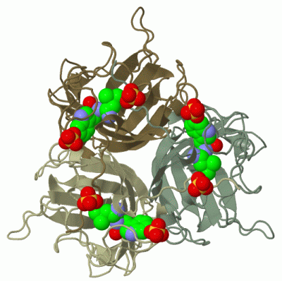
Chains, Units
Summary Information (see also Sequences/Alignments below) |
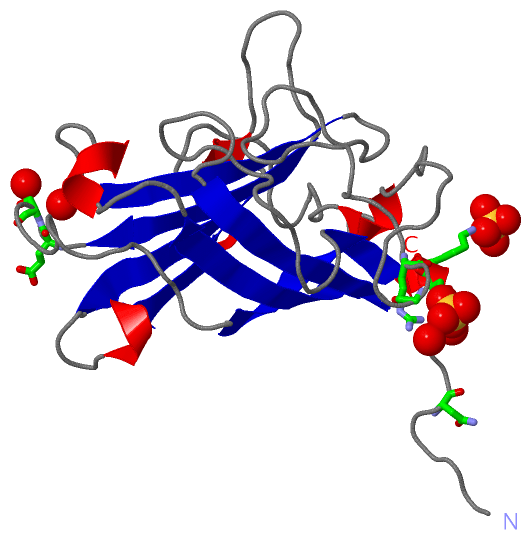
Ligands, Modified Residues, Ions (1, 2)
Asymmetric Unit (1, 2)
|
Sites (2, 2)
Asymmetric Unit (2, 2)
|
SS Bonds (0, 0)| (no "SS Bond" information available for 1QHV) |
Cis Peptide Bonds (0, 0)| (no "Cis Peptide Bond" information available for 1QHV) |
SAPs(SNPs)/Variants (0, 0)| (no "SAP(SNP)/Variant" information available for 1QHV) |
PROSITE Motifs (0, 0)| (no "PROSITE Motif" information available for 1QHV) |
Exons (0, 0)| (no "Exon" information available for 1QHV) |
Sequences/Alignments
Asymmetric UnitChain A from PDB Type:PROTEIN Length:195 aligned with Q96590_ADE02 | Q96590 from UniProtKB/TrEMBL Length:582 Alignment length:195 397 407 417 427 437 447 457 467 477 487 497 507 517 527 537 547 557 567 577 Q96590_ADE02 388 AITIGNKNDDKLTLWTTPDPSPNCRIHSDNDCKFTLVLTKCGSQVLATVAALAVSGDLSSMTGTVASVSIFLRFDQNGVLMENSSLKKHYWNFRNGNSTNANPYTNAVGFMPNLLAYPKTQSQTAKNNIVSQVYLHGDKTKPMILTITLNGTSESTETSEVSTYSMSFTWSWESGKYTTETFATNSYTFSYIAQE 582 SCOP domains d1qhva_ A: Adenovirus fiber protein knob domain SCOP domains CATH domains 1qhvA00 A:388-582 Adenovirus Type 5 Fiber Protein (Receptor Binding Domain) CATH domains Pfam domains --------------------------------------------------------------------------------------------------------------------------------------------------------------------------------------------------- Pfam domains SAPs(SNPs) --------------------------------------------------------------------------------------------------------------------------------------------------------------------------------------------------- SAPs(SNPs) PROSITE --------------------------------------------------------------------------------------------------------------------------------------------------------------------------------------------------- PROSITE Transcript --------------------------------------------------------------------------------------------------------------------------------------------------------------------------------------------------- Transcript 1qhv A 388 AITIGNKNDDKLTLWTTPDPSPNCRIHSDNDCKFTLVLTKCGSQVLATVAALAVSGDLSSMTGTVASVSIFLRFDQNGVLMENSSLKKHYWNFRNGNSTNANPYTNAVGFMPNLLAYPKTQSQTAKNNIVSQVYLHGDKTKPMILTITLNGTSESTETSEVSTYSMSFTWSWESGKYTTETFATNSYTFSYIAQE 582 397 407 417 427 437 447 457 467 477 487 497 507 517 527 537 547 557 567 577 Chain A from PDB Type:PROTEIN Length:195 aligned with SPIKE_ADE02 | P03275 from UniProtKB/Swiss-Prot Length:582 Alignment length:195 397 407 417 427 437 447 457 467 477 487 497 507 517 527 537 547 557 567 577 SPIKE_ADE02 388 AITIGNKNDDKLTLWTTPDPSPNCRIHSDNDCKFTLVLTKCGSQVLATVAALAVSGDLSSMTGTVASVSIFLRFDQNGVLMENSSLKKHYWNFRNGNSTNANPYTNAVGFMPNLLAYPKTQSQTAKNNIVSQVYLHGDKTKPMILTITLNGTSESTETSEVSTYSMSFTWSWESGKYTTETFATNSYTFSYIAQE 582 SCOP domains d1qhva_ A: Adenovirus fiber protein knob domain SCOP domains CATH domains 1qhvA00 A:388-582 Adenovirus Type 5 Fiber Protein (Receptor Binding Domain) CATH domains Pfam domains --------------------------------------------------------------------------------------------------------------------------------------------------------------------------------------------------- Pfam domains SAPs(SNPs) --------------------------------------------------------------------------------------------------------------------------------------------------------------------------------------------------- SAPs(SNPs) PROSITE --------------------------------------------------------------------------------------------------------------------------------------------------------------------------------------------------- PROSITE Transcript --------------------------------------------------------------------------------------------------------------------------------------------------------------------------------------------------- Transcript 1qhv A 388 AITIGNKNDDKLTLWTTPDPSPNCRIHSDNDCKFTLVLTKCGSQVLATVAALAVSGDLSSMTGTVASVSIFLRFDQNGVLMENSSLKKHYWNFRNGNSTNANPYTNAVGFMPNLLAYPKTQSQTAKNNIVSQVYLHGDKTKPMILTITLNGTSESTETSEVSTYSMSFTWSWESGKYTTETFATNSYTFSYIAQE 582 397 407 417 427 437 447 457 467 477 487 497 507 517 527 537 547 557 567 577
|
||||||||||||||||||||
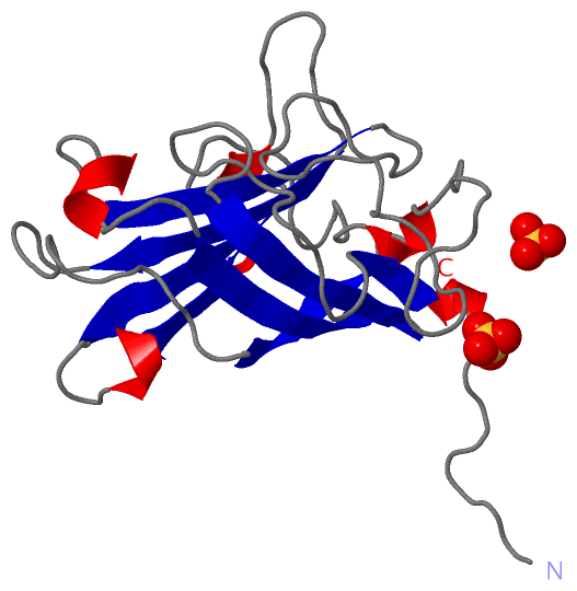
SCOP Domains (1, 1)
Asymmetric Unit
 |
CATH Domains (1, 1)
Asymmetric Unit
|
Pfam Domains (0, 0)| (no "Pfam Domain" information available for 1QHV) |
Gene Ontology (9, 14)|
Asymmetric Unit(hide GO term definitions) Chain A (Q96590_ADE02 | Q96590)
Chain A (SPIKE_ADE02 | P03275)
|
||||||||||||||||||||||||||||||||||||||||||||||||||||||||||||||||||||||||||||||||||||||||||||||||||||||||||||
Interactive Views
|
|||||||||||||||||||||||||||||||||||||||||||||||||||||||||||||||||||||||||||||||||||||||||||||||||||||||||||||||||||||||||||||||||||||||||||||||
Still Images
|
||||||||||||||||
Databases
|
||||||||||||||||||||||||||||||||||||||||||||||||||||||||||||||||||||||||||||||||||||||||||||||||||||||||||||||||||||||||||||||||||||||||||||||||||||||||||||||||||||||||||||||||||||||||||
Analysis Tools
|
||||||||||||||||||||||||||||||||||||||||||||||||||||||||||||||||||||||||
Entries Sharing at Least One Protein Chain (UniProt ID)
Related Entries Specified in the PDB File
|
|