|
|
|
|
 Description
|
|
Compounds
|
||||||||||||||||||||||||||||||||||||||||||||
Chains, Units
Summary Information (see also Sequences/Alignments below) |
Ligands, Modified Residues, Ions (1, 1)
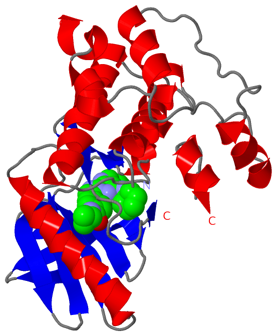
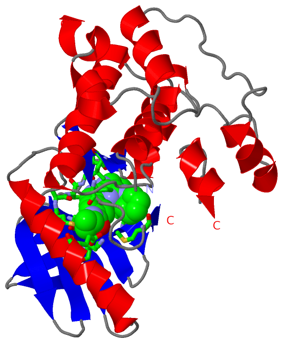
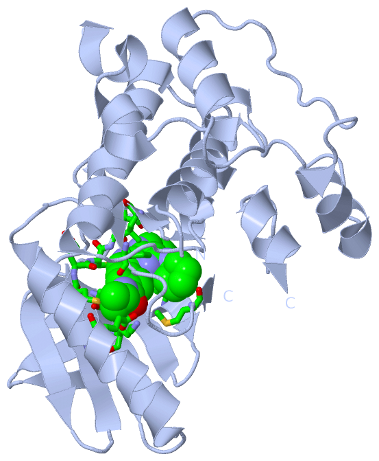
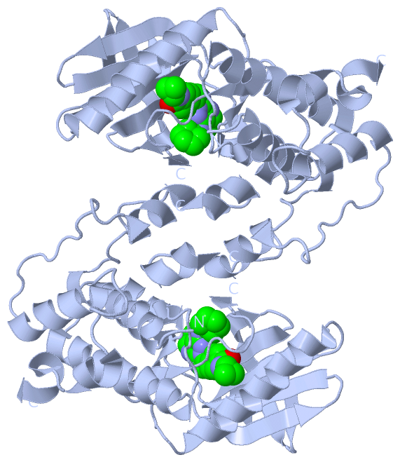
Asymmetric Unit (1, 1)
|
Sites (1, 1)
Asymmetric Unit (1, 1)
|
SS Bonds (0, 0)| (no "SS Bond" information available for 3WZJ) |
Cis Peptide Bonds (2, 2)
Asymmetric Unit
|
||||||||||||
SAPs(SNPs)/Variants (0, 0)| (no "SAP(SNP)/Variant" information available for 3WZJ) |
PROSITE Motifs (0, 0)| (no "PROSITE Motif" information available for 3WZJ) |
Exons (0, 0)| (no "Exon" information available for 3WZJ) |
Sequences/Alignments
Asymmetric Unit
Chain A from PDB Type:PROTEIN Length:257
SCOP domains ----------------------------------------------------------------------------------------------------------------------------------------------------------------------------------------------------------------------------------------------------------------- SCOP domains
CATH domains ----------------------------------------------------------------------------------------------------------------------------------------------------------------------------------------------------------------------------------------------------------------- CATH domains
Pfam domains ----------------------------------------------------------------------------------------------------------------------------------------------------------------------------------------------------------------------------------------------------------------- Pfam domains
SAPs(SNPs) ----------------------------------------------------------------------------------------------------------------------------------------------------------------------------------------------------------------------------------------------------------------- SAPs(SNPs)
PROSITE ----------------------------------------------------------------------------------------------------------------------------------------------------------------------------------------------------------------------------------------------------------------- PROSITE
Transcript ----------------------------------------------------------------------------------------------------------------------------------------------------------------------------------------------------------------------------------------------------------------- Transcript
3wzj A 516 ECISVKGRIYSILKQIGSGGSSKVFQVLNEKKQIYAIKYVNLEEADNQTLDSYRNEIAYLNKLQQHSDKIIRLYDYEITDQYIYMVMECGNIDLNSWLKKKKSIDPWERKSYWKNMLEAVHTIHQHGIVHSDLKPANFLIVDGMLKLIDFGIANQMQPDGTVNYMPPEAIKDMISPKSDVWSLGCILYYMTYGKTPFQQIINQISKLHAIIDPNHEIEFPDIPEKDLQDVLKCCLKRDPKQRISIPELLAHPYVQIQ 794
525 535 545 555 565 575 585 595 605 615 625 635 645 655 665 685 695 || 717 727 737 747 757 767 777 787
674| 698|
685 711
|
||||||||||||||||||||
SCOP Domains (0, 0)| (no "SCOP Domain" information available for 3WZJ) |
CATH Domains (0, 0)| (no "CATH Domain" information available for 3WZJ) |
Pfam Domains (0, 0)| (no "Pfam Domain" information available for 3WZJ) |
Gene Ontology (21, 21)|
Asymmetric Unit(hide GO term definitions) |
Interactive Views
|
|||||||||||||||||||||||||||||||||||||||||||||||||||||||||||||||||||||||||||||||||||||||||||||||||||||||||||||||||||||||||||||||||||||||||||||||||||||
Still Images
|
||||||||||||||||
Databases
|
||||||||||||||||||||||||||||||||||||||||||||||||||||||||||||||||||||||||||||||||||||||||||||||||||||||||||||||||||||||||||||||||||||||||||||||||||||||||||||||||
Analysis Tools
|
|||||||||||||||||||||||||||||||||||||||||||||||||||||||||||||
Entries Sharing at Least One Protein Chain (UniProt ID)
Related Entries Specified in the PDB File
|
|