Show PDB file:   
         Plain Text   HTML   (compressed file size)
QuickSearch:   
by PDB,NDB,UniProt,PROSITE Code or Search Term(s)  
(-)Asymmetric Unit
(-)Asym. Unit - sites
(-)Biological Unit 1
(-)Biol. Unit 1 - sites
(-)Biological Unit 2
(-)Biological Unit 3
collapse expand < >
Image Asymmetric Unit
Asymmetric Unit  (Jmol Viewer)
Image Asym. Unit - sites
Asym. Unit - sites  (Jmol Viewer)
Image Biological Unit 1
Biological Unit 1  (Jmol Viewer)
Image Biol. Unit 1 - sites
Biol. Unit 1 - sites  (Jmol Viewer)
Image Biological Unit 2
Biological Unit 2  (Jmol Viewer)
Image Biological Unit 3
Biological Unit 3  (Jmol Viewer)

(-) Description

Title :  CRYSTAL STRUCTURE OF A0-DOMAIN OF P5 FROM H. SAPIENS
 
Authors :  K. Inaba, M. Suzuki
Date :  03 Sep 12  (Deposition) - 04 Sep 13  (Release) - 04 Sep 13  (Revision)
Method :  X-RAY DIFFRACTION
Resolution :  1.93
Chains :  Asym. Unit :  A,B
Biol. Unit 1:  A  (1x)
Biol. Unit 2:  B  (1x)
Biol. Unit 3:  A,B  (1x)
Keywords :  Pdi Family Member, Thioredoxin Fold, Protein Disulfide Isomerase, Isomerase (Keyword Search: [Gene Ontology, PubMed, Web (Google))
 
Reference :  Y. Sato, R. Kojima, M. Okumura, M. Hagiwara, S. Masui, K. Maegawa, M. Saiki, T. Horibe, M. Suzuki, K. Inaba
Synergistic Cooperation Of Pdi Family Members In Peroxiredoxin 4-Driven Oxidative Protein Folding
Sci Rep V. 3 2456 2013
PubMed-ID: 23949117  |  Reference-DOI: 10.1038/SREP02456

(-) Compounds

Molecule 1 - PROTEIN DISULFIDE-ISOMERASE A6
    ChainsA, B
    EC Number5.3.4.1
    EngineeredYES
    Expression SystemESCHERICHIA COLI
    Expression System PlasmidPET15B
    Expression System StrainBL21(DE3)
    Expression System Taxid562
    Expression System Vector TypePLASMID
    FragmentA0 DOMAIN, UNP RESIDUES 25-140
    GenePDIA6, ERP5, P5, TXNDC7
    Organism CommonHUMAN
    Organism ScientificHOMO SAPIENS
    Organism Taxid9606
    SynonymENDOPLASMIC RETICULUM PROTEIN 5, ER PROTEIN 5, ERP5, PROTEIN DISULFIDE ISOMERASE P5, THIOREDOXIN DOMAIN-CONTAINING PROTEIN 7

 Structural Features

(-) Chains, Units

  12
Asymmetric Unit AB
Biological Unit 1 (1x)A 
Biological Unit 2 (1x) B
Biological Unit 3 (1x)AB

Summary Information (see also Sequences/Alignments below)

(-) Ligands, Modified Residues, Ions  (2, 4)

Asymmetric Unit (2, 4)
No.NameCountTypeFull Name
1MLY2Mod. Amino AcidN-DIMETHYL-LYSINE
2PO42Ligand/IonPHOSPHATE ION
Biological Unit 1 (2, 2)
No.NameCountTypeFull Name
1MLY1Mod. Amino AcidN-DIMETHYL-LYSINE
2PO41Ligand/IonPHOSPHATE ION
Biological Unit 2 (2, 2)
No.NameCountTypeFull Name
1MLY1Mod. Amino AcidN-DIMETHYL-LYSINE
2PO41Ligand/IonPHOSPHATE ION
Biological Unit 3 (2, 4)
No.NameCountTypeFull Name
1MLY2Mod. Amino AcidN-DIMETHYL-LYSINE
2PO42Ligand/IonPHOSPHATE ION

(-) Sites  (2, 2)

Asymmetric Unit (2, 2)
No.NameEvidenceResiduesDescription
1AC1SOFTWAREHIS A:59 , PRO A:101 , ARG A:120 , HOH A:368 , HOH A:380 , SER B:35BINDING SITE FOR RESIDUE PO4 A 201
2AC2SOFTWAREHIS A:59 , ARG A:62 , ASN B:38 , GLN B:43BINDING SITE FOR RESIDUE PO4 B 201

(-) SS Bonds  (0, 0)

(no "SS Bond" information available for 3VWW)

(-) Cis Peptide Bonds  (2, 2)

Asymmetric Unit
No.Residues
1Phe A:100 -Pro A:101
2Phe B:100 -Pro B:101

 Sequence-Structure Mapping

(-) SAPs(SNPs)/Variants  (0, 0)

(no "SAP(SNP)/Variant" information available for 3VWW)

(-) PROSITE Motifs  (1, 2)

Asymmetric Unit (1, 2)
 PROSITEUniProtKBPDB
No.IDACDescriptionIDLocationCountLocation
1THIOREDOXIN_1PS00194 Thioredoxin family active site.PDIA6_HUMAN47-65
 
182-200
  2A:49-67
B:49-67
-
Biological Unit 1 (1, 1)
 PROSITEUniProtKBPDB
No.IDACDescriptionIDLocationCountLocation
1THIOREDOXIN_1PS00194 Thioredoxin family active site.PDIA6_HUMAN47-65
 
182-200
  1A:49-67
-
-
Biological Unit 2 (1, 1)
 PROSITEUniProtKBPDB
No.IDACDescriptionIDLocationCountLocation
1THIOREDOXIN_1PS00194 Thioredoxin family active site.PDIA6_HUMAN47-65
 
182-200
  1-
B:49-67
-
Biological Unit 3 (1, 2)
 PROSITEUniProtKBPDB
No.IDACDescriptionIDLocationCountLocation
1THIOREDOXIN_1PS00194 Thioredoxin family active site.PDIA6_HUMAN47-65
 
182-200
  2A:49-67
B:49-67
-

(-) Exons   (4, 8)

Asymmetric Unit (4, 8)
 ENSEMBLUniProtKBPDB
No.Transcript IDExonExon IDGenome LocationLengthIDLocationLengthCountLocationLength
1.8aENST000002722278aENSE00001401250chr2:10952970-10952804167PDIA6_HUMAN1-770--
1.10ENST0000027222710ENSE00000996575chr2:10942766-10942625142PDIA6_HUMAN7-54482A:27-56
B:27-56
30
30
1.11ENST0000027222711ENSE00000962411chr2:10937883-1093782658PDIA6_HUMAN54-73202A:56-75
B:56-75
20
20
1.12bENST0000027222712bENSE00000962412chr2:10937333-10937207127PDIA6_HUMAN74-116432A:76-118
B:76-118
43
43
1.13ENST0000027222713ENSE00000962413chr2:10933328-10933222107PDIA6_HUMAN116-151362A:118-142
B:118-142
25
25
1.14ENST0000027222714ENSE00000962414chr2:10932051-10931921131PDIA6_HUMAN152-195440--
1.15ENST0000027222715ENSE00000962415chr2:10930959-10930845115PDIA6_HUMAN195-233390--
1.16ENST0000027222716ENSE00000962416chr2:10930015-10929875141PDIA6_HUMAN234-280470--
1.17ENST0000027222717ENSE00000962418chr2:10929107-1092902385PDIA6_HUMAN281-309290--
1.18ENST0000027222718ENSE00000962419chr2:10928895-1092882373PDIA6_HUMAN309-333250--
1.19ENST0000027222719ENSE00000962420chr2:10927565-10927407159PDIA6_HUMAN333-386540--
1.20ENST0000027222720ENSE00000962421chr2:10925156-1092506097PDIA6_HUMAN386-418330--
1.21cENST0000027222721cENSE00001893291chr2:10924452-10923517936PDIA6_HUMAN419-440220--

(-) Sequences/Alignments

Asymmetric Unit
   Reformat: Number of residues per line =  ('0' or empty: single-line sequence representation)
  Number of residues per labelling interval =   
  UniProt sequence: complete  aligned part    
   Show mapping: SCOP domains CATH domains Pfam domains Secondary structure (by author)
SAPs(SNPs) PROSITE motifs Exons
(details for a mapped element are shown in a popup box when the mouse pointer rests over it)
Chain A from PDB  Type:PROTEIN  Length:116
 aligned with PDIA6_HUMAN | Q15084 from UniProtKB/Swiss-Prot  Length:440

    Alignment length:116
                                    34        44        54        64        74        84        94       104       114       124       134      
          PDIA6_HUMAN    25 DDVIELTPSNFNREVIQSDSLWLVEFYAPWCGHCQRLTPEWKKAATALKDVVKVGAVDADKHHSLGGQYGVQGFPTIKIFGSNKNRPEDYQGGRTGEAIVDAALSALRQLVKDRLG 140
               SCOP domains d3vwwa_ A: automated matches                                                                                         SCOP domains
               CATH domains -------------------------------------------------------------------------------------------------------------------- CATH domains
               Pfam domains -------------------------------------------------------------------------------------------------------------------- Pfam domains
         Sec.struct. author ...ee....hhhhhh......eeeeee...hhhhhhhhhhhhhhhhhh....eeeeee...hhhhhhhh......eeeee.......eee....hhhhhhhhhhhhhhhhhhhhhh Sec.struct. author
                 SAPs(SNPs) -------------------------------------------------------------------------------------------------------------------- SAPs(SNPs)
                    PROSITE ----------------------THIOREDOXIN_1      --------------------------------------------------------------------------- PROSITE
           Transcript 1 (1) Exon 1.10  PDB: A:27-56       -------------------Exon 1.12b  PDB: A:76-118 UniProt: 74-116  ------------------------ Transcript 1 (1)
           Transcript 1 (2) -----------------------------Exon 1.11           ------------------------------------------Exon 1.13  PDB: A:118-142 Transcript 1 (2)
                 3vww A  27 DDVIELTPSNFNREVIQSDSLWLVEFYAPWCGHCQRLTPEWKKAATALKDVVKVGAVDADKHHSLGGQYGVQGFPTIkIFGSNKNRPEDYQGGRTGEAIVDAALSALRQLVKDRLG 142
                                    36        46        56        66        76        86        96       106       116       126       136      
                                                                                                       104-MLY                                  

Chain B from PDB  Type:PROTEIN  Length:116
 aligned with PDIA6_HUMAN | Q15084 from UniProtKB/Swiss-Prot  Length:440

    Alignment length:116
                                    34        44        54        64        74        84        94       104       114       124       134      
          PDIA6_HUMAN    25 DDVIELTPSNFNREVIQSDSLWLVEFYAPWCGHCQRLTPEWKKAATALKDVVKVGAVDADKHHSLGGQYGVQGFPTIKIFGSNKNRPEDYQGGRTGEAIVDAALSALRQLVKDRLG 140
               SCOP domains d3vwwb_ B: automated matches                                                                                         SCOP domains
               CATH domains -------------------------------------------------------------------------------------------------------------------- CATH domains
               Pfam domains -------------------------------------------------------------------------------------------------------------------- Pfam domains
         Sec.struct. author ...eee...hhhhhh......eeeeee...hhhhhhhhhhhhhhhhhh....eeeeee...hhhhhhhh......eeeee.......ee.....hhhhhhhhhhhhhhhhhhhhhh Sec.struct. author
                 SAPs(SNPs) -------------------------------------------------------------------------------------------------------------------- SAPs(SNPs)
                    PROSITE ----------------------THIOREDOXIN_1      --------------------------------------------------------------------------- PROSITE
           Transcript 1 (1) Exon 1.10  PDB: B:27-56       -------------------Exon 1.12b  PDB: B:76-118 UniProt: 74-116  ------------------------ Transcript 1 (1)
           Transcript 1 (2) -----------------------------Exon 1.11           ------------------------------------------Exon 1.13  PDB: B:118-142 Transcript 1 (2)
                 3vww B  27 DDVIELTPSNFNREVIQSDSLWLVEFYAPWCGHCQRLTPEWKKAATALKDVVKVGAVDADKHHSLGGQYGVQGFPTIkIFGSNKNRPEDYQGGRTGEAIVDAALSALRQLVKDRLG 142
                                    36        46        56        66        76        86        96       106       116       126       136      
                                                                                                       104-MLY                                  

   Legend:   → Mismatch (orange background)
  - → Gap (green background, '-', border residues have a numbering label)
    → Modified Residue (blue background, lower-case, 'x' indicates undefined single-letter code, labelled with number + name)
  x → Chemical Group (purple background, 'x', labelled with number + name, e.g. ACE or NH2)
  extra numbering lines below/above indicate numbering irregularities and modified residue names etc., number ends below/above '|'

 Classification and Annotation

(-) SCOP Domains  (1, 2)

Asymmetric Unit

(-) CATH Domains  (0, 0)

(no "CATH Domain" information available for 3VWW)

(-) Pfam Domains  (0, 0)

(no "Pfam Domain" information available for 3VWW)

(-) Gene Ontology  (17, 17)

Asymmetric Unit(hide GO term definitions)
Chain A,B   (PDIA6_HUMAN | Q15084)
molecular function
    GO:0016853    isomerase activity    Catalysis of the geometric or structural changes within one molecule. Isomerase is the systematic name for any enzyme of EC class 5.
    GO:0005515    protein binding    Interacting selectively and non-covalently with any protein or protein complex (a complex of two or more proteins that may include other nonprotein molecules).
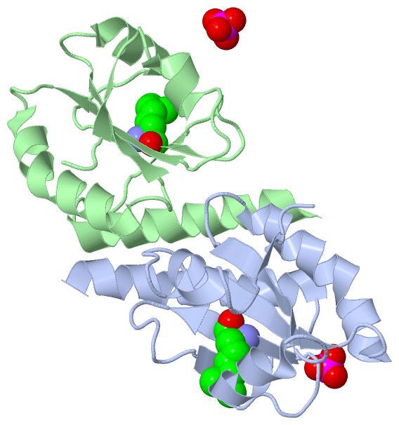
    GO:0003756    protein disulfide isomerase activity    Catalysis of the rearrangement of both intrachain and interchain disulfide bonds in proteins.
biological process
    GO:0036498    IRE1-mediated unfolded protein response    A series of molecular signals mediated by the endoplasmic reticulum stress sensor IRE1 (Inositol-requiring transmembrane kinase/endonuclease). Begins with activation of IRE1 in response to endoplasmic reticulum (ER) stress, and ends with regulation of a downstream cellular process, e.g. transcription. One target of activated IRE1 is the transcription factor HAC1 in yeast, or XBP1 in mammals; IRE1 cleaves an intron of a mRNA coding for HAC1/XBP1 to generate an activated HAC1/XBP1 transcription factor, which controls the up regulation of UPR-related genes. At least in mammals, IRE1 can also signal through additional intracellular pathways including JNK and NF-kappaB.
    GO:0043277    apoptotic cell clearance    The recognition and removal of an apoptotic cell by a neighboring cell or by a phagocyte.
    GO:0045454    cell redox homeostasis    Any process that maintains the redox environment of a cell or compartment within a cell.
    GO:0006457    protein folding    The process of assisting in the covalent and noncovalent assembly of single chain polypeptides or multisubunit complexes into the correct tertiary structure.
    GO:0034976    response to endoplasmic reticulum stress    Any process that results in a change in state or activity of a cell (in terms of movement, secretion, enzyme production, gene expression, etc.) as a result of a stress acting at the endoplasmic reticulum. ER stress usually results from the accumulation of unfolded or misfolded proteins in the ER lumen.
cellular component
    GO:0005783    endoplasmic reticulum    The irregular network of unit membranes, visible only by electron microscopy, that occurs in the cytoplasm of many eukaryotic cells. The membranes form a complex meshwork of tubular channels, which are often expanded into slitlike cavities called cisternae. The ER takes two forms, rough (or granular), with ribosomes adhering to the outer surface, and smooth (with no ribosomes attached).
    GO:0034663    endoplasmic reticulum chaperone complex    A protein complex that is located in the endoplasmic reticulum and is composed of chaperone proteins, including BiP, GRP94; CaBP1, protein disulfide isomerase (PDI), ERdj3, cyclophilin B, ERp72, GRP170, UDP-glucosyltransferase, and SDF2-L1.
    GO:0005788    endoplasmic reticulum lumen    The volume enclosed by the membranes of the endoplasmic reticulum.
    GO:0005789    endoplasmic reticulum membrane    The lipid bilayer surrounding the endoplasmic reticulum.
    GO:0005793    endoplasmic reticulum-Golgi intermediate compartment    A complex system of membrane-bounded compartments located between endoplasmic reticulum (ER) and the Golgi complex, with a distinctive membrane protein composition; involved in ER-to-Golgi and Golgi-to-ER transport.
    GO:0070062    extracellular exosome    A vesicle that is released into the extracellular region by fusion of the limiting endosomal membrane of a multivesicular body with the plasma membrane. Extracellular exosomes, also simply called exosomes, have a diameter of about 40-100 nm.
    GO:0042470    melanosome    A tissue-specific, membrane-bounded cytoplasmic organelle within which melanin pigments are synthesized and stored. Melanosomes are synthesized in melanocyte cells.
    GO:0016020    membrane    A lipid bilayer along with all the proteins and protein complexes embedded in it an attached to it.
    GO:0005886    plasma membrane    The membrane surrounding a cell that separates the cell from its external environment. It consists of a phospholipid bilayer and associated proteins.

 Visualization

(-) Interactive Views

Asymmetric Unit
  Complete Structure
    Jena3D(integrated viewing of ligand, site, SAP, PROSITE, SCOP information)
    WebMol | AstexViewer[tm]@PDBe
(Java Applets, require no local installation except for Java; loading may be slow)
    STRAP
(Java WebStart application, automatic local installation, requires Java; full application with system access!)
    RasMol
(require local installation)
    Molscript (VRML)
(requires installation of a VRML viewer; select preferred view via VRML and generate a mono or stereo PDF format file)
 
  Ligands, Modified Residues, Ions
    MLY  [ RasMol | Jena3D ]  +environment [ RasMol | Jena3D ]
    PO4  [ RasMol | Jena3D ]  +environment [ RasMol | Jena3D ]
 
  Sites
    AC1  [ RasMol ]  +environment [ RasMol ]
    AC2  [ RasMol ]  +environment [ RasMol ]
 
  Cis Peptide Bonds
    Phe A:100 - Pro A:101   [ RasMol ]  
    Phe B:100 - Pro B:101   [ RasMol ]  
 
Biological Units
  Complete Structure
    Biological Unit 1  [ Jena3D ]
    Biological Unit 2  [ Jena3D ]
    Biological Unit 3  [ Jena3D ]

(-) Still Images

Jmol
  protein: cartoon or spacefill or dots and stick; nucleic acid: cartoon and stick; ligands: spacefill; active site: stick
Molscript
  protein, nucleic acid: cartoon; ligands: spacefill; active site: ball and stick

 Databases and Analysis Tools

(-) Databases

Access by PDB/NDB ID
  3vww
    Family and Domain InformationProDom | SYSTERS
    General Structural InformationGlycoscienceDB | MMDB | NDB | OCA | PDB | PDBe | PDBj | PDBsum | PDBWiki | PQS | PROTEOPEDIA
    Orientation in MembranesOPM
    Protein SurfaceSURFACE
    Secondary StructureDSSP (structure derived) | HSSP (homology derived)
    Structural GenomicsGeneCensus
    Structural NeighboursCE | VAST
    Structure ClassificationCATH | Dali | SCOP
    Validation and Original DataBMRB Data View | BMRB Restraints Grid | EDS | PROCHECK | RECOORD | WHAT_CHECK
 
Access by UniProt ID/Accession number
  PDIA6_HUMAN | Q15084
    Comparative Protein Structure ModelsModBase
    Genomic InformationEnsembl
    Protein-protein InteractionDIP
    Sequence, Family and Domain InformationInterPro | Pfam | SMART | UniProtKB/SwissProt
 
Access by Enzyme Classificator   (EC Number)
  5.3.4.1
    General Enzyme InformationBRENDA | EC-PDB | Enzyme | IntEnz
    PathwayKEGG | MetaCyc
 
Access by Disease Identifier   (MIM ID)
  (no 'MIM ID' available)
    Disease InformationOMIM
 
Access by GenAge ID
  (no 'GenAge ID' available)
    Age Related InformationGenAge

(-) Analysis Tools

Access by PDB/NDB ID
    Domain InformationXDom
    Interatomic Contacts of Structural UnitsCSU
    Ligand-protein ContactsLPC
    Protein CavitiescastP
    Sequence and Secondary StructurePDBCartoon
    Structure AlignmentSTRAP(Java WebStart application, automatic local installation, requires Java; full application with system access!)
    Structure and Sequence BrowserSTING
 
Access by UniProt ID/Accession number
  PDIA6_HUMAN | Q15084
    Protein Disorder PredictionDisEMBL | FoldIndex | GLOBPLOT (for more information see DisProt)

 Related Entries

(-) Entries Sharing at Least One Protein Chain (UniProt ID)

UniProtKB/Swiss-Prot
        PDIA6_HUMAN | Q150841x5d 3w8j 4ef0 4gwr

(-) Related Entries Specified in the PDB File

2dml NMR STRUCTURE OF P5 A0-DOMAIN
3vwu
3vwv
3w8j