| molecular function |
|---|
| | GO:0016853 | | isomerase activity | | Catalysis of the geometric or structural changes within one molecule. Isomerase is the systematic name for any enzyme of EC class 5. |
| | GO:0003756 | | protein disulfide isomerase activity | | Catalysis of the rearrangement of both intrachain and interchain disulfide bonds in proteins. |
| biological process |
|---|
| | GO:0043277 | | apoptotic cell clearance | | The recognition and removal of an apoptotic cell by a neighboring cell or by a phagocyte. |
| | GO:0045454 | | cell redox homeostasis | | Any process that maintains the redox environment of a cell or compartment within a cell. |
| | GO:0030168 | | platelet activation | | A series of progressive, overlapping events triggered by exposure of the platelets to subendothelial tissue. These events include shape change, adhesiveness, aggregation, and release reactions. When carried through to completion, these events lead to the formation of a stable hemostatic plug. |
| | GO:0070527 | | platelet aggregation | | The adhesion of one platelet to one or more other platelets via adhesion molecules. |
| | GO:0006457 | | protein folding | | The process of assisting in the covalent and noncovalent assembly of single chain polypeptides or multisubunit complexes into the correct tertiary structure. |
| | GO:0034976 | | response to endoplasmic reticulum stress | | Any process that results in a change in state or activity of a cell (in terms of movement, secretion, enzyme production, gene expression, etc.) as a result of a stress acting at the endoplasmic reticulum. ER stress usually results from the accumulation of unfolded or misfolded proteins in the ER lumen. |
| cellular component |
|---|
| | GO:0005783 | | endoplasmic reticulum | | The irregular network of unit membranes, visible only by electron microscopy, that occurs in the cytoplasm of many eukaryotic cells. The membranes form a complex meshwork of tubular channels, which are often expanded into slitlike cavities called cisternae. The ER takes two forms, rough (or granular), with ribosomes adhering to the outer surface, and smooth (with no ribosomes attached). |
| | GO:0034663 | | endoplasmic reticulum chaperone complex | | A protein complex that is located in the endoplasmic reticulum and is composed of chaperone proteins, including BiP, GRP94; CaBP1, protein disulfide isomerase (PDI), ERdj3, cyclophilin B, ERp72, GRP170, UDP-glucosyltransferase, and SDF2-L1. |
| | GO:0005788 | | endoplasmic reticulum lumen | | The volume enclosed by the membranes of the endoplasmic reticulum. |
| | GO:0005793 | | endoplasmic reticulum-Golgi intermediate compartment | | A complex system of membrane-bounded compartments located between endoplasmic reticulum (ER) and the Golgi complex, with a distinctive membrane protein composition; involved in ER-to-Golgi and Golgi-to-ER transport. |
| | GO:0070062 | | extracellular exosome | | A vesicle that is released into the extracellular region by fusion of the limiting endosomal membrane of a multivesicular body with the plasma membrane. Extracellular exosomes, also simply called exosomes, have a diameter of about 40-100 nm. |
| | GO:0042470 | | melanosome | | A tissue-specific, membrane-bounded cytoplasmic organelle within which melanin pigments are synthesized and stored. Melanosomes are synthesized in melanocyte cells. |
| | GO:0016020 | | membrane | | A lipid bilayer along with all the proteins and protein complexes embedded in it an attached to it. |
| | GO:0005886 | | plasma membrane | | The membrane surrounding a cell that separates the cell from its external environment. It consists of a phospholipid bilayer and associated proteins. |
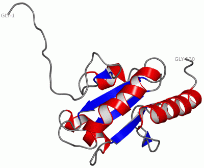
 Description
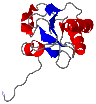