Show PDB file:   
         Plain Text   HTML   (compressed file size)
QuickSearch:   
by PDB,NDB,UniProt,PROSITE Code or Search Term(s)  
(-)Asymmetric Unit
(-)Asym. Unit - sites
(-)Biological Unit 1
(-)Biol. Unit 1 - sites
(-)Biological Unit 2
collapse expand < >
Image Asymmetric Unit
Asymmetric Unit  (Jmol Viewer)
Image Asym. Unit - sites
Asym. Unit - sites  (Jmol Viewer)
Image Biological Unit 1
Biological Unit 1  (Jmol Viewer)
Image Biol. Unit 1 - sites
Biol. Unit 1 - sites  (Jmol Viewer)
Image Biological Unit 2
Biological Unit 2  (Jmol Viewer)

(-) Description

Title :  MMP12 IN A COMPLEX WITH THE DIMERIC ADDUCT: 5-(5-PHENYLHYDANTOIN)-5-PHENYLHYDANTOIN
 
Authors :  D. J. Derbyshire, H. Danielson, S. Nystrum
Date :  29 Nov 11  (Deposition) - 02 Jan 13  (Release) - 02 Jan 13  (Revision)
Method :  X-RAY DIFFRACTION
Resolution :  1.30
Chains :  Asym. Unit :  A,B
Biol. Unit 1:  A  (1x)
Biol. Unit 2:  B  (1x)
Keywords :  Hydrolase, Metalloprotease (Keyword Search: [Gene Ontology, PubMed, Web (Google))
 
Reference :  H. Nordstrom, T. Gossas, D. J. Derbyshire, H. Danielson
Characterization Of Fragments Interacting With Mmp-12
To Be Published
PubMed: search

(-) Compounds

Molecule 1 - MACROPHAGE METALLOELASTASE
    ChainsA, B
    EC Number3.4.24.65
    EngineeredYES
    Expression SystemESCHERICHIA COLI
    Expression System PlasmidPET11A
    Expression System StrainBL21(DE3)
    Expression System Taxid469008
    Expression System Vector TypePLASMID
    FragmentCATALYTIC DOMAIN, RESIDUES 106-263
    Gene4321, HME, MMP12
    MutationYES
    Organism CommonHUMAN
    Organism ScientificHOMO SAPIENS
    Organism Taxid9606
    SynonymMME, MACROPHAGE ELASTASE, ME, HME, MATRIX METALLOPROTEINASE- 12, MMP-12

 Structural Features

(-) Chains, Units

  12
Asymmetric Unit AB
Biological Unit 1 (1x)A 
Biological Unit 2 (1x) B

Summary Information (see also Sequences/Alignments below)

(-) Ligands, Modified Residues, Ions  (6, 27)

Asymmetric Unit (6, 27)
No.NameCountTypeFull Name
10D21Ligand/Ion(4R,4'S)-4,4'-DIPHENYL-4,4'-BIIMIDAZOLIDINE-2,2',5,5'-TETRONE
2CA7Ligand/IonCALCIUM ION
3CL6Ligand/IonCHLORIDE ION
4EDO2Ligand/Ion1,2-ETHANEDIOL
5IMD5Ligand/IonIMIDAZOLE
6ZN6Ligand/IonZINC ION
Biological Unit 1 (3, 5)
No.NameCountTypeFull Name
10D21Ligand/Ion(4R,4'S)-4,4'-DIPHENYL-4,4'-BIIMIDAZOLIDINE-2,2',5,5'-TETRONE
2CA-1Ligand/IonCALCIUM ION
3CL-1Ligand/IonCHLORIDE ION
4EDO1Ligand/Ion1,2-ETHANEDIOL
5IMD3Ligand/IonIMIDAZOLE
6ZN-1Ligand/IonZINC ION
Biological Unit 2 (2, 3)
No.NameCountTypeFull Name
10D2-1Ligand/Ion(4R,4'S)-4,4'-DIPHENYL-4,4'-BIIMIDAZOLIDINE-2,2',5,5'-TETRONE
2CA-1Ligand/IonCALCIUM ION
3CL-1Ligand/IonCHLORIDE ION
4EDO1Ligand/Ion1,2-ETHANEDIOL
5IMD2Ligand/IonIMIDAZOLE
6ZN-1Ligand/IonZINC ION

(-) Sites  (27, 27)

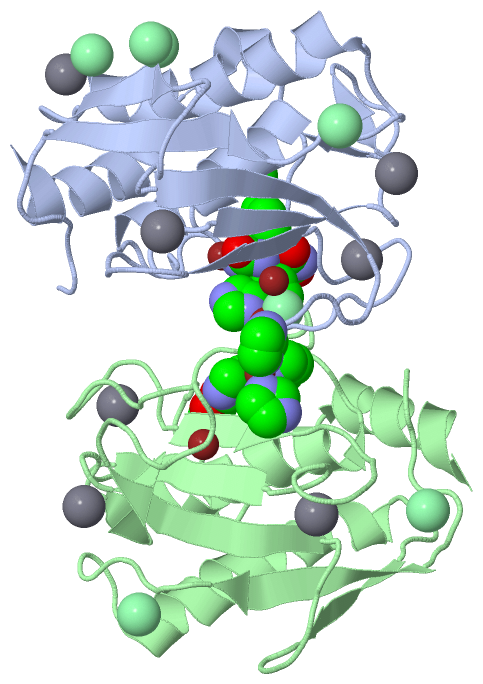
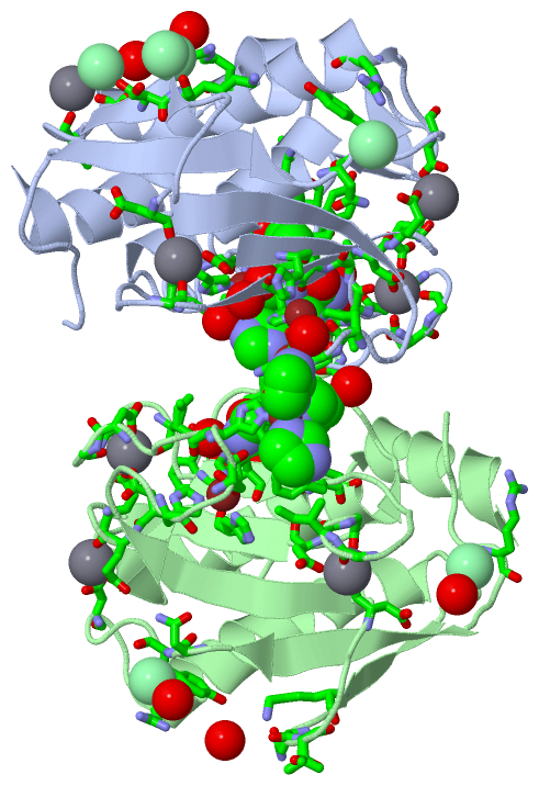
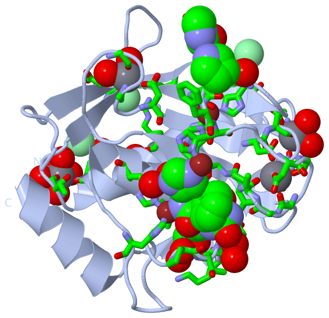
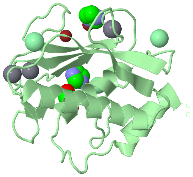
Asymmetric Unit (27, 27)
No.NameEvidenceResiduesDescription
01AC1SOFTWAREHIS A:218 , HIS A:222 , HIS A:228 , 0D2 A:314BINDING SITE FOR RESIDUE ZN A 301
02AC2SOFTWAREHIS A:168 , ASP A:170 , HIS A:183 , HIS A:196BINDING SITE FOR RESIDUE ZN A 302
03AC3SOFTWAREASP A:124 , GLU A:199 , GLU A:201 , HOH A:414 , HOH A:431BINDING SITE FOR RESIDUE CA A 303
04AC4SOFTWAREASP A:158 , GLY A:190 , GLY A:192 , ASP A:194 , HOH A:402 , HOH A:444BINDING SITE FOR RESIDUE CA A 304
05AC5SOFTWAREASP A:175 , GLY A:176 , GLY A:178 , ILE A:180 , ASP A:198 , GLU A:201BINDING SITE FOR RESIDUE CA A 305
06AC6SOFTWAREHIS A:172 , IMD A:307 , HIS B:228 , CL B:301BINDING SITE FOR RESIDUE ZN A 306
07AC7SOFTWAREHIS A:172 , ZN A:306 , 0D2 A:314 , HOH A:464 , HIS B:228 , PRO B:238 , CL B:301BINDING SITE FOR RESIDUE IMD A 307
08AC8SOFTWAREGLY A:169 , ASP A:170 , PHE A:171 , IMD A:313 , HOH A:418 , HIS B:172 , ZN B:308 , IMD B:309BINDING SITE FOR RESIDUE IMD A 312
09AC9SOFTWARESER A:142 , THR A:145 , HOH A:401 , HOH A:410 , HOH A:427 , HOH A:428 , HOH A:446BINDING SITE FOR RESIDUE CA A 308
10BC1SOFTWAREASN A:120 , TYR A:121 , ARG A:127BINDING SITE FOR RESIDUE CL A 309
11BC2SOFTWAREHOH A:609 , LYS B:151 , ASN B:153 , HOH B:557BINDING SITE FOR RESIDUE CL A 310
12BC3SOFTWAREARG A:135 , SER A:150 , LYS A:151 , ASN B:153 , THR B:154BINDING SITE FOR RESIDUE CL A 311
13BC4SOFTWAREGLY A:169 , IMD A:312 , HOH A:469 , HOH A:660 , HOH A:667 , HIS B:172 , ZN B:308 , IMD B:309 , HOH B:446BINDING SITE FOR RESIDUE IMD A 313
14BC5SOFTWAREGLY A:179 , ILE A:180 , LEU A:181 , ALA A:182 , HIS A:218 , HIS A:222 , HIS A:228 , PRO A:238 , TYR A:240 , ZN A:301 , IMD A:307 , HOH A:413 , HOH A:435 , HOH A:498 , SER B:229 , SER B:230 , PRO B:238BINDING SITE FOR RESIDUE 0D2 A 314
15BC6SOFTWARELEU A:214 , THR A:239 , TYR A:240 , LYS A:241 , HOH A:425 , HOH A:572BINDING SITE FOR RESIDUE EDO A 315
16BC7SOFTWAREHIS A:172 , ZN A:306 , IMD A:307 , HIS B:228BINDING SITE FOR RESIDUE CL B 301
17BC8SOFTWAREASN B:120 , TYR B:121 , ARG B:127 , HOH B:616BINDING SITE FOR RESIDUE CL B 302
18BC9SOFTWAREHIS B:218 , HIS B:222 , HIS B:228 , IMD B:311BINDING SITE FOR RESIDUE ZN B 303
19CC1SOFTWAREHIS B:168 , ASP B:170 , HIS B:183 , HIS B:196BINDING SITE FOR RESIDUE ZN B 304
20CC2SOFTWAREASP B:124 , GLU B:199 , GLU B:201 , HOH B:417 , HOH B:423BINDING SITE FOR RESIDUE CA B 305
21CC3SOFTWAREASP B:158 , GLY B:190 , GLY B:192 , ASP B:194 , HOH B:408 , HOH B:412BINDING SITE FOR RESIDUE CA B 306
22CC4SOFTWAREASP B:175 , GLY B:176 , GLY B:178 , ILE B:180 , ASP B:198 , GLU B:201BINDING SITE FOR RESIDUE CA B 307
23CC5SOFTWAREIMD A:312 , IMD A:313 , HOH A:469 , HIS B:172 , IMD B:309 , HOH B:446BINDING SITE FOR RESIDUE ZN B 308
24CC6SOFTWAREIMD A:312 , IMD A:313 , ASP B:170 , HIS B:172 , PHE B:185 , ILE B:191 , ZN B:308 , HOH B:446BINDING SITE FOR RESIDUE IMD B 309
25CC7SOFTWAREARG B:110 , HOH B:463BINDING SITE FOR RESIDUE CL B 310
26CC8SOFTWAREALA B:182 , HIS B:218 , HIS B:222 , HIS B:228 , ZN B:303 , EDO B:312 , HOH B:442BINDING SITE FOR RESIDUE IMD B 311
27CC9SOFTWAREHIS B:218 , PRO B:238 , TYR B:240 , IMD B:311 , HOH B:407 , HOH B:581BINDING SITE FOR RESIDUE EDO B 312

(-) SS Bonds  (0, 0)

(no "SS Bond" information available for 3UVC)

(-) Cis Peptide Bonds  (1, 1)

Asymmetric Unit
No.Residues
1Gly A:106 -Pro A:107

 Sequence-Structure Mapping

(-) SAPs(SNPs)/Variants  (0, 0)

(no "SAP(SNP)/Variant" information available for 3UVC)

(-) PROSITE Motifs  (1, 2)

Asymmetric Unit (1, 2)
 PROSITEUniProtKBPDB
No.IDACDescriptionIDLocationCountLocation
1ZINC_PROTEASEPS00142 Neutral zinc metallopeptidases, zinc-binding region signature.MMP12_HUMAN215-224
 
  2A:215-224
B:215-224
Biological Unit 1 (1, 1)
 PROSITEUniProtKBPDB
No.IDACDescriptionIDLocationCountLocation
1ZINC_PROTEASEPS00142 Neutral zinc metallopeptidases, zinc-binding region signature.MMP12_HUMAN215-224
 
  1A:215-224
-
Biological Unit 2 (1, 1)
 PROSITEUniProtKBPDB
No.IDACDescriptionIDLocationCountLocation
1ZINC_PROTEASEPS00142 Neutral zinc metallopeptidases, zinc-binding region signature.MMP12_HUMAN215-224
 
  1-
B:215-224

(-) Exons   (0, 0)

(no "Exon" information available for 3UVC)

(-) Sequences/Alignments

Asymmetric Unit
   Reformat: Number of residues per line =  ('0' or empty: single-line sequence representation)
  Number of residues per labelling interval =   
  UniProt sequence: complete  aligned part    
   Show mapping: SCOP domains CATH domains Pfam domains Secondary structure (by author)
SAPs(SNPs) PROSITE motifs Exons
(details for a mapped element are shown in a popup box when the mouse pointer rests over it)
Chain A from PDB  Type:PROTEIN  Length:158
 aligned with MMP12_HUMAN | P39900 from UniProtKB/Swiss-Prot  Length:470

    Alignment length:158
                                   115       125       135       145       155       165       175       185       195       205       215       225       235       245       255        
          MMP12_HUMAN   106 GPVWRKHYITYRINNYTPDMNREDVDYAIRKAFQVWSNVTPLKFSKINTGMADILVVFARGAHGDFHAFDGKGGILAHAFGPGSGIGGDAHFDEDEFWTTHSGGTNLFLTAVHEIGHSLGLGHSSDPKAVMFPTYKYVDINTFRLSADDIRGIQSLYG 263
               SCOP domains d3uvca_ A: automated matches                                                                                                                                   SCOP domains
               CATH domains -------------------------------------------------------------------------------------------------------------------------------------------------------------- CATH domains
               Pfam domains -------------------------------------------------------------------------------------------------------------------------------------------------------------- Pfam domains
         Sec.struct. author .......eeeeee.......hhhhhhhhhhhhhhhhhh....eeee.......eeeeee.................eee..........eeee....ee.....eehhhhhhhhhhhhh...................hhhhh..hhhhhhhhhhhhh Sec.struct. author
                 SAPs(SNPs) -------------------------------------------------------------------------------------------------------------------------------------------------------------- SAPs(SNPs)
                    PROSITE -------------------------------------------------------------------------------------------------------------ZINC_PROTE--------------------------------------- PROSITE
                 Transcript -------------------------------------------------------------------------------------------------------------------------------------------------------------- Transcript
                 3uvc A 106 GPVWRKHYITYRINNYTPDMNREDVDYAIRKAFQVWSNVTPLKFSKINTGMADILVVFARGAHGDFHAFDGKGGILAHAFGPGSGIGGDAHFDEDEFWTTHSGGTNLFLTAVHAIGHSLGLGHSSDPKAVMFPTYKYVDINTFRLSADDIRGIQSLYG 263
                                   115       125       135       145       155       165       175       185       195       205       215       225       235       245       255        

Chain B from PDB  Type:PROTEIN  Length:159
 aligned with MMP12_HUMAN | P39900 from UniProtKB/Swiss-Prot  Length:470

    Alignment length:159
                                   114       124       134       144       154       164       174       184       194       204       214       224       234       244       254         
          MMP12_HUMAN   105 GGPVWRKHYITYRINNYTPDMNREDVDYAIRKAFQVWSNVTPLKFSKINTGMADILVVFARGAHGDFHAFDGKGGILAHAFGPGSGIGGDAHFDEDEFWTTHSGGTNLFLTAVHEIGHSLGLGHSSDPKAVMFPTYKYVDINTFRLSADDIRGIQSLYG 263
               SCOP domains d3uvcb_ B: automated matches                                                                                                                                    SCOP domains
               CATH domains --------------------------------------------------------------------------------------------------------------------------------------------------------------- CATH domains
               Pfam domains --------------------------------------------------------------------------------------------------------------------------------------------------------------- Pfam domains
         Sec.struct. author ........eeeeee.......hhhhhhhhhhhhhhhhhh....eeee.......eeeeee.................eee..........eeee....ee.....eehhhhhhhhhhhhh..........................hhhhhhhhhhhhh Sec.struct. author
                 SAPs(SNPs) --------------------------------------------------------------------------------------------------------------------------------------------------------------- SAPs(SNPs)
                    PROSITE --------------------------------------------------------------------------------------------------------------ZINC_PROTE--------------------------------------- PROSITE
                 Transcript --------------------------------------------------------------------------------------------------------------------------------------------------------------- Transcript
                 3uvc B 105 MGPVWRKHYITYRINNYTPDMNREDVDYAIRKAFQVWSNVTPLKFSKINTGMADILVVFARGAHGDFHAFDGKGGILAHAFGPGSGIGGDAHFDEDEFWTTHSGGTNLFLTAVHAIGHSLGLGHSSDPKAVMFPTYKYVDINTFRLSADDIRGIQSLYG 263
                                   114       124       134       144       154       164       174       184       194       204       214       224       234       244       254         

   Legend:   → Mismatch (orange background)
  - → Gap (green background, '-', border residues have a numbering label)
    → Modified Residue (blue background, lower-case, 'x' indicates undefined single-letter code, labelled with number + name)
  x → Chemical Group (purple background, 'x', labelled with number + name, e.g. ACE or NH2)
  extra numbering lines below/above indicate numbering irregularities and modified residue names etc., number ends below/above '|'

 Classification and Annotation

(-) SCOP Domains  (1, 2)

Asymmetric Unit

(-) CATH Domains  (0, 0)

(no "CATH Domain" information available for 3UVC)

(-) Pfam Domains  (0, 0)

(no "Pfam Domain" information available for 3UVC)

(-) Gene Ontology  (21, 21)

Asymmetric Unit(hide GO term definitions)
Chain A,B   (MMP12_HUMAN | P39900)
molecular function
    GO:0005509    calcium ion binding    Interacting selectively and non-covalently with calcium ions (Ca2+).
    GO:0004175    endopeptidase activity    Catalysis of the hydrolysis of internal, alpha-peptide bonds in a polypeptide chain.
    GO:0016787    hydrolase activity    Catalysis of the hydrolysis of various bonds, e.g. C-O, C-N, C-C, phosphoric anhydride bonds, etc. Hydrolase is the systematic name for any enzyme of EC class 3.
    GO:0046872    metal ion binding    Interacting selectively and non-covalently with any metal ion.
    GO:0004222    metalloendopeptidase activity    Catalysis of the hydrolysis of internal, alpha-peptide bonds in a polypeptide chain by a mechanism in which water acts as a nucleophile, one or two metal ions hold the water molecule in place, and charged amino acid side chains are ligands for the metal ions.
    GO:0008237    metallopeptidase activity    Catalysis of the hydrolysis of peptide bonds by a mechanism in which water acts as a nucleophile, one or two metal ions hold the water molecule in place, and charged amino acid side chains are ligands for the metal ions.
    GO:0008233    peptidase activity    Catalysis of the hydrolysis of a peptide bond. A peptide bond is a covalent bond formed when the carbon atom from the carboxyl group of one amino acid shares electrons with the nitrogen atom from the amino group of a second amino acid.
    GO:0004252    serine-type endopeptidase activity    Catalysis of the hydrolysis of internal, alpha-peptide bonds in a polypeptide chain by a catalytic mechanism that involves a catalytic triad consisting of a serine nucleophile that is activated by a proton relay involving an acidic residue (e.g. aspartate or glutamate) and a basic residue (usually histidine).
    GO:0008270    zinc ion binding    Interacting selectively and non-covalently with zinc (Zn) ions.
biological process
    GO:0030574    collagen catabolic process    The proteolytic chemical reactions and pathways resulting in the breakdown of collagen in the extracellular matrix, usually carried out by proteases secreted by nearby cells.
    GO:0022617    extracellular matrix disassembly    A process that results in the breakdown of the extracellular matrix.
    GO:0050679    positive regulation of epithelial cell proliferation    Any process that activates or increases the rate or extent of epithelial cell proliferation.
    GO:0060054    positive regulation of epithelial cell proliferation involved in wound healing    Any process that activates or increases the rate or extent of epithelial cell proliferation, contributing to the restoration of integrity to a damaged tissue following an injury.
    GO:0006508    proteolysis    The hydrolysis of proteins into smaller polypeptides and/or amino acids by cleavage of their peptide bonds.
    GO:0042493    response to drug    Any process that results in a change in state or activity of a cell or an organism (in terms of movement, secretion, enzyme production, gene expression, etc.) as a result of a drug stimulus. A drug is a substance used in the diagnosis, treatment or prevention of a disease.
    GO:0014070    response to organic cyclic compound    Any process that results in a change in state or activity of a cell or an organism (in terms of movement, secretion, enzyme production, gene expression, etc.) as a result of an organic cyclic compound stimulus.
    GO:0042060    wound healing    The series of events that restore integrity to a damaged tissue, following an injury.
    GO:0035313    wound healing, spreading of epidermal cells    The migration of an epidermal cell along or through a wound gap that contributes to the reestablishment of a continuous epidermis.
cellular component
    GO:0031012    extracellular matrix    A structure lying external to one or more cells, which provides structural support for cells or tissues.
    GO:0005576    extracellular region    The space external to the outermost structure of a cell. For cells without external protective or external encapsulating structures this refers to space outside of the plasma membrane. This term covers the host cell environment outside an intracellular parasite.
    GO:0005578    proteinaceous extracellular matrix    A layer consisting mainly of proteins (especially collagen) and glycosaminoglycans (mostly as proteoglycans) that forms a sheet underlying or overlying cells such as endothelial and epithelial cells. The proteins are secreted by cells in the vicinity. An example of this component is found in Mus musculus.

 Visualization

(-) Interactive Views

Asymmetric Unit
  Complete Structure
    Jena3D(integrated viewing of ligand, site, SAP, PROSITE, SCOP information)
    WebMol | AstexViewer[tm]@PDBe
(Java Applets, require no local installation except for Java; loading may be slow)
    STRAP
(Java WebStart application, automatic local installation, requires Java; full application with system access!)
    RasMol
(require local installation)
    Molscript (VRML)
(requires installation of a VRML viewer; select preferred view via VRML and generate a mono or stereo PDF format file)
 
  Ligands, Modified Residues, Ions
    0D2  [ RasMol | Jena3D ]  +environment [ RasMol | Jena3D ]
    CA  [ RasMol | Jena3D ]  +environment [ RasMol | Jena3D ]
    CL  [ RasMol | Jena3D ]  +environment [ RasMol | Jena3D ]
    EDO  [ RasMol | Jena3D ]  +environment [ RasMol | Jena3D ]
    IMD  [ RasMol | Jena3D ]  +environment [ RasMol | Jena3D ]
    ZN  [ RasMol | Jena3D ]  +environment [ RasMol | Jena3D ]
 
  Sites
    AC1  [ RasMol ]  +environment [ RasMol ]
    AC2  [ RasMol ]  +environment [ RasMol ]
    AC3  [ RasMol ]  +environment [ RasMol ]
    AC4  [ RasMol ]  +environment [ RasMol ]
    AC5  [ RasMol ]  +environment [ RasMol ]
    AC6  [ RasMol ]  +environment [ RasMol ]
    AC7  [ RasMol ]  +environment [ RasMol ]
    AC8  [ RasMol ]  +environment [ RasMol ]
    AC9  [ RasMol ]  +environment [ RasMol ]
    BC1  [ RasMol ]  +environment [ RasMol ]
    BC2  [ RasMol ]  +environment [ RasMol ]
    BC3  [ RasMol ]  +environment [ RasMol ]
    BC4  [ RasMol ]  +environment [ RasMol ]
    BC5  [ RasMol ]  +environment [ RasMol ]
    BC6  [ RasMol ]  +environment [ RasMol ]
    BC7  [ RasMol ]  +environment [ RasMol ]
    BC8  [ RasMol ]  +environment [ RasMol ]
    BC9  [ RasMol ]  +environment [ RasMol ]
    CC1  [ RasMol ]  +environment [ RasMol ]
    CC2  [ RasMol ]  +environment [ RasMol ]
    CC3  [ RasMol ]  +environment [ RasMol ]
    CC4  [ RasMol ]  +environment [ RasMol ]
    CC5  [ RasMol ]  +environment [ RasMol ]
    CC6  [ RasMol ]  +environment [ RasMol ]
    CC7  [ RasMol ]  +environment [ RasMol ]
    CC8  [ RasMol ]  +environment [ RasMol ]
    CC9  [ RasMol ]  +environment [ RasMol ]
 
  Cis Peptide Bonds
    Gly A:106 - Pro A:107   [ RasMol ]  
 
Biological Units
  Complete Structure
    Biological Unit 1  [ Jena3D ]
    Biological Unit 2  [ Jena3D ]

(-) Still Images

Jmol
  protein: cartoon or spacefill or dots and stick; nucleic acid: cartoon and stick; ligands: spacefill; active site: stick
Molscript
  protein, nucleic acid: cartoon; ligands: spacefill; active site: ball and stick

 Databases and Analysis Tools

(-) Databases

Access by PDB/NDB ID
  3uvc
    Family and Domain InformationProDom | SYSTERS
    General Structural InformationGlycoscienceDB | MMDB | NDB | OCA | PDB | PDBe | PDBj | PDBsum | PDBWiki | PQS | PROTEOPEDIA
    Orientation in MembranesOPM
    Protein SurfaceSURFACE
    Secondary StructureDSSP (structure derived) | HSSP (homology derived)
    Structural GenomicsGeneCensus
    Structural NeighboursCE | VAST
    Structure ClassificationCATH | Dali | SCOP
    Validation and Original DataBMRB Data View | BMRB Restraints Grid | EDS | PROCHECK | RECOORD | WHAT_CHECK
 
Access by UniProt ID/Accession number
  MMP12_HUMAN | P39900
    Comparative Protein Structure ModelsModBase
    Genomic InformationEnsembl
    Protein-protein InteractionDIP
    Sequence, Family and Domain InformationInterPro | Pfam | SMART | UniProtKB/SwissProt
 
Access by Enzyme Classificator   (EC Number)
  3.4.24.65
    General Enzyme InformationBRENDA | EC-PDB | Enzyme | IntEnz
    PathwayKEGG | MetaCyc
 
Access by Disease Identifier   (MIM ID)
  (no 'MIM ID' available)
    Disease InformationOMIM
 
Access by GenAge ID
  (no 'GenAge ID' available)
    Age Related InformationGenAge

(-) Analysis Tools

Access by PDB/NDB ID
    Domain InformationXDom
    Interatomic Contacts of Structural UnitsCSU
    Ligand-protein ContactsLPC
    Protein CavitiescastP
    Sequence and Secondary StructurePDBCartoon
    Structure AlignmentSTRAP(Java WebStart application, automatic local installation, requires Java; full application with system access!)
    Structure and Sequence BrowserSTING
 
Access by UniProt ID/Accession number
  MMP12_HUMAN | P39900
    Protein Disorder PredictionDisEMBL | FoldIndex | GLOBPLOT (for more information see DisProt)

 Related Entries

(-) Entries Sharing at Least One Protein Chain (UniProt ID)

UniProtKB/Swiss-Prot
        MMP12_HUMAN | P399001jiz 1jk3 1os2 1os9 1rmz 1ros 1utt 1utz 1y93 1ycm 1z3j 2hu6 2jxy 2k2g 2k9c 2krj 2mlr 2mls 2n8r 2oxu 2oxw 2oxz 2poj 2w0d 2wo8 2wo9 2woa 2z2d 3ba0 3ehx 3ehy 3f15 3f16 3f17 3f18 3f19 3f1a 3lik 3lil 3lir 3ljg 3lk8 3lka 3n2u 3n2v 3nx7 3rts 3rtt 3ts4 3tsk 4efs 4gql 4gr0 4gr3 4gr8 4guy 4h30 4h49 4h76 4h84 4i03 4ijo 5cxa 5czm 5d2b 5d3c 5i0l 5i2z 5i3m 5i43 5i4o 5l79 5l7f 5lab

(-) Related Entries Specified in the PDB File

2w0d DOES A FAST NUCLEAR MAGNETIC RESONANCE SPECTROSCOPY- AND X- RAY CRYSTALLOGRAPHY HYBRID APPROACH PROVIDE RELIABLE STRUCTURAL INFORMATION OF LIGAND-PROTEIN COMPLEXES? A CASE STUDY OF METALLOPROTEINASES