|
|
|
|
 Description
|
|
Compounds
|
||||||||||||||||||||||||||||||||||||||||||||||||||||||||||||
Chains, Units
Summary Information (see also Sequences/Alignments below) |
Ligands, Modified Residues, Ions (0, 0)| (no "Ligand,Modified Residues,Ions" information available for 3TB7) |
Sites (0, 0)| (no "Site" information available for 3TB7) |
SS Bonds (0, 0)| (no "SS Bond" information available for 3TB7) |
Cis Peptide Bonds (1, 1)
Asymmetric/Biological Unit
|
||||||||
SAPs(SNPs)/Variants (0, 0)| (no "SAP(SNP)/Variant" information available for 3TB7) |
PROSITE Motifs (0, 0)| (no "PROSITE Motif" information available for 3TB7) |
Exons (0, 0)| (no "Exon" information available for 3TB7) |
Sequences/Alignments
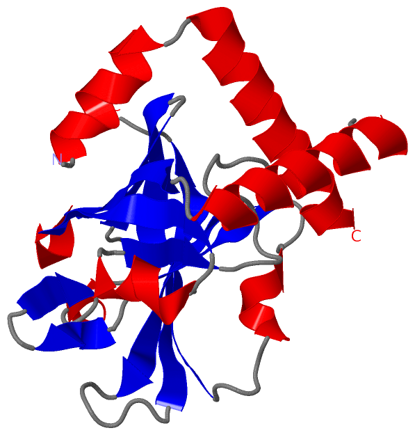
Asymmetric/Biological UnitChain A from PDB Type:PROTEIN Length:188 aligned with Q8E0S7_STRA5 | Q8E0S7 from UniProtKB/TrEMBL Length:305 Alignment length:200 55 65 75 85 95 105 115 125 135 145 155 165 175 185 195 205 215 225 235 245 Q8E0S7_STRA5 46 NINAFKEAVTKIDRVEINRRLELAYAYNASIAGAKTNGEYPALKDPYSAEQKQAGVVEYARMLEVKEQIGHVIIPRINQDIPIYAGSAEENLQRGVGHLEGTSLPVGGESTHAVLTAHRGLPTAKLFTNLDKVTVGDRFYIEHIGGKIAYQVDQIKVIAPDQLEDLYVIQGEDHVTLLTCTPYMINSHRLLVRGKRIPYV 245 SCOP domains -------------------------------------------------------------------------------------------------------------------------------------------------------------------------------------------------------- SCOP domains CATH domains -------------------------------------------------------------------------------------------------------------------------------------------------------------------------------------------------------- CATH domains Pfam domains -------------------------------------------------------------------------------------------------------------------------------------------------------------------------------------------------------- Pfam domains SAPs(SNPs) -------------------------------------------------------------------------------------------------------------------------------------------------------------------------------------------------------- SAPs(SNPs) PROSITE -------------------------------------------------------------------------------------------------------------------------------------------------------------------------------------------------------- PROSITE Transcript -------------------------------------------------------------------------------------------------------------------------------------------------------------------------------------------------------- Transcript 3tb7 A 5 NINAFKEAVTKIDRVEINRRLELAYAYNASIAG------------PYSAEQKQAGVVEYARMLEVKEQIGHVIIPRINQDIPIYAGSAEENLQRGVGHLEGTSLPVGGESTHAVLTAHRGLPTAKLFTNLDKVTVGDRFYIEHIGGKIAYQVDQIKVIAPDQLEDLYVIQGEDHVTLLTCTPYMINSHRLLVRGKRIPYV 204 14 24 34 | - | 54 64 74 84 94 104 114 124 134 144 154 164 174 184 194 204 37 50
|
||||||||||||||||||||
SCOP Domains (0, 0)| (no "SCOP Domain" information available for 3TB7) |
CATH Domains (0, 0)| (no "CATH Domain" information available for 3TB7) |
Pfam Domains (0, 0)| (no "Pfam Domain" information available for 3TB7) |
Gene Ontology (2, 2)|
Asymmetric/Biological Unit(hide GO term definitions) Chain A (Q8E0S7_STRA5 | Q8E0S7)
|
||||||||||||||||||
Interactive Views
|
|||||||||||||||||||||||||||||||||||||||||||||||||||||||||||||||||||||||||||||||||||||||||||||||||||||||||||||||||||||
Still Images
|
||||||||||||||||
Databases
|
||||||||||||||||||||||||||||||||||||||||||||||||||||||||||||||||||||||||||||||||||||||||||||||||||||||||||||||||||||||||||||||||||||||||||||||||||||||||||||||||
Analysis Tools
|
|||||||||||||||||||||||||||||||||||||||||||||||||||||||||||||
Entries Sharing at Least One Protein Chain (UniProt ID)
Related Entries Specified in the PDB File
|
|